Skip to main page content
U.S. flag

An official website of the United States government

Dot gov

The . gov means it’s official. Federal government websites often end in . gov or . mil. Before sharing sensitive information, make sure you’re on a federal government site. VSports app下载.

Https

The site is secure V体育官网. The https:// ensures that you are connecting to the official website and that any information you provide is encrypted and transmitted securely. .

. 2018 Jul 24;9(8):812.
doi: 10.1038/s41419-018-0855-8.

"V体育安卓版" Oxidative stress and autophagy-related changes during retinal degeneration and development

Affiliations

V体育ios版 - Oxidative stress and autophagy-related changes during retinal degeneration and development

Laura Trachsel-Moncho et al. Cell Death Dis. .

Abstract (VSports)

Retinitis pigmentosa (RP) is an inherited retinopathy that leads to photoreceptor loss. RP has been related to oxidative stress, autophagy, and inflammation. This study aimed to identify changes in the levels of oxidative stress and autophagy markers in the retina of control and rd10 mice during different phases of retinal development. Changes in the retinal oxidation system were investigated by measuring the levels of oxidized and reduced glutathione (GSH/GSSG), retinal avidin-positive cells, and 4-hydroxynonenal (4-HNE) staining intensity. Autophagy characterization was explored by measuring the levels of microtubule-associated protein 1 light chain 3 (LC3), beclin, autophagy-related proteins 5 and 7 (Atg5 and Atg7), and lysosomal associated membrane protein-2A (LAMP-2A). At P28 retinal GSH concentrations decreased in rd10 mice compared to the controls. No differences were found in retinal GSSG concentrations between the control and rd10 mice. There was an increase in retinal GSSG concentrations and a decrease in the GSH/GSSG ratio in the control and rd10 mice at P21 and P28 compared to P13. We observed an increase in avidin-positive cells in rd10 retinas. 4-HNE was increased in rd10 retinas at P13, and it also increased in control mice with age. We did not observe any differences in the retinal levels of LC3II/I ratio, Beclin, Atg5, or Atg7 in the rd10 mice compared to the controls. There was an increase in the LAMP-2A concentrations in the control and rd10 mice with development age (P28 concentrations vs VSports手机版. P13). Although only slight differences were found in the oxidative stress and autophagy markers between the control and rd10 mice, there were increases in the GSSG, 4-HNE, and LAMP-2A with age. This increase in the oxidative stress and chaperone-mediated autophagy has not been described before and occurred just after the mice opened their eyes, potentially indicating a retinal response to light exposure. .


"V体育官网入口" Conflict of interest statement

The authors declare that they have no conflict of interest.

Figures
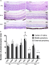
Fig. 1
Fig. 1. The number of photoreceptor rows in different areas of the retina.
a An illustration of the degradation of the retina at P5, P13, and P21 for the C57 (control) on the left, and the rd10 mice on the right. b The mean number of photoreceptor rows in the control and rd10 mice; error bars indicate the standard deviation (*p < 0.03 vs every other group; #p < 0.05 vs P13 rd10; &p < 0.05 vs P5 C57 and P13 C57)
Fig. 2
Fig. 2. Retinal cell death.
a Representative images of TUNEL staining in green, accompanied by DAPI staining in blue. The GCL, INL and ONL layers are indicated. The picture in the leftmost group is the control mice, while the rightmost is the rd10 mice (*p < 0.03 vs every group; #p < 0.05 vs P13 C57 and P21 C57; &p < 0.05 vs P5 C57; $p < 0.05 vs P21 rd10 and P28 rd10). b The mean number of TUNEL positive cells in the control and rd10 mice retina, error bars indicate the standard deviation. (#p < 0.03 vs every other group; #p < 0.05 vs P13 rd10; &p < 0.05 vs P5 C57 y P13 C57).
Fig. 3
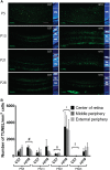
Fig. 3. Oxidative stress markers in control and rd10 retinas.
a Mean retinal levels of GSH at different ages. b Mean retinal levels of GSSG at different ages. c Mean retinal levels of glutamate at different ages. d The retinal GSH:GSSG ratio in the retina at different ages (*p < 0.01 vs P13). e Representative images of the avidin staining in red in different retinal sections. f Mean avidin-positive cell number in the control and rd10 different mice types, where error bars indicate the standard deviation (*p < 0.03 vs. each control). g Mean 4-HNE retinal intensity staining in the control and rd10 different mice types, where error bars indicate the standard deviation (*p < 0.05 vs. C57 P13). h Mean 4-HNE intensity staining in the photoreceptor outer segments in the control and rd10 different mice types, where error bars indicate the standard deviation. i Representative images of the 4-HNE staining in green in different retinal sections
Fig. 4
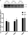
Fig. 4. Detection of the LC3 protein by western blot in the different age groups.
a Image of the western-blot bands for both the control and rd10 at different ages. b The graph represents the optical density quantification of the bands (ratio LC3 II-I) for each experimental group
Fig. 5
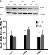
Fig. 5. Detection of the Beclin protein by western blot in the different age groups.
a Image of the western-blot bands for both the control and rd10 mice at different ages. b The graph represents the optical density quantification of the bands for each experimental group
Fig. 6
Fig. 6. Detection of the Atg5 and Atg7 proteins by western blot in the different age groups.
a Image of the western-blot bands for both the control and rd10 mice at different ages. b The graph represents the optical density quantification of the Atg5 bands for each experimental group. c The graph represents the optical density quantification of the Atg7 bands for each experimental group
Fig. 7
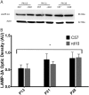
Fig. 7. Detection of the LAMP-2A protein by western blot in the different age groups.
a Image of the western-blot bands for both the control and rd10 mice at different ages. b The graph represents the optical density quantification of the bands for each experimental group (*p < 0.003 for differences between P13 and P28 in control and rd10 mice)

References

    1. Hartong DT, Berson EL, Dryja TP. Retinitis pigmentosa. Lancet. 2006;368:1795–1809. doi: 10.1016/S0140-6736(06)69740-7. - "VSports在线直播" DOI - PubMed
    1. Mitamura Y, et al. Diagnostic imaging in patients with retinitis pigmentosa. J. Med. Investig. 2012;59:1–11. doi: 10.2152/jmi.59.1. - V体育2025版 - DOI - PubMed
    1. Farrar GJ, Kenna PF, Humphries P. On the genetics of retinitis pigmentosa and on mutation-independent approaches to therapeutic intervention. EMBO J. 2002;21:857–864. doi: 10.1093/emboj/21.5.857. - DOI - PMC - PubMed
    1. Usui S, et al. Increased expression of catalase and superoxide dismutase 2 reduces cone cell death in retinitis pigmentosa. Mol. Ther. 2009;17:778–786. doi: 10.1038/mt.2009.47. - "V体育官网" DOI - PMC - PubMed
    1. Komeima K, Rogers BS, Campochiaro PA. Antioxidants slow photoreceptor cell death in mouse models of retinitis pigmentosa. J. Cell. Physiol. 2007;213:809–815. doi: 10.1002/jcp.21152. - DOI - PubMed

"V体育安卓版" Publication types

"V体育安卓版" MeSH terms

Substances

LinkOut - more resources (VSports注册入口)