High expression of CD44v9 and xCT in chemoresistant hepatocellular carcinoma: Potential targets by sulfasalazine
- PMID: 29981246
- PMCID: PMC6125439
- DOI: 10.1111/cas.13728
High expression of CD44v9 and xCT in chemoresistant hepatocellular carcinoma: Potential targets by sulfasalazine
Abstract
CD44v9 is expressed in cancer stem cells (CSC) and stabilizes the glutamate-cystine transporter xCT on the cytoplasmic membrane, thereby decreasing intracellular levels of reactive oxygen species (ROS). This mechanism confers ROS resistance to CSC and CD44v9-expressing cancer cells. The aims of the present study were to assess: (i) expression status of CD44v9 and xCT in hepatocellular carcinoma (HCC) tissues, including those derived from patients treated with hepatic arterial infusion chemoembolization (HAIC) therapy with cisplatin (CDDP); and (ii) whether combination of CDDP with sulfasalazine (SASP), an inhibitor of xCT, was more effective on tumor cells than CDDP alone by inducing ROS-mediated apoptosis. Twenty non-pretreated HCC tissues and 7 HCC tissues administered HAIC therapy with CDDP before surgical resection were subjected to immunohistochemistry analysis of CD44v9 and xCT expression. Human HCC cell lines HAK-1A and HAK-1B were used in this study; the latter was also used for xenograft experiments in nude mice to assess in vivo efficacy of combination treatment. CD44v9 positivity was significantly higher in HAIC-treated tissues (5/7) than in non-pretreated tissues (2/30), suggesting the involvement of CD44v9 in the resistance to HAIC. xCT was significantly expressed in poorly differentiated HCC tissues. Combination treatment effectively killed the CD44v9-harboring HAK-1B cells through ROS-mediated apoptosis and significantly decreased xenografted tumor growth. In conclusion, the xCT inhibitor SASP augmented ROS-mediated apoptosis in CDDP-treated HCC cells, in which the CD44v9-xCT system functioned. As CD44v9 is typically expressed in HAIC-resistant HCC cells, combination treatment with SASP with CDDP may overcome such drug resistance. VSports手机版.
Keywords: anticancer drug resistance; cancer stem cell; combinational therapy; oxidative stress; sphere formation V体育安卓版. .
© 2018 The Authors. Cancer Science published by John Wiley & Sons Australia, Ltd on behalf of Japanese Cancer Association V体育ios版. .
"V体育官网入口" Figures

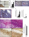
 P < 0.01; C, Limited number of
  P < 0.01; n.s., not significant. F, (left upper panel)
  P < 0.01

 P < 0.01). B, A synergistic inhibitory effect was seen even in sphere‐forming
  P < 0.01
 P < 0.001. B, Decrease in intracellular glutathione (
 P < 0.01. C, In western blotting, the apoptosis markers cleaved (Cl) poly (ADP‐ribose) polymerase (
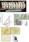
 P < 0.001). C, Consistent with the tumor growth inhibition, immunohistochemistry analysis of cleaved caspase‐3 showed significantly highest positivity in tumor tissues of mice treated with both
  means P < 0.001 or P < 0.01). D,
  P < 0.05;
  P < 0.001)
References
-
- White DL, Thrift AP, Kanwal F, Davila J, El‐Serag HB. Incidence of hepatocellular carcinoma in all 50 United States, from 2000 through 2012. Gastroenterology. 2017;152:812‐820. - PMC (VSports最新版本) - PubMed
-
- Forner A, Llovet JM, Bruix J. Hepatocellular carcinoma. Lancet. 2012;379:1245‐1255. - PubMed
-
- Nagamatsu H, Sumie S, Niizeki T, et al. Hepatic arterial infusion chemoembolization therapy for advanced hepatocellular carcinoma: multicenter phase II study. Cancer Chemother Pharmacol. 2016;77:243‐250. - PubMed
-
- Pang RW, Poon RT. Cancer stem cell as a potential therapeutic target in hepatocellular carcinoma. Curr Cancer Drug Targets. 2012;12:1081‐1094. - PubMed
MeSH terms (VSports在线直播)
- VSports最新版本 - Actions
- "VSports手机版" Actions
- V体育2025版 - Actions
- Actions (V体育2025版)
- V体育官网 - Actions
- Actions (V体育官网入口)
- Actions (V体育2025版)
- V体育平台登录 - Actions
- "VSports手机版" Actions
- "VSports app下载" Actions
- VSports手机版 - Actions
- "VSports" Actions
- Actions (VSports在线直播)
- V体育官网入口 - Actions
- "V体育2025版" Actions
VSports在线直播 - Substances
- "VSports最新版本" Actions
- Actions (VSports最新版本)
- "V体育平台登录" Actions
LinkOut - more resources
Full Text Sources
"VSports" Other Literature Sources
Medical
VSports在线直播 - Research Materials

