Skip to main page content
U.S. flag

An official website of the United States government

Dot gov

The . gov means it’s official. Federal government websites often end in VSports app下载. gov or . mil. Before sharing sensitive information, make sure you’re on a federal government site. .

Https

The site is secure. The https:// ensures that you are connecting to the official website and that any information you provide is encrypted and transmitted securely V体育官网. .

. 2018 Aug 1;128(8):3341-3355.
doi: 10.1172/JCI99032. Epub 2018 Jun 25.

"VSports注册入口" Nano-targeted induction of dual ferroptotic mechanisms eradicates high-risk neuroblastoma

Affiliations

Nano-targeted induction of dual ferroptotic mechanisms eradicates high-risk neuroblastoma

Behrouz Hassannia et al. J Clin Invest. .

VSports - Abstract

High-risk neuroblastoma is a devastating malignancy with very limited therapeutic options VSports手机版. Here, we identify withaferin A (WA) as a natural ferroptosis-inducing agent in neuroblastoma, which acts through a novel double-edged mechanism. WA dose-dependently either activates the nuclear factor-like 2 pathway through targeting of Kelch-like ECH-associated protein 1 (noncanonical ferroptosis induction) or inactivates glutathione peroxidase 4 (canonical ferroptosis induction). Noncanonical ferroptosis induction is characterized by an increase in intracellular labile Fe(II) upon excessive activation of heme oxygenase-1, which is sufficient to induce ferroptosis. This double-edged mechanism might explain the superior efficacy of WA as compared with etoposide or cisplatin in killing a heterogeneous panel of high-risk neuroblastoma cells, and in suppressing the growth and relapse rate of neuroblastoma xenografts. Nano-targeting of WA allows systemic application and suppressed tumor growth due to an enhanced accumulation at the tumor site. Collectively, our data propose a novel therapeutic strategy to efficiently kill cancer cells by ferroptosis. .

Keywords: Drug therapy; Nanotechnology; Neurological disorders; Neuroscience; Oncology V体育安卓版. .


Conflict of interest statement (V体育官网)

Conflict of interest: The authors have declared that no conflict of interest exists.

Figures

Figure 1
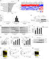
Figure 1. WA acts as a class II ferroptosis-inducing agent to kill etoposide- and cisplatin-resistant neuroblastoma cells.
(A) Heatmap representing cell death sensitivity of various high-risk neuroblastoma cell lines after exposure to WA, etoposide (Eto), and cisplatin (Cis). See also Supplemental Table 1. SG, Sytox Green. (B) Snapshots from a live cell imaging of IMR-32 and SK-N-SH cells treated with WA. See also Supplemental Videos 1 and 2. Green fluorescent staining represents SytoxGreen. (C) Heatmap representing the cell death sensitivity of IMR-32 and SK-N-SH cells after exposure to 1 μM or 10 μM WA, respectively, in the absence or presence of different ferroptosis inhibitors, including the iron chelator ciclopirox olamine (CPX, 5 M), the lipid peroxidation inhibitor ferrostatin-1 (Fer1, 500 nM), the lipophilic antioxidant α-tocopherol (αToco, 100 μM), and the kinase inhibitors U0126 (10 μM) and Flt3 inhibitor (500nM). See also Supplemental Table 3. (D) Flow cytometric analysis of the lipid peroxidation sensor (C11-BODIPY-581/591 dye) on live-gated cells (SytoxBlue-negative cells) after treatment of IMR-32 cells with WA. (E) Western blot analysis revealing GPX4 and β-tubulin expression in IMR-32 cells treated with a high dose (10 μM) and a medium dose (1 μM) of WA. (F) Western blot analysis revealing GPX4 and actin after immunoprecipitation of biotin-WA in lysates of IMR-32 cells treated with a high dose (10 μM) and a medium dose (1 μM) of biotin-WA. (G) Western blot analysis revealing GPX4 expression in GPX4-depleted mouse embryonic fibroblasts (MEFs) reconstituted with GPX4 or GPX4AllCys/Ser 5 hours after treatment with WA (10 μM). (H) Molecular modeling simulation of WA docked on Cys107 of human GPX4 crystal structure (2OBI). See also Supplemental Table 4 and Supplemental Video 3. **P < 0.01, ****P < 0.0001, 2-way ANOVA test (A).
Figure 2
Figure 2. WA increases intracellular labile Fe(II) upon excessive activation of HMOX1, which is sufficient to induce ferroptosis.
(A) IPA of genes differentially expressed after treatment with WA (1μM) in IMR-32 cells. (B) Gene expression profiles of Ctrl, WA-treated, and WN-treated IMR-32 cells. Color key legend represents Z score values. (C) Relative HMOX1 mRNA expression in IMR-32 cells after WA-treatment. (D) Western blot revealing KEAP1, NRF2, and HMOX1 in IMR-32 cells treated with WA. (E) Western blot revealing KEAP1 after immunoprecipitation of biotin-WA in lysates of IMR-32 cells treated with biotin-WA (1 μM). (F) Percentage of cell death induced by WA in the presence/absence of HMOX1 inhibitor (ZnPP). (G) Western blot revealing HMOX1 in response to WA (1 μM). (H) Percentage of cell death induced by WA upon RNAi-mediated knockdown of HMOX1. The results are representative of 2 independent experiments. (I) Percentage of cell death induced by WA in the presence/absence of hemin. (J) Western blot revealing HMOX1 in response to hemin (10 μM). (K) Heatmap representing the sensitivity of IMR-32 cells in response to hemin in the presence of ferroptosis inhibitors. See also Supplemental Table 5. (L) Cellular levels of labile Fe(II), using RhoNox-1, in function of time in response to WA (1 μM). (M) Cellular levels of labile Fe(II), using RhoNox-1, 8 hours after treatment with WA (1 μM), hemin (10 μM), or the combination. MFI, mean fluorescence intensity. (N) Heatmap representing cell death sensitivity of IMR-32 cells in response to (NH4)2Fe(SO4)2 in the absence/presence of ferroptosis inhibitors. See also Supplemental Table 6. The combined results of 2 or 3 independent experiments are shown for F, I, K, and N. Error bars represent SEM. *P < 0.05, ***P < 0.001, ****P < 0.0001, 2-way ANOVA test (F, I, L,and M). NDGA, nordihydroguaiaretic acid.
Figure 3
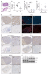
Figure 3. WA-eradicated high-risk neuroblastoma tumors show decreased GPX4 expression, increased HMOX1 expression, and decreased relapse rates.
(A and B) Light microscopic pictures of H&E staining of tumor sections (A) and quantification of tumor size area (B) representing effect of WA and vehicle (Ctrl) on SH-EP–derived tumors in a chicken chorioallantoic membrane (CAM) model. The combined results of 2 independent experiments are shown. Error bars represent SEM; n = 10 (Ctrl), n = 10 (WA). (C) Quantification of tumor growth rates after therapeutic treatment regime (100–200 mm3 at the start of treatment) with vehicle (Ctrl), WA, or etoposide (Eto). The results of 1 experiment for etoposide and combined results of 2 independent experiments for WA are shown. Each point indicates an individual mouse. Error bars represent SEM; n = 11 (Ctrl), n = 13 (WA), n = 6 (Eto). (D) Quantification of tumor growth rates in mice after termination of treatment with WA and Eto (tumor size cutoff below 180 mm3 at time of termination of the treatment). (EI) Immunohistochemical staining revealing proliferation using Ki67 antibody (E), DNA fragmentation using TUNEL staining (F), caspase-3 activation using anti–active caspase-3 antibodies (G), GPX4 (H), and HMOX1 expression level (I) in tumor sections after therapeutic treatment regime with vehicle (Ctrl) or WA. (J) Western blot revealing GPX4 and HMOX1 in lysates of tumor sections after therapeutic treatment regime with vehicle (Ctrl) or WA. ****P < 0.0001, 2-tailed t test (B), 1-way ANOVA (C).
Figure 4
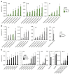
Figure 4. Oxidative lipidomics profiles show massive lipid peroxidation in neuroblastoma tumors treated with WA.
(A and B) Levels of oxygenated phospholipid species, with significantly higher levels (33 species) detected in neuroblastoma tumors from mice treated with WA compared with vehicle-treated mice (Ctrl). Error bars represent SEM; n = 3. (C) Levels of lysophospholipids, with significant increase (22 species) in neuroblastoma tumors treated with WA. Error bars represent SEM; n = 3. PL, phospholipids; PG, phosphatidylglycerol; PI, phosphatidylinositol; PC, phosphatidylcholine; PE, phosphatidylethanolamine; PS, phosphatidylserine; L, lyso; MLCL, monolyso-cardiolipin. *P < 0.05, 2-tailed t test (AC).
Figure 5
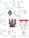
Figure 5. Nano-targeting of WA avoids systemic side effects and suppresses tumor growth.
(A) Schematic representation of the amphiphilic degradable pH-sensitive nanoparticle encapsulating WA (WA-NP). (B) Histogram indicating the size of empty (Empty-NP) and WA-encapsulated nanoparticles (WA-NP). d;nm means diameter of nanoparticles in nanometer. (C) Percentage of cell death in function of time triggered by WA-NPs, Empty-NPs, and WA in IMR-32 cells. (D) Percentage of body weight change in mice 4 days after daily i.p. injection of WA and vehicle. (E) In vivo fluorescence images of neuroblastoma tumor xenografts in nude mice injected i.p. with Cy5 and Cy5-loaded nanoparticles (Cy5-NPs). (F) Percentage of body weight change in mice 9 days after daily i.p. injection of empty (Empty-NP) or WA-encapsulated nanoparticles (WA-NP). (G) Quantification of tumor growth rates after therapeutic treatment regime (60–70 mm3 at the start of injection) with empty (Empty-NP) or WA-encapsulated nanoparticles (WA-NP). Each point indicates an individual mouse. Error bars represents SEM; n = 8 per group. (H) PL•, phospholipid radical; PLOO•, phospholipid peroxyradical; LOOH, phospholipid hydroperoxide; PLOH, phospholipid alcohol. Schematic representation of a double-edged ferroptosis induction mechanism upon WA exposure. WA induces uncontrolled lipid peroxidation and ferroptosis through inactivation of GPX4 (canonical ferroptosis induction) and excessive activation of HMOX1 followed by an increase in the labile Fe(II) pool (noncanonical ferroptosis induction). Inhibiting the lipid detoxification process, by inactivating GPX4, as well as fueling the reactive free radical chain process by increasing the labile Fe(II) pool effectively boosts ferroptosis. *P < 0.05; **P < 0.001, 2-tailed t test (D and G).

References

    1. Maris JM. Recent advances in neuroblastoma. N Engl J Med. 2010;362(23):2202–2211. doi: 10.1056/NEJMra0804577. - DOI (VSports) - PMC - PubMed
    1. Cheung NK, Dyer MA. Neuroblastoma: developmental biology, cancer genomics and immunotherapy. Nat Rev Cancer. 2013;13(6):397–411. doi: 10.1038/nrc3526. - DOI - PMC - PubMed
    1. Holohan C, Van Schaeybroeck S, Longley DB, Johnston PG. Cancer drug resistance: an evolving paradigm. Nat Rev Cancer. 2013;13(10):714–726. doi: 10.1038/nrc3599. - DOI - PubMed
    1. Aaes TL, et al. Vaccination with necroptotic cancer cells induces efficient anti-tumor immunity. Cell Rep. 2016;15(2):274–287. doi: 10.1016/j.celrep.2016.03.037. - DOI (VSports手机版) - PubMed
    1. Yatim N, et al. RIPK1 and NF-κB signaling in dying cells determines cross-priming of CD8+ T cells. Science. 2015;350(6258):328–334. doi: 10.1126/science.aad0395. - DOI - PMC - PubMed

Publication types