<acronym id="QQtRv08"></acronym>
VSports在线直播 - Skip to main page content
U.S. flag

An official website of the United States government

Dot gov

The . gov means it’s official. Federal government websites often end in VSports app下载. gov or . mil. Before sharing sensitive information, make sure you’re on a federal government site. .

Https

The site is secure. The https:// ensures that you are connecting to the official website and that any information you provide is encrypted and transmitted securely. V体育官网.

. 2018 Jun 19;48(6):1258-1270.e6.
doi: 10.1016/j.immuni.2018.04.015. Epub 2018 Jun 5.

A Single-Cell Transcriptomic Atlas of Thymus Organogenesis Resolves Cell Types and Developmental Maturation (VSports在线直播)

Affiliations

A Single-Cell Transcriptomic Atlas of Thymus Organogenesis Resolves Cell Types and Developmental Maturation

Eric M Kernfeld et al. Immunity. .

VSports app下载 - Abstract

Thymus development is critical to the adaptive immune system, yet a comprehensive transcriptional framework capturing thymus organogenesis at single-cell resolution is still needed. We applied single-cell RNA sequencing (RNA-seq) to capture 8 days of thymus development, perturbations of T cell receptor rearrangement, and in vitro organ cultures, producing profiles of 24,279 cells. We resolved transcriptional heterogeneity of developing lymphocytes, and genetic perturbation confirmed T cell identity of conventional and non-conventional lymphocytes. We characterized maturation dynamics of thymic epithelial cells in vivo, classified cell maturation state in a thymic organ culture, and revealed the intrinsic capacity of thymic epithelium to preserve transcriptional regularity despite exposure to exogenous retinoic acid. Finally, by integrating the cell atlas with human genome-wide association study (GWAS) data and autoimmune-disease-related genes, we implicated embryonic thymus-resident cells as possible participants in autoimmune disease etiologies. This resource provides a single-cell transcriptional framework for biological discovery and molecular analysis of thymus organogenesis. VSports手机版.

Keywords: Drop-seq; cell atlas; development; lymphocytes; lymphoid organ; single-cell RNA-seq; thymic epithelium; thymus; thymus organogenesis; transcriptomics. V体育安卓版.


Conflict of interest statement

Declaration of Interests

The authors declare no competing interests.

Figures
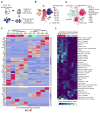
Figure 1
Figure 1
Transcriptomic diversity in the thymus is driven by cell identity and maturation. (A) Schematic of procedures for extraction, sequencing, and single-cell analysis of the thymus. (B, C) Two-dimensional representation of cells via tSNE, colored by developmental day (B) and cluster identity (C); each dot represents one cell. tSNE was performed after quality control and cell cycle correction (methods). (D) Row-standardized heatmap of hand-curated and data-driven marker genes for each cluster. Expression is measured in units of log2 (1 + transcripts per 10,000). Each row has mean zero and standard deviation one. Clusters are ordered by Seurat dendrogram fitted to cluster means. (E) DEPICT-based intersection of cluster-specific gene expression and GWAS studies. Data include n=3 biological replicates at each of E12.5, E13.5 and E16.5 and n=2 biological replicates at each of E14.5, E15.5, E17.5, and E18.5. Data from P0 consist of two technical replicates (two lobes from a single thymus, processed separately following dissection). The number of independent experiments performed was: 1 for each of days E15.5, E17.5, E18.5, and P0; 2 at each of days E13.5, E14.5, and E16.5; and 3 at E12.5. See also Figures S1 and S2.
Figure 2
Figure 2
Single-cell data reveal molecular details and subsets of thymocytes. (A) Schematic showing that this figure includes the cells from BLD1-3. (B,C) tSNE colored by developmental day (B) or cluster identity (C); each dot represents one cell. (D) Row-standardized heatmap of genes curated from the literature. Entries display average lognormalized expression. Clusters are ordered manually based on developmental status. (E-G) TACS display of thymocytes showing maturation and Notch signaling preceding linage commitment. Panel (E) shows Il2ra versus Cd44 by cluster. Panel (F) shows Il2ra versus Cd44 by embryonic day with Tconv5 excluded. Panel (G) shows Bcl11b versus Dtx1 by cluster. The number of independent experiments performed was 1 for each day E15.5, E17.5, E18.5, and P0; 2 at each day E13.5, E14.5, and E16.5; and 3 at E12.5. See also Figure S3.
Figure 3
Figure 3
Single-cell data reveal molecular details and subsets of non-conventional lymphocytes. (A) Schematic showing that this figure includes the cells from BLD5. (B,C) tSNE colored by developmental day (B) or cluster identity (C); each dot represents one cell. (D) Row-standardized heatmaps of genes showing the strongest expression differences across clusters (left panel) and genes curated from the literature (right panel). Entries display average log normalized expression. Seurat dendrogram is fitted to cluster means. ). The number of independent experiments performed was: 1 for each of days E15.5, E17.5, E18.5, and P0; 2 at each of days E13.5, E14.5, and E16.5; and 3 at E12.5. See also Fig. S4.
Figure 4
Figure 4
Rag1−/− mouse thymi demonstrate that clusters labeled as thymocytes depend on antigen receptor rearrangement. (A,B) tSNE colored by cell type (A) and replicate (B); each dot represents one cell. E16.5 Rag1−/− cells were assigned to clusters via supervised machine learning (methods). WT data are the same three E16.5 replicates used for the WT cell atlas. Rag1−/− data include two biological replicates. (C) TACS plot depicting composition of WT and Rag1−/− E16.5 thymocytes. Contours outline regions of successively higher cell density. (D) Volcano plot for differentially expressed transcripts within Cd44loIl2ralo between Rag1−/− and WT thymocytes at E16.5. (E) Composition of E16.5 NCL compartment by genotype. (F) tSNE of NCL cells from E16.5 WT and Rag1−/− colored by atlas-based classification. P-values in (C) and (E) were computed on cell-count tables by Fisher’s exact test, with the 4014 cells from panel (C) grouped by TACS quadrant. Rag1−/− replicates were processed together in the same experiment.
Figure 5
Figure 5
Heterogeneity within thymic epithelium. (A) Schematic showing that this figure includes the cells from TEC1-3. (B–D) tSNE colored by developmental day (B), cluster identity (C), and Cd74 expression (D); each dot represents one cell. (E) Dendrogram relating TEC subsets, and heatmap of genes curated from the literature. Log-normalized expression was averaged within clusters and each row was centered and scaled to have zero mean and unit variance. Seurat dendrogram was computed using cluster means. (F,I) Cells included for pseudotime inference: mTEC (F); cTEC (I). (G,J) Embryonic day displayed by diffusion components and pseudotime: mTEC (G); cTEC (J). (H,K) Selected genes smoothed on pseudotime: mTEC (H); cTEC (K). Trend shows a mean estimate, and shaded region is a point-wise 95% CI (does not account for uncertainty in pseudotime estimation). The number of independent experiments performed was: 1 for each of days E15.5, E17.5, E18.5, and P0; 2 at each of days E13.5, E14.5, and E16.5; and 3 at E12.5. See also Fig. S5.
Figure 6
Figure 6
Fetal thymic organ cultures allow for maturation of thymic cell populations. (A) Schematic of organ culture model assessment. (B) Cell atlas-based classification of FTOC cell populations. (D) Display of thymocyte maturation (Il2ra versus Cd44) in cell atlas compared to FTOC endpoint via TACS (upper panel) and unbiased fractional identity estimation (lower panel). (E) Display of TEC maturation (Epcam versus MHCII) in cell atlas compared to FTOC endpoint via TACS (upper panel) and unbiased measurement of epithelial cell maturation using a fractional identity estimation method (lower panel). Tests compare WT E13.5 samples to the FTOC, corresponding to a null hypothesis of no maturation after dissection. P-values were computed by Fisher’s exact test with cells and grouped by TACS quadrant. Tests involve 3664 T cells and 2623 TECs. FTOC samples represent n=3 biological replicates and 3 independent experiments. See also Fig. S6 and S7.
Figure 7
Figure 7
TEC maturation and heterogeneity are protected from exposure to retinoic acid. (A) Schematic of organ culture perturbation experiment. (B) Schematic of methodology for applying atlas cluster labels to FTOC TECs. (C, D) tSNE of control and RA-perturbed TECs from FTOC colored by cluster identity (C) and treatment (D). (E) Volcano plot showing genes differentially expressed between control and RA treatment. (F) tSNE showing candidate transcripts Cyp26b1, Dhrs3, and Akap12. RA FTOC samples represent n=3 biological replicates and 3 independent experiments. See also Fig. S7.

VSports最新版本 - References

    1. Abramson J, Anderson G. Thymic Epithelial Cells. Annu Rev Immunol. 2017;35:85–118. - PubMed
    1. Akiyama N, Takizawa N, Miyauchi M, Yanai H, Tateishi R, Shinzawa M, Yoshinaga R, Kurihara M, Demizu Y, Yasuda H, et al. Identification of embryonic precursor cells that differentiate into thymic epithelial cells expressing autoimmune regulator. J Exp Med. 2016;213:1441–1458. - PMC - PubMed
    1. Alves NL, Ribeiro AR. Thymus medulla under construction: Time and space oddities. Eur J Immunol. 2016;46:829–833. - PubMed
    1. Anderson P, Caligiuri M, Ritz J, Schlossman SF. CD3-negative natural killer cells express zeta TCR as part of a novel molecular complex. Nature. 1989;341:159–162. - PubMed
    1. Baik S, Jenkinson EJ, Lane PJL, Anderson G, Jenkinson WE. Generation of both cortical and Aire(+) medullary thymic epithelial compartments from CD205(+) progenitors. Eur J Immunol. 2013;43:589–594. - PMC - PubMed

Publication types

MeSH terms