Skip to main page content
U.S. flag

An official website of the United States government

Dot gov

The . gov means it’s official. Federal government websites often end in . gov or . mil VSports app下载. Before sharing sensitive information, make sure you’re on a federal government site. .

Https

The site is secure. The https:// ensures that you are connecting to the official website and that any information you provide is encrypted and transmitted securely. V体育官网.

. 2018 Jun;15(6):8536-8544.
doi: 10.3892/ol.2018.8368. Epub 2018 Mar 28.

Suppression of tumor cell proliferation and migration by human umbilical cord mesenchymal stem cells: A possible role for apoptosis and Wnt signaling

Affiliations

Suppression of tumor cell proliferation and migration by human umbilical cord mesenchymal stem cells: A possible role for apoptosis and Wnt signaling

Yin Yuan et al. Oncol Lett. 2018 Jun.

Abstract

Human umbilical cord-derived mesenchymal stem cells (hUCMSCs) represent potential therapeutic tools for solid tumors. However, there are numerous inconsistent results regarding the effects of hUCMSCs on tumors, and the mechanisms underlying this remain poorly understood. The present study further examined this controversial issue by analyzing the molecular mechanisms of the inhibitory effects of hUCMSCs on the proliferation and migration of the human lung cancer A549 cell line and the human hepatocellular carcinoma (HCC) BEL7402 cell line in vitro. Flow cytometric analysis demonstrated that hUCMSCs arrested tumor cells in specific phases of the cell cycle and induced the apoptosis of tumor cells by using the hUCMSC-conditioned medium (hUCMSC-CM). The hUCMSC-CM also attenuated the migratory abilities of the two tumor cell types. Furthermore, the expression of B-cell lymphoma 2 (Bcl-2), the pro-form of caspase-7 (pro-caspase-7), β-catenin and c-Myc was downregulated, while that of ephrin receptor (EphA5), a biomarker of cancer cell dormancy, was slightly increased in these two tumor cell lines treated with hUCMSC-CM. Specifically, when co-cultured via direct cell-to-cell contact, hUCMSCs were able to spontaneously fuse with any of the two types of solid tumor cells VSports手机版. These observations suggested that hUCMSCs may be a promising candidate for the biological therapy of lung cancer and HCC. Future studies should focus on detailed evidence for cell fusion, as well as other mechanisms proposed in the present study, by introducing additional experimental approaches and models. .

Keywords: Wnt signaling; apoptosis; cell fusion; mesenchymal stem cells; tumor. V体育安卓版.


Figures
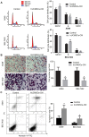
Figure 1.
Figure 1.
Effects of hUCMSC-CM on the proliferation, migration and survival of A549 and BEL7402 tumor cells. Serum-free culture medium was used instead of hUCMSC-CM in control groups. (A) Effect of hUCMSC-CM on the cell cycle of A549 and BEL7402 tumor cells. The percentage of cells in the G1, S and G2 phases is presented in the left panel and statistical analysis is presented in the right panel. **P<0.01 vs. control. (B) The migratory ability of tumor cells in the hUCMSC-CM group was analyzed by a Transwell assay. Representative images of migratory cells on the membrane (original magnification, ×400; left). Average number of migrated cells per field from three independent experiments (right). *P<0.05 vs. control. (C) Flow cytometry was performed to examine cell apoptosis in each group. Representative flow cytometric analysis (left). Quantification of flow cytometric analysis (right). *P<0.05 vs. control.
Figure 2.
Figure 2.
Molecular changes in A549 and BEL7402 tumor cells treated with hUCMSC-CM. The expression levels of EphA5, Bcl-2, pro caspase-7, β-catenin and c-Myc were detected by western blot analysis. Treatment with hUCMSC-CM decreased the protein expression of Bcl-2, pro caspase-7, β-catenin and c-Myc in A549 and BEL7402 tumor cells. Serum-free culture medium was used as control for hUCMSC-CM. (A) Representative western blot analysis images. (B) Quantitative data were acquired via densitometric analysis. Data are presented as the mean ± standard deviation of three independent experiments. *P<0.05 vs. control. EphA5, ephrin receptor A5; Bcl-2, B-cell lymphoma 2.
Figure 3.
Figure 3.
Confocal laser-scanning images of cell fusion between hUCMSCs and tumor cells. Tumor cells and hUCMSCs were stained with DiO (green) and DiD (red), respectively. All samples were counterstained with Hoechst 33342 (blue) to indicate the nucleus. After co-culture for 72 h, the hUCMSCs were revealed to have merged into A549 or BEL7402 tumor cells, as illustrated by the existence of hybrids with double nuclei and yellow cell membranes (DiO and DiD double-stained cells indicated by the white arrow) in the representative images. Scale bar, 20 µm; magnification, ×400.
Figure 4.
Figure 4.
Flow cytometric analysis of spontaneous cell fusion between DiO-labeled tumor cells and DiD-labeled hUCMSCs.

References

    1. Wang W, Zhang L, Liu L, Zheng Y, Zhang Y, Yang S, Shi R, Wang S. Chemosensitizing effect of shRNA-mediated ERCC1 silencing on a Xuanwei lung adenocarcinoma cell line and its clinical significance. Oncol Rep. 2017;37:1989–1997. doi: 10.3892/or.2017.5443. - DOI - PMC - PubMed
    1. Rojas A, Zhang P, Wang Y, Foo WC, Muñoz NM, Xiao L, Wang J, Gores GJ, Hung MC, Blechacz B. A positive TGF-β/c-KIT feedback loop drives tumor progression in advanced primary liver cancer. Neoplasia. 2016;18:371–386. doi: 10.1016/j.neo.2016.04.002. - "VSports app下载" DOI - PMC - PubMed
    1. Baskar R, Yap SP, Chua KL, Itahana K. The diverse and complex roles of radiation on cancer treatment: Therapeutic target and genome maintenance. Am J Cancer Res. 2012;2:372–382. - PMC (VSports最新版本) - PubMed
    1. Lee DH, Ahn Y, Kim SU, Wang KC, Cho BK, Phi JH, Park IH, Black PM, Carroll RS, Lee J, Kim SK. Targeting rat brainstem glioma using human neural stem cells and human mesenchymal stem cells. Clin Cancer Res. 2009;15:4925–4934. doi: 10.1158/1078-0432.CCR-08-3076. - DOI - PubMed
    1. Ciavarella S, Dominici M, Dammacco F, Silvestris F. Mesenchymal stem cells: A new promise in anticancer therapy. Stem Cells Dev. 2011;20:1–10. doi: 10.1089/scd.2010.0223. - DOI - PubMed

"V体育安卓版" LinkOut - more resources