VSports最新版本 - Skip to main page content
U.S. flag

An official website of the United States government

Dot gov

The . gov means it’s official. Federal government websites often end in . gov or . mil. Before sharing sensitive information, make sure you’re on a federal government site VSports app下载. .

Https

The site is secure. The https:// ensures that you are connecting to the official website and that any information you provide is encrypted and transmitted securely. V体育官网.

. 2018 Jul 4;52(1):1800236.
doi: 10.1183/13993003.00236-2018. Print 2018 Jul.

Fibroblast growth factor 23 and Klotho contribute to airway inflammation

Affiliations

Fibroblast growth factor 23 and Klotho contribute to airway inflammation

Stefanie Krick et al. Eur Respir J. .

Abstract

Circulating levels of fibroblast growth factor (FGF)23 are associated with systemic inflammation and increased mortality in chronic kidney disease. α-Klotho, a co-receptor for FGF23, is downregulated in chronic obstructive pulmonary disease (COPD). However, whether FGF23 and Klotho-mediated FGF receptor (FGFR) activation delineates a pathophysiological mechanism in COPD remains unclear. We hypothesised that FGF23 can potentiate airway inflammation via Klotho-independent FGFR4 activation. FGF23 and its effect were studied using plasma and transbronchial biopsies from COPD and control patients, and primary human bronchial epithelial cells isolated from COPD patients as well as a murine COPD model. Plasma FGF23 levels were significantly elevated in COPD patients. Exposure of airway epithelial cells to cigarette smoke and FGF23 led to a significant increase in interleukin-1β release via Klotho-independent FGFR4-mediated activation of phospholipase Cγ/nuclear factor of activated T-cells signalling. In addition, Klotho knockout mice developed COPD and showed airway inflammation and elevated FGFR4 expression in their lungs, whereas overexpression of Klotho led to an attenuation of airway inflammation. Cigarette smoke induces airway inflammation by downregulation of Klotho and activation of FGFR4 in the airway epithelium in COPD. Inhibition of FGF23 or FGFR4 might serve as a novel anti-inflammatory strategy in COPD. VSports手机版.


Conflict of interest statement

Conflict of interest: None declared.

Figures

Figure 1.
Figure 1.
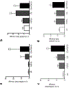
FGF23 levels are increased in patients with mild-to-moderate COPD who have goblet cell hyperplasia and increased levels of pro-inflammatory cytokines. (a) FGF23 plasma levels in non-COPD and COPD patients divided in mild-to-moderate and severe COPD and comparison of their FGF23 plasma levels. (b) Hematoxylin (upper row) and anti-FGF23 (lower row) staining of representative endobronchial biopsies from 1 control subject (left row) and 2 (middle and right row) subjects with mild-to-moderate COPD (20X magnification) with scale bar = 40 μm. (c) Quantification of goblet cells per field of view shows a significant increase in mild-to-moderate COPD when compared to subjects without COPD (n=6 of each group). (d) IL-6 plasma levels (left panel) and IL-8 levels in BALF (right panel) and dot blots showing their correlation with FGF23 levels in control subjects and patients with mild COPD (n = 6 of each group). Statistical analysis was done using Student’s t test showing mean ± S.E.M. or ANOVA with *P<0.05 and **P<0.01 and for correlation analysis the Pearson correlation coefficient.
Figure 2.
Figure 2.
Effect of cigarette smoke and FGF23 on expression of pro-inflammatory cytokines in COPD-BEC. (a) mRNA levels of IL-6 and (b) IL-8 in COPD-BEC 24h after stimulation with cigarette smoke (4 cigarettes) ± FGF23 (25 ng/ml). (c) IL-1β mRNA and (d) secreted protein levels in the basolateral media from COPD-BEC after stimulation with FGF23, CS or both. Data are represented as fold change in mRNA or protein expression with n = 3 independent experiments from 6 different lungs. All bar graphs are mean ± S.E.M. *P<0.05, **P<0.01 and ***P<0.005, compared to control (ctrl) group.
Figure 3.
Figure 3.
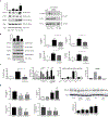
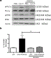
FGF23 induces activation of FGFR4/PLCγ/NFAT signaling in COPD-BEC. (a) Left panel: Representative immunoblot and densitometric analysis (p-PLCγ/total PLCγ/actin ratio with n = 3) shows FGF23 + CS-induced phosphorylation of PLCγ (FGF23- and smoke dependent) and of ERK (purely smoke dependent). Right panel: Immunoblot of co-immunoprecipitation with FGFR4 using an anti-PLCγ antibody shows activation of PLCγ after FGF23 stimulation in COPD-BEC, which could be inhibited by pre-incubation with anti-FGFR4. (b) CS + FGF23 induce PLCγ and ERK phosphorylation as seen above but anti-FGFR4 only inhibits PLCγ phosphorylation. Both anti-FGFR4 and cyclosporine A (CsA), a calcineurin inhibitor, inhibit CS + FGF23-mediated induction of IL-1β mRNA and secreted protein levels. (c) CsA also attenuates induction of COX2 mRNA. FGF23, CSE alone and both activate NFAT, as assessed by a luciferase-based reporter gene assay in 16HBE cells, which can be blocked by CsA. Anti-FGFR4 inhibits FGF23-induced NFAT activation. The right two bar diagrams indicate mRNA levels of NFAT isoforms in both HBEC from nonsmokers and COPD patients as well as 16HBE cells. (d) siRNA targeting NFAT isoforms NFAT2c and NFAT3c were transfected into 16HBE cells. siNFAT2c reduces the expression of NFAT2c and NFAT3c, whereas siNFAT3c is specific for NFAT3c (left panel). When 16HBE cells were stimulated with FGF23 (20 ng/ml), CSE (5%) or both for 24 hours, IL-1β increased (second to right panel). Finally, the effects of siNFAT2c (knockdown of both the 2c and 3c isoforms) and siNFAT3c transfection were assessed after stimulation with CSE+FGF23 (middle panel). CSE±FGF23 also led to significant upregulation of FGFR4 protein expression in 16HBE cells. All n = 3 independent experiments (from 3 different lungs in the case of COPD-BEC) showing mean ± S.E. with *P<0.05, **P<0.01 and ***P<0.005.
Figure 4.
Figure 4.
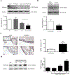
Cigarette smoke exposure regulates expression of klotho and FGFR4 in COPD airway epithelia. (a) Representative immunoblot showing downregulation of klotho protein and bar graphs indicating mRNA levels from COPD-BEC after treatment with CS, FGF23 or both stimuli for 24h. (b) Representative immunoblot and klotho mRNA levels of HBEC from nonsmokers compared to COPD-BEC. (n = 3 independent experiments from 6 different lungs. (c) Immunohistochemistry for FGFR4 and counterstaining with hematoxylin of endobronchial biopsies from 2 representative control patients and 2 COPD patients, compared to a negative control (upper left corner, 20X magnification). Bar graphs showing increased FGFR4 mRNA expression in COPD-BECs compared to control lungs. (d) Immunoblot analysis of FGFR4 protein levels (representative blot from 1 COPD patient, tested in 3 different COPD lungs) and quantitative real time PCR of FGFR4 mRNA levels in COPD-BECs 24h after exposure to 2, 4 and 6 cigarettes (1 cigarette = 6 puffs). (All n = 3 independent experiments from 3 different lungs showing mean ± S.E.M. with **P<0.01 and ***P<0.001).
Figure 5.
Figure 5.
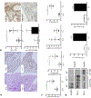
Loss of klotho expression causes increased airway inflammation in mice. (a) Hematoxylin and anti-FGF23 staining of lung tissue from wild type (kl+/+) and hypomorphic klotho (kl−/−) mice (4X magnification). (b) Mean linear intercept analysis demonstrates widened airspaces in the kl−/− lung samples and FGF23 mRNA levels are upregulated in whole lung tissue from kl−/− when compared to kl+/+ mice (6 mice for each group) (left panel). Immunohistochemical staining using anti-FGF23 in paraffin embedded mouse lung tissue sections indicates a signal in the bronchial epithelium (right panel). (c) Assessment of total cells in BALF from kl+/+ versus kl−/− mice (4 mice for each group). Also shown are BALF macrophage/monocyte count, BALF lymphocyte count × 104 per ml BAL fluid and IL-6 fold changes in BALF from kl+/+ versus kl−/− mice. (d) Co-immunoprecipitation of endogenous FGFR4 by using anti-PLC-γ or anti-GFP (control) in lung tissue lysates from either kl−/− or kl+/+ mice indicates increased fgfr4 in kl−/− lungs. qRT-PCR for FGFR4 mRNA in kl−/− MTEC showed a significant upregulation when compared to wild type MTEC and IL-6 protein levels in basolateral media from kl+/+ and kl−/− MTEC. (Shown are means ± S.E.M. with *P<0.05, **P<0.01 and ***P<0.001; for mouse experiments, three different independent experiments were done; for each experiment, cells were pooled from 3-4 animals and morphometry and BALF analysis was done from 4 mice in each group).
Figure 6.
Figure 6.
Supplementation of human recombinant klotho inhibits CS and FGF23-induced IL-ϊβ secretion. (a) Representative immunoblots show FGF23 (25 ng/ml for 30 min) induced phosphorylation of PLCγ. Inhibition of PLCγ phosphorylation occurs by pre-incubation with klotho (1 µg /ml, lane 3 and 0.1 µg /m, lane 4). (b) Soluble klotho inhibits CS+FGF23-mediated increases of IL-1β protein levels, assessed by ELISA from basolateral media. (All n = 3 independent experiments from 3 different lungs showing mean ± S.E. with **P<0.01).
Figure 7.
Figure 7.
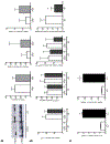
Effect of klotho overexpression and klotho deficiency on FGF23 signaling in mice. (a) Representative immunoblots show FGF23 protein levels in total lung lysates from control mice and mice acutely exposed to cigarette smoke (CS). Bar graphs demonstrating serum FGF23 levels, total lung klotho and FGFR4 mRNA levels in both mouse groups (b) Klotho mRNA levels, serum FGF23 levels, BAL fluid total cell count and CS-induced fold increase in total mouse lung IL-6 expression after exposure to acute CS and compared to non-exposed control mice (both wild type and klotho overexpressing mice). c) Bar graphs indicate IL-6 and FGFR4 transcript levels of MTECs isolated from kl+/+ and kl−/− mice (Means are shown ± SEM with **p<0.01 and ***p<0.005).
Figure 8.
Figure 8.
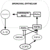
Possible signaling mechanism for cigarette smoke and FGF23-mediated regulation of FGFR4/PLCγ/calcineurin/NFAT pathway. Diagram illustrating the direct effects of FGF23 and cigarette smoke on the COPD airway epithelium: cigarette smoke induces upregulation of FGFR4 and decreases klotho expression in the bronchial epithelium. In combination with elevated FGF23 plasma levels in COPD activates FGFR4/PLCγ/calcineurin/NFAT signaling to release IL-1β thereby inducing inflammation. Definition of Abbreviations: IL=interleukin, FGF=fibroblast growth factor, FGFR= FGF receptor, NFAT= Nuclear factor of activated T-cells, PLC=phospholipase C.

References

    1. Samet JM (2013) Tobacco smoking: the leading cause of preventable disease worldwide. Thorac Surg Clin 23, 103–112 - PubMed
    1. de Boer WI, Sont JK, van Schadewijk A, Stolk J, van Krieken JH, and Hiemstra PS (2000) Monocyte chemoattractant protein 1, interleukin 8, and chronic airways inflammation in COPD. J Pathol 190, 619–626 - PubMed
    1. Polosukhin VV (2001) Ultrastructural of the bronchial epithelium in chronic inflammation. Ultrastruct Pathol 25, 119–128 - PubMed (V体育平台登录)
    1. Mortaz E, Henricks PA, Kraneveld AD, Givi ME, Garssen J, and Folkerts G (2011) Cigarette smoke induces the release of CXCL-8 from human bronchial epithelial cells via TLRs and induction of the inflammasome. Biochimica et biophysica acta 1812, 1104–1110 - PubMed
    1. Schiavi SC, and Kumar R (2004) The phosphatonin pathway: new insights in phosphate homeostasis. Kidney Int 65, 1–14 - PubMed

"V体育ios版" Publication types

"VSports最新版本" MeSH terms

LinkOut - more resources