Skip to main page content
U.S. flag

An official website of the United States government

Dot gov

The VSports app下载. gov means it’s official. Federal government websites often end in . gov or . mil. Before sharing sensitive information, make sure you’re on a federal government site. .

Https

The site is secure. The https:// ensures that you are connecting to the official website and that any information you provide is encrypted and transmitted securely V体育官网. .

. 2018 Apr 27;9(1):1685.
doi: 10.1038/s41467-018-03966-7.

"V体育平台登录" Snail promotes ovarian cancer progression by recruiting myeloid-derived suppressor cells via CXCR2 ligand upregulation

Affiliations

Snail promotes ovarian cancer progression by recruiting myeloid-derived suppressor cells via CXCR2 ligand upregulation

Mana Taki et al. Nat Commun. .

"VSports在线直播" Abstract

Snail is a major transcriptional factor that induces epithelial-mesenchymal transition (EMT) VSports手机版. In this study, we explore the effect of Snail on tumor immunity. Snail knockdown in mouse ovarian cancer cells suppresses tumor growth in immunocompetent mice, associated with an increase of CD8+ tumor-infiltrating lymphocytes and a decrease of myeloid-derived suppressor cells (MDSCs). Snail knockdown reduces the expression of CXCR2 ligands (CXCL1 and CXCL2), chemokines that attract MDSCs to the tumor via CXCR2. Snail upregulates CXCR ligands through NF-kB pathway, and most likely, through direct binding to the promoters. A CXCR2 antagonist suppresses MDSC infiltration and delays tumor growth in Snail-expressing mouse tumors. Ovarian cancer patients show elevated serum CXCL1/2, which correlates with Snail expression, MDSC infiltration, and short overall survival. Thus, Snail induces cancer progression via upregulation of CXCR2 ligands and recruitment of MDSCs. Blocking CXCR2 represents an immunological therapeutic approach to inhibit progression of Snail-high tumors undergoing EMT. .


Conflict of interest statement

E. N. was involved in this study as effort outside of the endowed associate professor of DSK project, Medical Innovation Center, Kyoto University sponsored by Sumitomo Dainippon Pharma Co Ltd V体育安卓版. The remaining authors declare no competing interests.

Figures

Fig. 1
Fig. 1
Snail expression is related to epithelial-to-mesenchymal transition and poor prognosis in ovarian cancer. a Correlation between generic epithelial-to-mesenchymal transition (EMT) scores for 266 high-grade serous ovarian cancer (HGSOC) samples from TCGA dataset and Snail expression. The EMT signature was acquired from a previous report by Tan et al.. Generic EMT scores were calculated following the method of single-sample Gene Set Enrichment Analysis (ssGSEA). The correlation coefficient (R) and P-value were based on Pearson’s product-moment correlation analysis; R = 0.41 P < 0.0001. b Snail expression in HGSOC samples representing four subtypes from the TCGA datasets; ***P < 0.001 and ****P < 0.0001, mesenchymal vs. the other three subtypes based on one-way ANOVA with Tukey’s multiple comparisons test. Bars: mean and SEM. c Classification of HGSOC as determined by the intensity of nuclear Snail immunostaining. Cases with scores of 0/1 were assigned to the Snail-low group, whereas those with scores of 2/3 were assigned to the Snail-high group to be used in survival analysis. Scale bars, 100 μm. d Overall survival curves for patients with HGSOC in Kyoto University Hospital (n = 56; 48 stage III cases and 8 stage IV cases), based on Snail staining of disseminated tumors in the omentum. P = 0.021 by log-rank test
Fig. 2
Fig. 2
Snail suppresses tumor progression and is associated with tumor immunity. a Western blot of HM-1-control and HM-1-shSnail cells. CDH1: E-Cadherin, VIM vimentin. b Invasion of HM-1-control and HM-1-shSnail cells; n = 5. c Wound healing of HM-1-control and HM-1-shSnail cells. Data represent the percentage of wound width (24 h) relative to that at 0 h; n = 6. d Growth curves of HM-1-control and HM-1-shSnail subcutaneous tumors in immunocompetent mice. P-values represent significance between two groups at day 31; n = 6. e Growth curves of HM-1-control and HM-1-shSnail subcutaneous tumors from immunodeficient nude mice; n = 6; NS no significant difference between two groups at day 23. *P < 0.05, **P < 0.01, and ***P < 0.001 (one-way ANOVA with Tukey’s multiple comparisons test in be). Averaged data are presented as the mean ± SEM
Fig. 3
Fig. 3
Snail is associated with increased intratumoral myeloid-derived suppressor cells. a Immunostained cell count in HM-1-control and HM-1-shSnail (sh1) subcutaneous tumors from immunocompetent mice. HPF high power field; n = 5–6. CD8+ (left), Gr-1+ (middle), and CD11b+ (right). b Flow cytometry of HM-1-control and HM-1-shSnail (sh1) subcutaneous tumors from immunocompetent mice. The percentage of positive cells is plotted; n = 6. CD8+ (left; CD3+CD4CD8+), intracellular IFNγ (middle; IFNγ CD3+CD8+), and MDSC (right; CD45+Gr-1+CD11b+). c Representative image showing CD33 expression (a marker of human MDSC) in peritoneal dissemination of human high-grade serous ovarian cancer (HGSOC). Scale bar, 100 μm. d Correlation between infiltration of CD33+ cells and Snail expression in corresponding disseminated tumors in the omentum (R = 0.35, P = 0.0082) of HGSOC; n = 56; Pearson’s product-moment correlation analysis. *P < 0.05, **P < 0.01, and ***P < 0.001 (unpaired t-test in a and b). Averaged data are presented as the mean ± SEM
Fig. 4
Fig. 4
CXCR2 ligands are highly expressed in Snail-high ovarian tumors. a GO (Gene Ontology) term analysis of DNA microarray data obtained from HM-1-control (n = 3) and HM-1-shSnail (sh1; n = 2 and sh2; n = 2) cells. Genes that were significantly downregulated were used for GO term analysis. Genes are listed in Supplementary Table 2. b Snail expression was correlated with the expression of CXCR2 ligands (CXCL1; upper left, P < 0.0001, CXCL2; upper right, P < 0.001, and CXCL5; lower left, P < 0.05) in TCGA samples (n = 266). Pearson’s product-moment correlation analysis. c Reverse transcription polymerase chain reaction (RT-PCR) of human ovarian cancer cell lines, OVCAR8 and OVCAR8-shSnail (left), and OVCA433 and OVCA433-Snail (right); n = 4. d, e ELISA of cell supernatants of human ovarian cancer cell lines, d OVCAR8 and OVCAR8-shSnail, and e OVCA433 and OVCA433-Snail; n = 6. f RT-PCR of HM-1-control and HM-1-shSnail cells; n = 5. g, h Levels of CXCL1, CXCL2, and CXCL5 in subcutaneous tumors (g) and in blood (h) taken from mice bearing HM-1-control and HM-1-shSnail tumors; n = 6. *P < 0.05, **P < 0.01, and ***P < 0.001 (unpaired t-test in ce, g, h; one-way ANOVA with Tukey’s multiple comparisons test in f). Averaged data are presented as the mean ± SEM
Fig. 5
Fig. 5
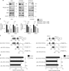
Snail induces CXCL1 and CXCL2 via the NF-κB pathway and possibly via the direct binding to their promoters. a Western blotting of nuclear Snail, phosphorylated p65 (phospho-p65), p65 and RelB from HM-1, OVCAR8 and OVCA433 cells. HDAC1 was used as control. b Reverse transcription polymerase chain reaction (RT-PCR) to analyze the expression of Cxcl1 and Cxcl2 in HM-1-control and HM-1-shSnail cells (left), and the expression of CXCL1 and CXCL2 in OVCAR8-control and OVCAR8-shSnail cells (right), treated with or without BAY11-7082 (NF-κB inhibitor) at 10 μM for 24 h; n = 4. c Schematic representation of CXCL1 and CXCL2 promoter organization, and the corresponding luciferase reporter constructs pGL-CXCL1 (1523 bp: −1523 to +110 bp, 984 bp: −984 to +110 bp, 300 bp: −300 to +110 bp) and pGL-CXCL2 (1606bp: −1606 to +104 bp, 942 bp: −942 to +104 bp, 457 bp: −457 to +104 bp). TSS transcriptional start site, E1 exon1, and Luc luciferase. The black bars indicate E-boxes (CANNTG), which are the binding sites of Snail. d Luciferase reporter assays to analyze the activity of the pGL-CXCL1 and pGL-CXCL2 promoter constructs in 293FT-control and 293FT-shSnail cells. Relative luciferase activities are shown; n = 5. *P < 0.05, **P < 0.01, and ***P < 0.001 (one-way ANOVA with Tukey’s multiple comparisons test in b; unpaired t-test in d). Averaged data are presented as the mean ± SEM
Fig. 6
Fig. 6
CXCR2 ligands induce myeloid-derived suppressor cell infiltration. a Chemotaxis of mouse MDSCs, from subcutaneous tumor of HM-1 tumor-bearing mice, in response to CXCL1, CXCL2, and CXCL5; n = 4. b Chemotaxis of mouse MDSCs, from subcutaneous tumor of HM-1 tumor-bearing mice, pre-treated with SB265610 (CXCR2 antagonist) at each concentration in the presence of each CXCR2 ligands (at 100 ng/mL); n = 4. c Chemotaxis response of MDSCs, from human ovarian cancer ascites, to CXCL1, CXCL2, and CXCL5. The chemotaxis index is shown; n = 4. d Chemotaxis response of human MDSCs treated with SB265610 at each concentration in the presence of each CXCR2 ligands; n = 4. *P < 0.05, **P < 0.01, and ***P < 0.001 (one-way ANOVA with Tukey’s multiple comparisons test in ad)
Fig. 7
Fig. 7
A CXCR2 antagonist inhibits tumor progression by Snail. a Tumor growth in mice subcutaneously injected with HM-1-control cells or HM-1-shSnail cells, treated with anti-Ly6G antibody (400 µg/body) or IgG twice a week from day 1 after tumor inoculation. P-values represent significance between two groups at day 19; n = 5–6. b Flow cytometric analyses of subcutaneous HM-1-control tumors treated with anti-Ly6G antibody or IgG at day 19. Percentage of positive cells relative to total cells is plotted. MDSCs (left) and CD8+T cells/MDSCs (right); n = 6. c Flow cytometric analyses of subcutaneous HM-1-shSnail tumors treated with anti-Ly6G antibody or IgG at day 19. The percentage of positive cells relative to total cell count is plotted. MDSCs (left) and CD8+T cells/MDSCs (right); n = 5–6. d Tumor growth in mice subcutaneously injected with HM-1-control cells or HM-1-shSnail cells, treated with SB265610 (2 mg/kg body weight) or PBS six times a week from day 1 after tumor inoculation. P-values represent significance between two groups at day 21; n = 4–6. e Flow cytometric analyses of subcutaneous HM-1-control tumors treated with SB265610 or PBS at day 21. Percentage of positive cells relative to total cells is plotted. MDSCs (left) and CD8+T cells/MDSCs (right); n = 5-6. f Flow cytometric analyses of subcutaneous HM-1-shSnail tumors treated with SB265610 or PBS at day 21. The percentage of positive cells relative to total cell count is plotted. MDSCs (left) and CD8+T cells/MDSCs (right); n = 4–5. *P < 0.05, **P < 0.01 and, ***P < 0.001 (unpaired t-test in af)
Fig. 8
Fig. 8
Elevated levels of serum CXCL1 and CXCL2 reflect intratumoral MDSCs and are associated with poor prognosis in ovarian cancer patients. a Serum concentration of CXCL1 (left) and CXCL2 (right) in ovarian cancer patients (n = 26) vs. that in healthy donors (n = 8); mean ± SEM; ***P < 0.001 by unpaired t-test. b Correlation between CXCL1 and CXCL2 serum concentrations in ovarian cancer patients (n = 26); P = 0.0002, R = 0.67. Pearson’s product-moment correlation analysis. c Correlation between serum CXCL1 (left; P = 0.018) and CXCL2 (right; P = 0.09) levels and infiltration of CD33+ cells in peritoneal disseminations; n = 12; Pearson’s product-moment correlation analysis. d Correlation between serum CXCL1 (left; P = 0.049) and CXCL2 (right; P = 0.080) levels and Snail staining scores; n = 12; Pearson’s product-moment correlation analysis. e Overall survival of ovarian cancer patients, comparing the high-serum CXCL1 group (n = 12) to the low-serum CXCL1 group (n = 14). Cut-off levels, 42.83 pg/ml; AUC = 0.6842. Hazard ratio, 15.08; 95% confidence interval (CI), 3.859–102.9; P = 0.0005 by log-rank test. f Overall survival of ovarian cancer patients, comparing the high-serum CXCL2 group (n = 13) to the low-serum CXCL2 group (n = 13). Cut-off levels, 93.57 pg/ml; AUC = 0.6767. Hazard ratio, 13.87; 95% confidence interval (CI), 3.56–89.72; P = 0.0008 by log-rank test

"V体育安卓版" References

    1. Cancer Genome Atlas Research N. Integrated genomic analyses of ovarian carcinoma. Nature. 2011;474:609–615. doi: 10.1038/nature10166. - DOI - PMC - PubMed
    1. Davidson B, Trope CG, Reich R. Epithelial-mesenchymal transition in ovarian carcinoma. Front Oncol. 2012;2:33. doi: 10.3389/fonc.2012.00033. - "VSports手机版" DOI - PMC - PubMed
    1. Tan TZ, et al. Epithelial-mesenchymal transition spectrum quantification and its efficacy in deciphering survival and drug responses of cancer patients. EMBO Mol. Med. 2014;6:1279–1293. doi: 10.15252/emmm.201404208. - "VSports" DOI - PMC - PubMed
    1. Murakami R, et al. Establishment of a novel histopathological classification of high-grade serous ovarian carcinoma correlated with prognostically distinct gene expression subtypes. Am. J. Pathol. 2016;186:1103–1113. doi: 10.1016/j.ajpath.2015.12.029. - DOI - PubMed
    1. Hanahan D, Weinberg RA. Hallmarks of cancer: the next generation. Cell. 2011;144:646–674. doi: 10.1016/j.cell.2011.02.013. - DOI - PubMed

Publication types

MeSH terms