Skip to main page content (VSports app下载)
U.S. flag

An official website of the United States government

Dot gov

The VSports app下载. gov means it’s official. Federal government websites often end in . gov or . mil. Before sharing sensitive information, make sure you’re on a federal government site. .

Https

The site is secure V体育官网. The https:// ensures that you are connecting to the official website and that any information you provide is encrypted and transmitted securely. .

. 2018 May;20(5):586-596.
doi: 10.1038/s41556-018-0084-5. Epub 2018 Apr 25.

Loss of KLHL6 promotes diffuse large B-cell lymphoma growth and survival by stabilizing the mRNA decay factor roquin2 (VSports在线直播)

Affiliations

Loss of KLHL6 promotes diffuse large B-cell lymphoma growth and survival by stabilizing the mRNA decay factor roquin2

VSports app下载 - Jaewoo Choi et al. Nat Cell Biol. 2018 May.

Abstract

Kelch-like protein 6 (KLHL6) is an uncharacterized gene mutated in diffuse large B-cell lymphoma (DLBCL). Here we report that KLHL6 assembles with cullin3 to form a functional cullin-RING ubiquitin ligase. Mutations in KLHL6 inhibit its ligase activity by disrupting the interaction with cullin3. Loss of KLHL6 favours DLBCL growth and survival both in vitro and in xenograft models VSports手机版. We further established that the mRNA decay factor roquin2 is a substrate of KLHL6. Degradation of roquin2 is dependent on B-cell receptor activation, and requires the integrity of the Tyr691 residue in roquin2 that is essential for its interaction with KLHL6. A non-degradable roquin2(Y691F) mutant requires its RNA-binding ability to phenocopy the effect of KLHL6 loss. Stabilization of roquin2 promotes mRNA decay of the tumour suppressor and NF-κB pathway inhibitor, tumour necrosis factor-α-inducible gene 3. Collectively, our findings uncover the tumour suppressing mechanism of KLHL6. .


Conflict of interest statement

Author Information. The authors declare no competing financial interests V体育安卓版.

Figures (VSports手机版)
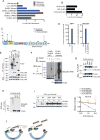
Figure 1
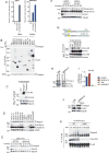
Figure 1. KLHL6 mutations in human DLBCL abolish its catalytic function as CRL3
(a) Percentage of KLHL6 mutations in patients with diffuse large B-cell lymphoma (DLBCL) [University of Nebraska Medical Center (UNMC), n=140 patients, The Cancer Genome Atlas (TCGA), n=48, Canada’s Michael Smith Genome Sciences Centre (CMSGSC), n= 96, and Broad Institute (Broad), n=58, Duke Cancer Institute (DCI), n= 1001], Chronic Lymphocytic Leukemia (CLL) [Departamento de Bioquímica y Biología Molecular, Instituto Universitario de Oncología (IUOPA), n=586] and Multiple Myeloma (MM) (Broad), n=205]. (b) Percentage of KLHL6 mutations in DLBCL subtypes. Cohorts from UNMC and CMSGSC were pooled and sub-classified as Activated B-cell-like (ABC)-, Germinal Center B-cell-like (GC)- and Uncharacterized-DLBCL. (c) Schematic representation of KLHL6 mutations (BTB, Broad-Complex, Tramtrack and Bric-a-brac; BACK, BTB and C-terminal Kelch plant homeodomain). (d) Proteomic analysis of KLHL6 complex. Spectral counts for CULLIN3 are shown. EV, empty vector. This analysis was performed once in HEK293T and ARP-1 cells. (e) Immunoblot analysis of immunoprecipitated FLAG-tagged E3 ligases in HEK293T cells. WCL, whole cell lysates. EV, empty vector. (f) In vitro ubiquitylation reaction of immunopurified FLAG-KLHL6 and ΔKelch mutant. (g) Immunoblot analysis of immunoprecipitated FLAG-tagged KLHL6 wild-type (WT), BTB-mutants (L65P, S94I and F97L), or empty vector (EV) in HEK293T cells. (h) In vitro ubiquitylation reaction of immunopurified FLAG-KLHL6 wild-type (WT) and BTB-mutants (L65P, S94I and F97L). (i) Immunoblot analysis of whole cell lysates from GFP+ OCI-Ly10 KLHL6−/− cells (clone-derived) retrovirally transduced with cDNAs encoding an empty vector (EV), KLHL6(WT) or BTB-mutants (L65P, S94I and F97L) and carrying a GFP marker (left panel). Cells were treated with cycloheximide (CHX) for the indicated times. Right panel shows quantification of KLHL6 protein levels (mean±s.d., n=3 independent experiments, two-way ANOVA, *** P value≤0.001; ****P value≤0.0001). (j) Schematic model of Cullin3-Ring-Ligase (CRL3)-KLHL6 complex. Unprocessed original scan of immunoblots for (e,f,g,h,i) are shown in Supplementary Fig. 8, and statistical source data and exact P values for (i) can be found in Supplementary Table 6. Unless otherwise noted, immunoblots are representative of three independent experiments.
Figure 2
Figure 2. KLHL6 interacts and promotes ubiquitylation and degradation of Roquin2
(a) Proteomic analysis of KLHL6 immunoprecipitations. Spectral counts for Roquin2 proteins are shown. EV, empty vector. The analysis was performed once in two different cell lines (HEK293T and ARP-1). (b) Immunoblot analysis of immunoprecipitated FLAG-F-box proteins (FBPs) or BTB proteins (BTBPs) in HEK293T cells. Lane 1 shows whole cell lysates (WCL) from cells transfected with an empty vector. (c) Immunoblot analysis of immunoprecipitated FLAG-Roquin1 or FLAG-Roquin2 in HEK293T cells. (d) Immunoblot analysis of human DLBCL cell lysates. A low exposure (l.e.) and high exposure (h.e.) are shown. A representative blot from two independent experiments is shown. (e) Immunoblot analysis of whole cell lysates from OCI-LY10 cells electroporated with indicated siRNAs and treated with cycloheximide (CHX). Quantification and statistical analysis is shown in Fig. S2d. (f) KLHL6+/+ and KLHL6−/− U2932 cells(clone-derived) were processed as in (e). A low exposure (l.e.) and high exposure (h.e.) are shown. Quantification and statistical analysis is shown in Fig. S2d. (g) Schematic representation of KLHL6 protein displaying endogenous mutations in VAL and SUDHL10 (top panel). Bottom panel shows immunoblot analysis from immunoprecipitated FLAG-tagged KLHL6 wild-type (WT), KLHL6 mutants (L24R, A25E, N60T, and T72R), or empty vector (EV) in HEK293T cells. (h) Immunoblot analysis of whole cell lysates from VAL and SUDHL10 cells electroporated with siRNA scramble (siCTRL) or targeting KLHL6 (siKLHL6). (i) Analysis of level of KLHL6 mRNA by quantitative PCR(qPCR). The value for the PCR product from U2932 cells was set as 1. A representative graph from two independent experiments is shown. (j) Immunoblot analysis of whole cell lysates from VAL and SUDHL10 cells stably expressing KLHL6 under a doxycycline (DOX) inducible promoter with a puryomycin cassette after 12h of DOX treatment. (k) In vitro ubiquitylation reaction of immunopurified FLAG-KLHL6 and HA-Roquin2. Unprocessed original scan of immunoblots for (b,c,d,e,f,g,h,j,k) are shown in Supplementary Fig. 8, and source data for (i) can be found in Supplementary Table 6. Unless otherwise noted, immunoblots are representative of three independent experiments.
Figure 3
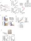
Figure 3. KLHL6 functions as a tumor suppressor in ABC-DLBCL by regulating its growth and survival
(a) Kaplan–Meier analysis based on gene expression data for ABC-DLBCL tumors (GSE10846, GSE34171 and GSE3131239-41) is shown (n=367 patients). Censored subjects are indicated on the Kaplan-Meier cure as tick mark. Statistical analysis was performed using the Log-rank (Mantel-Cox), two-sided test, 95% confidence interval. (b) Cell counts of U2932-, OCI-LY10- and TMD8-Cas9 cells expressing the indicated gRNAs and carrying a puromycin cassette (mean±s.d., n=3 independent experiments, two-way ANOVA, *P value≤0.05; **P value≤0.01; *** P value≤0.001; ****P value≤0.0001) (left panel). Cells were grown in media containing 1μg/ml (U2932- and OCI-LY10) or 4μg/ml (TMD8) of F(ab′)2-IgM. Right panel shows immunoblot analysis of whole cell lysates. (c) Apoptosis analysis of U2932-, OCI-LY10-, and TMD8-Cas9 cells expressing the indicated gRNAs and carrying a GFP marker. Cells were grown as in (b). Apoptosis was quantified on GFP+ and Annexin V+ cells (mean±s.d., n=3 independent experiments, one-way ANOVA, **P value≤0.01; *** P value≤0.001). (d) Left panel shows cell counts of GFP-sorted U2932 KLHL6−/− (clone-derived) cells expressing empty vector (EV), KLHL6(WT) or BTB-mutants (L65P, S94I and F97L) and carrying a GFP marker (mean±s.d., n=3 independent experiments, two-way ANOVA, **P value≤0.01; *** P value≤0.001; ****P value≤0.0001). Right panel shows the immunoblot analysis of whole cell lysates. (e) Xenograft experiments with GFP-sorted U2932 KLHL6−/− cells expressing an empty vector (EV), KLHL6(WT) or KLHL6(S94I) and carrying a GFP marker. Top panel shows the tumors at the experimental endpoint. Tumor volume (mean±s.d., n=5 mice per group, one-way ANOVA, *P value≤0.05; n.s, not significant) and tumor weight (mean±s.d., n=5 mice per group, one-tailed t-test, *P value ≤0.05; **P value ≤0.01; n.s, not significant) are shown in the bottom left and right, respectively. Unprocessed original scan of immunoblots for (b,d) are shown in Supplementary Fig. 8, and statistical source data and exact P values for (a,b,c,d,e) can be found in Supplementary Table 6. Unless otherwise noted, immunoblots are representative of three independent experiments.
Figure 4
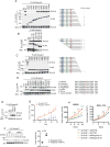
Figure 4. A non-degradable Roquin2 mutant phenocopies loss of KLHL6
(a) Left panel shows immunoblot analysis of immunoprecipitated FLAG-tagged Roquin2 wild type (WT) or mutants in HEK293T cells stably expressing KLHL6. EV, Empty vector. Right panel shows a schematic representation of Roquin2 mutants. Roquin2 mutants that interact (+) or do not interact (-) with KLHL6 are shown. A representative blot from two independent experiments is shown. Asterisk indicates non-specific bands. (b–d) Same as in (a). (e) Immunoblot analysis of immunoprecipitated FLAG-tagged Roquin2 wild type (WT) or mutants, as indicated, in HEK293T cells stably expressing KLHL6. EV, Empty vector. (f) Immunoblot analysis of whole cell lysates from a DLBCL cell line, BJAB, retrovirally transduced with cDNAs encoding Roquin2(WT) or Roquin2(Y691F) carrying a puromycin cassette. Cells were treated with cycloheximide (CHX) for the indicated times. Quantification and statistical analysis is shown in Fig. S4e. (g) Top panel shows tumor volume from subcutaneously injected NSG mice with U2932 cells stably expressing retroviruses encoding HA-Roquin2(WT) or HA-Roquin2(Y691F) carrying a puromycin cassette (mean±s.d., n=3 mice per group, two-way ANOVA, *P value≤0.05; **P value≤0.01; *** P value≤0.001; ****P value≤0.0001). Bottom panel shows tumor weight (mean±s.d., n=3 mice per group, two-tailed t-test, *** P value≤0.001). (h) Cell counts of GFP-sorted U2932 (left panel) or OCI-LY10 (right panel) KLHL6+/+ and KLHL6−/− (clone-derived) cells infected with scramble shRNA(shCTRL) or shRNA targeting Roquin2 (ShRoquin2#1 or #2) carrying a GFP marker. GFP+ cells were grown in media containing 1μg/ml of F(ab′)2-IgM. (mean±s.d., n=3 independent experiments, two-way ANOVA, ****P value≤0.0001). Unprocessed original scan of immunoblots for (a,b,c,d,e,f) are shown in Supplementary Fig. 8, and statistical source data and exact P values for (g,h) can be found in Supplementary Table 6. Unless otherwise noted, immunoblots are representative of three independent experiments.
Figure 5
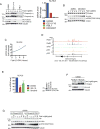
Figure 5. KLHL6 is a BCR/NF-κB target gene that links Roquin2 degradation to BCR signaling
(a) Immunoblot analysis of whole cell lysates from OCI-LY10, U2932, and HBL1 cells stimulated with 10 μg/ml F(ab′)2-IgM for 3 and 6 hours (left panel). A low exposure (l.e.) and high exposure (h.e.) are shown. Right panel shows level of KLHL6 mRNA analyzed by qPCR. The value for the PCR product from U2932 was set as 1. A representative graph from two independent experiments is shown. (b) Immunoblot analysis of whole cell lysates from U2932 cells treated with 10 μg/ml of F(ab′)2-IgM for the indicated times. Where indicated, cells were pre-treated with 5μM MLN4924 for 1 hour. (c) Analysis of level of KLHL6 mRNA by qPCR in U2932 cells treated with 10 μg/ml of F(ab′)2-IgM for the indicated times. A representative graph from two independent experiments is shown. The value for PCR product without treatment was set as 1. (d) Visualization of ChIP-seq peaks using the University of California Santa Cruz (UCSD) Genome browser (GEO Series accession GSE55105). RPM, reads per million mapped. (e) Same as in (c) except that U2932 cells were treated with DMSO, 10μM of IKK inhibitor (IKK-16) or 5μM of BTK inhibitor (Ibrutinib) for 6 hours. The value for PCR product present without treatment (DMSO) was set as 100% (mean±s.d., n=3 independent experiments, one-way ANOVA, *** P value≤0.001; ****P value≤0.0001). The right panel shows immunoblot analysis of whole cell lysates for the indicated proteins. (f) Immunoblot analysis of whole cell lysates from U2932 KLHL6+/+ and KLHL6−/− (clone-derived) cells treated with increasing concentrations of F(ab′)2-IgM for 6 hours. (g) Immunoblot analysis of whole cell lysates from U2932 cells stably expressing HA-Roquin2(WT) or HA-Roquin2(Y691F) treated with F(ab′)2-IgM for 6 hours. Unprocessed original scan of immunoblots for (a,b,e,f,g) are shown in Supplementary Fig. 8, and source data for (a,c) and statistical source data and exact P values for (e) can be found in Supplementary Table 6. Unless otherwise noted, immunoblots are representative of three independent experiments.
Figure 6
Figure 6. Stabilization of Roquin2 down-regulates BCR responsive genes
(a) Representative image of U2932 cells expressing HA-Roquin2(WT), HA-Roquin2(Y691F) or HA-Roquin2(Y691FΔROQ) with puromycin cassette plated into a matrigel (Left panel). Middle panel shows immunoblot analysis of whole cell lysates and right panel shows cell counts from the matrigel (mean±s.d., n=4 independent experiments, one-way ANOVA, ****P value≤0.0001, n.s, not significant). Scale bar 150μm. (b) Volcano plot (left panel) showing down-regulated mRNAs (blue) in U2932 cells expressing Roquin2(Y691F) vs Roquin2(WT) upon 12 hours treatment with 10 μg/ml of F(ab′)2-IgM [log2(fold-change)<–0.9]. Down-regulated mRNAs were further plotted in an MA (log ratio, mean average)-plot (right panel). The mRNAs up-regulated upon treatment with F(ab′)2-IgM in cells expressing Roquin2(WT) [log2(fold-change)>1] are shown in red. (n=3 independent experiments, DEseq2, P value<0.001). NT, non treated. (c) Venn diagram showing the overlap between genes down-regulated by expression of Roquin2(Y691F) and BCR responsive genes. (d) qPCR analysis of the indicated mRNAs in U2932 cells expressing HA-Roquin2(WT, Y691F or Y691FΔROQ) treated with 10 μg/ml of F(ab′)2-IgM for indicated times. Value for PCR product present at time 0 hour was set as 1 for each condition. A representative graph from two independent experiments is shown. (e) qPCR analysis of TNFAIP3 mRNA in U2932 cells electroporated with indicated siRNAs (left panel) and treated with actinomycinD for the indicated times. The value for PCR product present at time 0 hour was arbitrarily set as 100% (mean±s.d., n=3 independent experiments, two-way ANOVA, **P value ≤0.01, ****P value≤0.0001, n.s, not significant). Same analysis was performed in VAL cells expressing KLHL6 under a DOX-inducible promoter (Right panel). Cells were pre-treated with DOX for 12 hours and actinomycinD for the indicated times (mean±s.d., n=3 independent experiments, two-way ANOVA, *P value ≤0.05; *** P value≤0.001). Unprocessed original scan of immunoblots for (a) are shown in Supplementary Fig. 8, and source data for (d) and statistical source data and exact P values for (a,e) and (b) can be found in Supplementary Table 6 and 3, respectively. Unless otherwise noted, immunoblots are representative of three independent experiments.
Figure 7
Figure 7. KLHL6-Roquin2 axis controls NF-κB activation
(a) Immunoblot analysis of whole cell lysates from U2932 cells expressing HA-Roquin2(WT) or HA-Roquin2(Y691F) treated with 10μg/ml of F(ab′)2-IgM for the indicated times. (b) Immunoblot analysis of whole cell lysates from U2932 KLHL6+/+ or KLHL6−/− cells (clone-derived) treated with 10μg/ml of F(ab′)2-IgM for the indicated times. (c) Immunoblot analysis of whole cell lysates from HBL1 cells electroporated with indicated siRNAs and treated as in (a) for the indicated times. (d) Immunoblot analysis of whole cell lysates from U2932 KLHL6+/+ or KLHL6−/− cells (clone-derived) treated with 10μg/ml of F(ab′)2-IgM for the indicated times. (e) Immunoblot analysis of fractionated U2932 KLHL6+/+ or KLHL6−/− (clone-derived) cells. (f) RelA ChIP-qPCR for the NFBKIA promoter in U2932 KLHL6+/+, KLHL6−/− or KLHL6−/− cells infected with indicated shRNAs. Data are displayed as fold enrichment relative to IgG control. A representative graph from two independent experiments is shown. (g) Overlap of BTB-associated mutations of KLHL6 withTNFAIP3 alterations in DLBCLs. Top panel shows tumors sequenced at UNMC and DCI (n=1175) with deleterious mutations of KLHL6 in the BTB-domain and TNFAIP3 mutations. In the bottom panel, the TNFAIP3 subset is shown as biallelic and monoallelic deletions. (h) Cell counts of GFP-sorted RCK8 cells expressing Cas9, the indicated gRNAs and a GFP marker. Cells were grown in media containing 1μg/ml of F(ab′)2-IgM (mean±s.d., n=3 independent experiments, two-way ANOVA, n.s, not significant). Right panel shows immunoblot analysis of whole cell lysates. (i) Apoptosis analysis of cells from (h) is shown (mean±s.d., n=3 independent experiments, one-way ANOVA, n.s, not significant). (j) The percentage of GFP+ and AnnexinV+ HBL1 and HLY-1 cells expressing the indicated shRNAs is shown (mean±s.d., n=3 independent experiments, one-way ANOVA, *P value≤0.05; *** P value≤0.001, n.s, not significant). (k) Model of KLHL6-Roquin2 axis in ABC-DLBCL. Unprocessed original scan of immunoblots for (a,b,c,d,e,h) are shown in Supplementary Fig. 8, and source data for (f) and statistical source data for (h,i,j) and exact P values for (j) can be found in Supplementary Table 6. Unless otherwise noted, immunoblots are representative of three independent experiments.

"V体育官网" References

    1. Yang Y, Staudt LM. Protein ubiquitination in lymphoid malignancies. Immunol Rev. 2015;263:240–256. - PMC - PubMed
    1. Gupta-Rossi N, et al. Specific over-expression of deltex and a new Kelch-like protein in human germinal center B cells. Mol Immunol. 2003;39:791–799. - PubMed
    1. Kroll J, et al. The BTB-kelch protein KLHL6 is involved in B-lymphocyte antigen receptor signaling and germinal center formation. Mol Cell Biol. 2005;25:8531–8540. - PMC - PubMed
    1. Morin RD, et al. Frequent mutation of histone-modifying genes in non-Hodgkin lymphoma. Nature. 2011 - PMC - PubMed
    1. Lohr JG, et al. Discovery and prioritization of somatic mutations in diffuse large B-cell lymphoma (DLBCL) by whole-exome sequencing. Proc Natl Acad Sci U S A. 2012;109:3879–3884. - "VSports最新版本" PMC - PubMed

Publication types

MeSH terms