Skip to main page content
U.S. flag

An official website of the United States government

Dot gov

The VSports app下载. gov means it’s official. Federal government websites often end in . gov or . mil. Before sharing sensitive information, make sure you’re on a federal government site. .

Https

The site is secure. The https:// ensures that you are connecting to the official website and that any information you provide is encrypted and transmitted securely. V体育官网.

. 2018 May 14;33(5):890-904.e5.
doi: 10.1016/j.ccell.2018.03.017. Epub 2018 Apr 12.

Multi-stage Differentiation Defines Melanoma Subtypes with Differential Vulnerability to Drug-Induced Iron-Dependent Oxidative Stress (VSports app下载)

Affiliations

"VSports app下载" Multi-stage Differentiation Defines Melanoma Subtypes with Differential Vulnerability to Drug-Induced Iron-Dependent Oxidative Stress

Jennifer Tsoi et al. Cancer Cell. .

Abstract

Malignant transformation can result in melanoma cells that resemble different stages of their embryonic development. Our gene expression analysis of human melanoma cell lines and patient tumors revealed that melanoma follows a two-dimensional differentiation trajectory that can be subclassified into four progressive subtypes. This differentiation model is associated with subtype-specific sensitivity to iron-dependent oxidative stress and cell death known as ferroptosis VSports手机版. Receptor tyrosine kinase-mediated resistance to mitogen-activated protein kinase targeted therapies and activation of the inflammatory signaling associated with immune therapy involves transitions along this differentiation trajectory, which lead to increased sensitivity to ferroptosis. Therefore, ferroptosis-inducing drugs present an orthogonal therapeutic approach to target the differentiation plasticity of melanoma cells to increase the efficacy of targeted and immune therapies. .

Keywords: combination therapy; differentiation; ferroptosis; immunotherapy; kinase inhibitor therapy; melanoma; pharmacogenomics; systems biology V体育安卓版. .


VSports手机版 - Figures

Figure 1
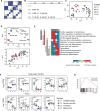
Figure 1. Identification of four melanoma subtypes related by progressive differentiation
(A) Matrix of consensus indices from hierarchical clustering of melanoma cell lines showing differences in clustering stability when grouped from k=2 to k=5 clusters. (B) Pairwise comparisons showing statistically significant cluster delineations. (C) PCA of melanoma cell line expression profiles annotated by identified clusters. (D) PCA of gene expression profiles from an in vitro embryonic stem cell (ESC) to melanocyte multi-stage differentiation system (top) and projection of melanoma cell line expression profiles into melanocyte differentiation stage PCA space (bottom). (E) Heatmap summary of rank-based enrichment analysis p values of each individual cluster vs. the remaining clusters in differentiation-associated gene sets. (F) Boxplots of select transcription factors and receptor tyrosine kinase (RTK) genes. Boxplot lines reflect lower quartile, median, and upper quartile. Whiskers reflect 1.5 times above or below the interquartile range, with points outside reflecting outliers. (U: Undifferentiated, N: Neural crest-like, T: Transitory, M: Melanocytic; number in each group: U=10, N=42, T=12, M=17; Kruskal-Wallis ANOVA and Dunn’s post hoc two-tailed test p values: * ≤ 0.05, ** ≤ 0.01, *** ≤ 0.001). (G) Analysis of Melanocytic (vs. Transitory) subtype shows enrichment of MITF targets. See also Tables S1 and S2.
Figure 2
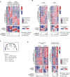
Figure 2. Treatment-induced dedifferentiation in the context of the four-stage differentiation model
(A, B) Heatmap of signature genes, average signature z-scores, and differentiation trajectory position changes for matched parental and resistant cell lines (A) and for a vemurafenib treatment timecourse in M229 and M397 melanoma cell lines compared to DMSO vehicle control (0 days treatment) (B). (C) Schematic representing progressive dedifferentiation along the two-dimensional trajectory model with increased time under vemurafenib treatment. (D) Heatmap of signature genes, average signature z-scores, and differentiation trajectory position changes for murine HCmel3 tumors or cell lines with treatment control or relapse from adoptive transfer of antigen specific T cells. (C: control, R: relapse). Dark grey arrows represent increased differentiation state and the light grey arrow indicates the treatment induced dedifferentiation direction. See also Figure S1 and Table S3.
Figure 3
Figure 3. Melanoma classifier identifies consistent subtypes in cell lines and tumors
(A) Schematic of the melanoma subtype classifier pipeline. (B) PCA of GDSC, CCLE and TCGA datasets annotated by the cluster prediction assignment. For the TCGA dataset, immune- and keratin-associated genes were removed to provide melanoma cell-specific analysis. See also Figures S2, S3, and S4, and Table S4.
Figure 4
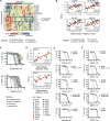
Figure 4. Integration of pharmacogenomics drug sensitivity profiles reveals subtype-specific sensitivity to ferroptosis inducing drugs
(A) Hierarchical clustering of the CTRP pharmacogenomics database AUC values across differentiation subtypes. (B) Plot of AUC values vs. the differentiation trajectory score for all ferroptosis inducing drugs from the CTRP. Low AUC values indicate increased sensitivity. (C) Dose response curves across indicated M series melanoma cell lines for erastin and RSL3. (D) Corresponding plot of log IC50 concentration values for erastin and RSL3 treatment versus the differentiation trajectory score. Black dashes indicate mean within the subtype group. (E, F) Dose-response curves showing increased sensitivity to erastin (E) and RSL3 (F) in cell lines with vemurafenib-induced dedifferentiation including both acquired resistance lines (P: parental, R: resistant) or long-term (LT) adaptive resistance (44 days). Percent viable cells are calculated relative to DMSO. Drug response curves are shown as mean ± sem of two replicates and representative of at least three independent experiments. See also Figure S5.
Figure 5
Figure 5. Erastin treatment induces ferroptosis in dedifferentiated melanoma cells
(A) Measurement of percent viable cells compared to DMSO control with erastin treatment alone or in combination with DFO or Trolox. Data shown represent mean ± sem of three replicates, and representative of at least three independent experiments. (B, C) ROS measurements after 10 hr erastin treatment across cell lines by flow cytometry using BODIPY-C11 probe to measure lipid ROS (B) and CM-H2DCFDA probe to measure cytosolic ROS (C). See also Figure S6.
Figure 6
Figure 6. Lower basal levels of glutathione in dedifferentiated melanoma increase sensitivity to ferroptosis induction
(A, B) Relative amounts of reduced glutathione GSH (A) and oxidized GSSG (B) measured by mass spectrometry-based metabolomics after 8 hr erastin treatment compared to untreated parental control for the indicated isogenic cell lines. (P: Erastin-insensitive parental cell lines; R: erastin-sensitive BRAFi-resistant cell lines.) (C) Barplot of reduced glutathione (GSH) levels measured by Ellman’s reagent of the isogenic cell line pairs (left). GSH levels vs. the differentiation trajectory score across panel of melanoma cell lines (right). Data shown represent mean ± sem of three independent experiments; p values: * ≤ 0.05, ** ≤ 0.01, *** ≤ 0.001. Subtype group means indicated by black dashes (right). (D) Trypan blue exclusion viability assay of 24 hr erastin or RSL3 treatment with or without supplementation of 5mM GSH in the culture medium. Data shown represent mean ± sem of three independent experiments; one-tailed t-test p values: * ≤ 0.05, ** ≤ 0.01, *** ≤ 0.001. See also Figure S6 and Table S5.
Figure 7
Figure 7. Reduction in persistent dedifferentiated melanoma cells upon combination treatment with ferroptosis inducing drugs
(A) Measurement of percent viable cells compared to vehicle control (DMSO) of erastin treatment combined with increasing concentration of vemurafenib for 72 hours. Data shown in barplots represent mean ± sem of three replicates. (B) Immunoblots of differentiation and signaling markers in cell lines treated with long-term (21 days) vemurafenib treatment. (C) Crystal violet staining assays of long-term combination treatment of erastin (E= 2 µM for M229 and M397, 5 µM for M249) and vemurafenib (V= 1 µM) for 16 days (M229), 24 days (M397), or 21 days (M249). DMSO treated cells were stained when confluent (7 days). (D) Immunoblots of cell lines treated with the indicated cytokines. (E) Crystal violet staining assays of erastin treatment for 7 days with cytokine exposure for the initial 3 days (M229 and M249) or 7 days (M397). IFNγ=100 U/mL, TNFα=1000 U/mL Data shown is representative of three independent experiments. See also Figure S7.

Comment in

"V体育官网入口" References

    1. Anders S, Pyl PT, Huber W. HTSeq—a Python framework to work with high-throughput sequencing data. Bioinformatics. 2015;31:166–169. - PMC - PubMed
    1. Caramel J, Papadogeorgakis E, Hill L, Browne GJ, Richard G, Wierinckx A, Saldanha G, Osborne J, Hutchinson P, Tse G, et al. A Switch in the Expression of Embryonic EMT-Inducers Drives the Development of Malignant Melanoma. Cancer Cell. 2013;24:466–480. - "VSports手机版" PubMed
    1. Chen Y, Lemire M, Choufani S, Butcher DT, Grafodatskaya D, Zanke BW, Gallinger S, Hudson TJ, Weksberg R. Discovery of cross-reactive probes and polymorphic CpGs in the Illumina Infinium HumanMethylation450 microarray. Epigenetics. 2013;8:203–209. - PMC (VSports手机版) - PubMed
    1. Denat L, Kadekaro AL, Marrot L, Leachman SA, Abdel-Malek ZA. Melanocytes as Instigators and Victims of Oxidative Stress. J. Invest. Dermatol. 2014;134:1512–1518. - PMC - PubMed
    1. Denecker G, Vandamme N, Akay Ö, Koludrovic D, Taminau J, Lemeire K, Gheldof A, De Craene B, Van Gele M, Brochez L, et al. Identification of a ZEB2-MITF-ZEB1 transcriptional network that controls melanogenesis and melanoma progression. Cell Death Differ. 2014;21:1250–1261. - PMC - PubMed

Publication types

MeSH terms