Skip to main page content
U.S. flag

An official website of the United States government

Dot gov

The . gov means it’s official. Federal government websites often end in VSports app下载. gov or . mil. Before sharing sensitive information, make sure you’re on a federal government site. .

Https

The site is secure. The https:// ensures that you are connecting to the official website and that any information you provide is encrypted and transmitted securely V体育官网. .

. 2018 May 1;128(5):1919-1936.
doi: 10.1172/JCI91646. Epub 2018 Apr 9.

Recipient mucosal-associated invariant T cells control GVHD within the colon

Affiliations

V体育ios版 - Recipient mucosal-associated invariant T cells control GVHD within the colon

VSports手机版 - Antiopi Varelias et al. J Clin Invest. .

Abstract

Mucosal-associated invariant T (MAIT) cells are a unique innate-like T cell subset that responds to a wide array of bacteria and yeast through recognition of riboflavin metabolites presented by the MHC class I-like molecule MR1. Here, we demonstrate using MR1 tetramers that recipient MAIT cells are present in small but definable numbers in graft-versus-host disease (GVHD) target organs and protect from acute GVHD in the colon following bone marrow transplantation (BMT). Consistent with their preferential juxtaposition to microbial signals in the colon, recipient MAIT cells generate large amounts of IL-17A, promote gastrointestinal tract integrity, and limit the donor alloantigen presentation that in turn drives donor Th1 and Th17 expansion specifically in the colon after BMT. Allogeneic BMT recipients deficient in IL-17A also develop accelerated GVHD, suggesting MAIT cells likely regulate GVHD, at least in part, by the generation of this cytokine. Indeed, analysis of stool microbiota and colon tissue from IL-17A-/- and MR1-/- mice identified analogous shifts in microbiome operational taxonomic units (OTU) and mediators of barrier integrity that appear to represent pathways controlled by similar, IL-17A-dependent mechanisms. Thus, MAIT cells act to control barrier function to attenuate pathogenic T cell responses in the colon and, given their very high frequency in humans, likely represent an important population in clinical BMT VSports手机版. .

Keywords: Bone marrow transplantation; Immunology; Transplantation. V体育安卓版.


Conflict of interest statement (V体育ios版)

Conflict of interest: LKN, JR, JM, and DPF are named inventors on a patent application (PCT/AU2013/000742, WO2014005194) V体育ios版. LKN, JYWM, JR, JM, and DPF are named inventors on a patent application (PCT/AU2015/050148, WO2015149130) involving MR1 ligands for MR1-restricted MAIT cells owned by University of Queensland, Monash University, and University of Melbourne.

Figures
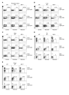
Figure 1
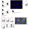
Figure 1. MAIT cells are present in GVHD target organs.
Lymphocytes from naive mouse peripheral blood (A), liver (B), lung (C), SI (D), and colon (E) were stained with the MR1 tetramer (5-OP-RU) or control tetramer (6-FP) and analyzed by flow cytometry. Gates were set based on the control tetramer and cells from MR1–/– mice. Mouse MAIT cells were defined as CD3+. Representative dot plots are shown.
Figure 2
Figure 2. Recipient MAIT cells provide protection from GVHD.
(A and B) G-CSF–mobilized BALB/c.WT splenocytes (25 × 106) were transplanted to lethally irradiated B6.WT or B6.MR1–/– mice and survival and clinical scores monitored. Data pooled from 2 independent experiments. n = 12 per group; TCD group, n = 3. (C and D) Grafts composed of B6.WT BM (5 × 10⁶) and B6.WT T cells (2 or 5 × 10⁶ as indicated) or B6.MR1–/– BM and B6.MR1–/– T cells were transplanted into lethally irradiated B6D2F1 recipients and survival and clinical scores determined. Data combined from 2 independent experiments are shown. n = 16 per group; TCD group, n = 7. (E and F) Lethally irradiated B6D2F1 recipients were transplanted with G-CSF–mobilized splenocytes (10 × 106) from B6.WT or B6.MR1–/– donors. Data combined from 2 replicate experiments are shown. n = 16 per group; TCD group, n = 6 mice. Survival represented by Kaplan-Meier analysis.
Figure 3
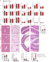
Figure 3. Recipient MAIT cells attenuate acute GVHD within the GI tract.
B6.WT and B6.MR1–/– mice were transplanted with G-CSF–mobilized BALB/c.WT splenocytes or TCD splenocytes. (A) Serum cytokine analysis was conducted on days 4, 7, and 14 after transplant. Day 4 data from 1–2 experiments. n = 5–10 per group; day 7 data pooled from 2 independent experiments, n = 11–12 per group; day 14 data from 1 experiment. n = 6–7 per group. (BE) Semiquantitative histopathology of liver (B), lung (C), SI (D), and colon (E) from B6.WT and B6.MR1–/– recipients at days 13–14 after SCT. Images were captured on a Nikon ECLIPSE Ci microscope fitted with a DS-Fi2 camera. Original magnification, ×4. Data combined from 2 independent experiments, n = 9–12 per group; TCD group, n = 6. (F) Perturbed intestinal barrier integrity as determined by FITC-dextran levels in serum on day 13 after SCT. All data analyzed using the Mann-Whitney U test except histology data, which was analyzed using the unpaired t test with Welch’s correction.
Figure 4
Figure 4. Recipient MAIT cells within the GI tract persist after BMT.
B6.WT mice were lethally irradiated and transplanted with 10 × 106 BALB/c BM and 3 × 10⁶ BALB/c T cells. Recipient lamina propria lymphocytes from the SI (A) and colon (B) isolated at day 3 and day 7 after transplant were stained with control and MR1 tetramer and analyzed by flow cytometry. Representative dot plots of host CD45+CD3+CD4negCD8neg cells additionally stained with γδ TCR and MR1 tetramer are shown. Quantification of MAIT cell numbers before and after transplant for the SI and colon is shown below plots. Enumeration of recipient γδ T cells from the same samples is shown for comparison. Numbers on graph show average percentage of reduction in absolute numbers compared with that in naive mice. Data from 1 or 2 experiments. n = 3–5 mice per time point.
Figure 5
Figure 5. Recipient MAIT cells attenuate proinflammatory donor CD4+ T cell expansion in the colon.
(A) B6.WT and B6.MR1–/– mice were lethally irradiated, transplanted with BALB/c.WT BM (10 × 106) and BALB/c.luciferase+ T cells (3 × 10⁶) and organs imaged at days 7 and 14 after transplant. Representative BLI images of the spleen, liver, lung, and GI tract (including mLNs) are shown. (B) Quantitated BLI at days 7 (top) and 14 (bottom) is shown. Data are pooled from 2 independent experiments for each time point. Day 7, n = 5–7 per group; day 14, n = 11 per group. (C) Frequency of proliferating donor CD4+ T cells in the colon lamina propria at day 14, as determined by intracellular Ki-67 expression. (D) Frequency of apoptotic donor CD4+ T cells in the colon lamina propria at day 14, as determined by intracellular active caspase-3 expression. Data combined from 2 replicate experiments are shown. n = 5 – 9 per group. Data analyzed using the Mann-Whitney U test.
Figure 6
Figure 6. Recipient MAIT cells attenuate proinflammatory donor CD4+ T cell expansion in the colon and secrete IL-17.
B6.WT and B6.MR1–/– mice were transplanted as in Figure 5A. Colon-derived lamina propria lymphocytes at day 14 after transplant were isolated and stimulated in vitro with PMA/ionomycin, followed by assessment of IFN-γ and IL-10 production (A) and IL-17A and TNF-α production (B) by donor CD4+ T cells using intracellular flow cytometry. Quantification of frequency (top) and absolute number (bottom) of cytokine-producing donor CD4+ T cells. Each point represents cells from pooled colons of 2 mice. Data from 2 independent experiments. n = 4–6 mice per group. (C) CD3+CD4negCD8neg (DN), CD3+CD4+CD8neg, and CD3+CD8+CD4neg cells were sort purified from B6.WT and B6.MAIT Tg LNs, stimulated in vitro with α-CD3/α-CD28 for 72 hours, and cytokines determined in culture supernatant. Data from 2 independent experiments with 2–3 WT and 6–8 MR1–/– mice pooled per group. Data analyzed using the Mann-Whitney U test.
Figure 7
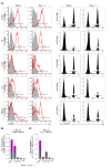
Figure 7. MAIT cells in the GI tract are constitutively activated in steady state and preferentially express IL-17 in the colon.
(A) Representative histograms showing CD69 (red overlay; isotype, gray histogram) and IL-17eYFP (solid black) expression on recipient MAIT cells in the colon, SI, lung, liver, and spleen in steady-state and after allogeneic SCT (d+1). (B and C) Number of CD69+ and IL-17eYFP+ MAIT cells in tissues was quantified. Data pooled from 4 replicate experiments are shown. Colon, 3 experiments (n = 6–12); SI, 3 experiments (n = 6–10); liver, 2 experiments (n = 4–8); lung, 2 experiments (n = 4–8); spleen, 2 experiments (n = 3–4).
Figure 8
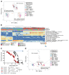
Figure 8. MAIT cells appear to shape the GI microbiome.
(A) PCA plot of 16S rRNA-seq data generated from fecal samples collected from B6.WT and B6.MR1–/– mice either before and after cohousing or when separately housed. Each dot represents an individual mouse, with symbols and colors corresponding to the genetic background and time point. Data combined from 3 independent experiments. Experiment 1, n = 6 mice per group; experiments 2 and 3, n = 5 mice per group. (B) Heatmap showing significantly different OTUs between before and after cohousing samples in either B6.WT or B6.MR1–/– mice. OTUs displaying consistent movement in all 3 experiments are shown. Details contained in Supplemental Tables 1 and 2. (C) Separately housed or cohoused B6.WT and B6.MR1–/– mice were transplanted and clinical scores and survival monitored. Data pooled from 3 independent experiments. n = 15 mice per group; TCD group, n = 9. Survival data analyzed with a log-rank test. ****P < 0.0001; **P = 0.0016 (cohoused B6.WT versus cohoused B6.MR1–/–); **P = 0.0012 (cohoused B6.WT versus separate B6.MR1–/–). (D) PCA plot combining data from A with sequencing data from fecal samples collected after transplant. Data combined from 2 independent experiments. Experiments 2 and 3, n = 5–10 mice per group. 16S rRNA-seq data analyzed using DESeq2.
Figure 9
Figure 9. Analogous alterations in the microbiome in the absence of MAIT cells and IL-17A.
(A) Separately housed B6.WT and B6.IL-17A–/– mice were transplanted with 25 × 106 G-CSF–mobilized BALB/c splenocytes, and clinical scores and survival were monitored. Data pooled from 2 independent experiments (n = 10 per group; TCD group, n = 3; B6.IL-17A–/– TCD group, n = 5). Survival data analyzed with a log-rank test. P < 0.0001, B6.WT versus B6.IL-17A–/–. (B) Fecal samples collected from naive B6.WT and B6.IL-17A–/– mice were profiled by 16S rRNA-seq. Principal component analysis of B6.WT, B6.IL-17A–/–, and B6.MR1–/– (Figure 7A data set) data demonstrates distinct OTU profiles. Each dot represents an individual mouse, with symbols and colors corresponding to the genetic background and experiment. Data from 4 experiments are shown. Experiment 1, WT versus MR1–/–, n = 6 mice per group; experiments 2 and 3, WT versus MR1–/–, n = 4-5 mice per group; experiment 4, WT versus IL-17A–/–, n = 8-10 mice per group. (C) Heatmap showing significantly different OTUs between naive B6.IL-17A–/– and B6.MR1–/– mice in comparison with B6.WT mice from the same samples as used in B. Details contained in Supplemental Table 3. 16S rRNA sequencing data analyzed using DESeq2.
Figure 10
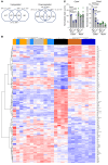
Figure 10. Regulation of host defense and metabolism genes by MAIT and IL-17A.
RNA-seq analysis was performed on tissue from proximal colon and distal ileum of naive WT, MR1–/–, and IL-17A–/– mice (n = 4 per group). (A) Comparison of the number of overlapping genes differentially expressed within the colon; upregulated (upper) and downregulated (lower) genes. (B) Heatmap depicting differentially expressed genes comparing MR1–/– and IL-17A–/– versus WT colon, but not ileum. log2 transformed, normalized read counts were used. (C) Normalized read count data for claudin 4 and claudin 8 expression are shown. Data analyzed using edgeR with a false discovery rate (FDR) < 0.05.
Figure 11
Figure 11. Recipient MAIT cells suppress alloantigen-specific Marilyn TCR Tg effector T cell expansion.
(AC) Lethally irradiated male B6.WT and B6.MR1–/– recipients were transplanted with TCD B6.CD11c-GCDL BM and B6.Marilyn TCR Tg T cells. Bacterial translocation in ileum tissue by FISH at day 14 after allogeneic SCT is shown in B. Individual organs were imaged on day 14 after allogeneic SCT, and BLI data combined from 3 replicate experiments are shown in C (n = 12–15 per group). (DH) Lethally irradiated male B6.WT and B6.MR1–/– recipients were transplanted with B6.WT BM and B6.Marilyn TCR Tg T cells, followed by transfer of Marilyn luc+ T cell on day 12 after allogeneic SCT. Individual organs were imaged 7 days after transfer, and BLI data combined from 3 replicate experiments are shown in E (n = 9–13 per group). Representative images are shown in F. Frequency of pathogenic IFN-γ+IL-17+ Marilyn luc+ T cells in mLN was determined, and data combined from 2 replicate experiments are shown in G (n = 5–10 per group). Representative FACS plots are shown in H. Data analyzed using the Mann-Whitney U test.

"VSports在线直播" References

    1. Markey KA, MacDonald KP, Hill GR. The biology of graft-versus-host disease: experimental systems instructing clinical practice. Blood. 2014;124(3):354–362. doi: 10.1182/blood-2014-02-514745. - DOI - PMC - PubMed
    1. Koyama M, Hill GR. Alloantigen presentation and graft-versus-host disease: fuel for the fire. Blood. 2016;127(24):2963–2970. doi: 10.1182/blood-2016-02-697250. - DOI - PubMed
    1. Koyama M, et al. Donor colonic CD103+ dendritic cells determine the severity of acute graft-versus-host disease. J Exp Med. 2015;212(8):1303–1321. doi: 10.1084/jem.20150329. - DOI - PMC - PubMed
    1. Hill GR, Ferrara JL. The primacy of the gastrointestinal tract as a target organ of acute graft-versus-host disease: rationale for the use of cytokine shields in allogeneic bone marrow transplantation. Blood. 2000;95(9):2754–2759. - PubMed
    1. Cua DJ, Tato CM. Innate IL-17-producing cells: the sentinels of the immune system. Nat Rev Immunol. 2010;10(7):479–489. doi: 10.1038/nri2800. - DOI (V体育官网) - PubMed

Publication types

MeSH terms