VSports最新版本 - Skip to main page content
U.S. flag

An official website of the United States government

Dot gov

The . gov means it’s official. Federal government websites often end in . gov or . mil VSports app下载. Before sharing sensitive information, make sure you’re on a federal government site. .

Https

The site is secure. The https:// ensures that you are connecting to the official website and that any information you provide is encrypted and transmitted securely V体育官网. .

. 2018 May 7;215(5):1315-1325.
doi: 10.1084/jem.20172063. Epub 2018 Mar 16.

Selective inhibition of the p38α MAPK-MK2 axis inhibits inflammatory cues including inflammasome priming signals

Affiliations

"V体育安卓版" Selective inhibition of the p38α MAPK-MK2 axis inhibits inflammatory cues including inflammasome priming signals

Chun Wang et al. J Exp Med. .

Abstract

p38α activation of multiple effectors may underlie the failure of global p38α inhibitors in clinical trials. A unique inhibitor (CDD-450) was developed that selectively blocked p38α activation of the proinflammatory kinase MK2 while sparing p38α activation of PRAK and ATF2 VSports手机版. Next, the hypothesis that the p38α-MK2 complex mediates inflammasome priming cues was tested. CDD-450 had no effect on NLRP3 expression, but it decreased IL-1β expression by promoting IL-1β mRNA degradation. Thus, IL-1β is regulated not only transcriptionally by NF-κB and posttranslationally by the inflammasomes but also posttranscriptionally by p38α-MK2. CDD-450 also accelerated TNF-α and IL-6 mRNA decay, inhibited inflammation in mice with cryopyrinopathy, and was as efficacious as global p38α inhibitors in attenuating arthritis in rats and cytokine expression by cells from patients with cryopyrinopathy and rheumatoid arthritis. These findings have clinical translation implications as CDD-450 offers the potential to avoid tachyphylaxis associated with global p38α inhibitors that may result from their inhibition of non-MK2 substrates involved in antiinflammatory and housekeeping responses. .


Figures

Figure 1.
Figure 1.
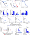
The selective inhibitor of p38α–MK2, CDD-450, inhibits IL-1β production by promoting IL-1β mRNA instability. (A–C) Effects of CDD-450 on p38α activation of MK2, PRAK, or ATF2. Activated p38α was added to inactive MK2, inactive PRAK, or ATF2 preincubated with various concentrations of CDD-450 or CDD-110. Phosphorylation of HSP27 peptide (for MK2 or PRAK activity) or ATF2 was determined. (D–F) qPCR analysis of IL-1β, NLRP3, and IL-18 mRNA expression. WT and NOMID BMMs were preincubated with CDD-450 or IKK2 inhibitor for 1 h and then with 100 ng/ml LPS for 3 or 8 h. (G–L) qPCR analysis of IL-1β, TNF-α, and NLRP3 mRNA stability. BMMs from WT and NOMID mice were stimulated with 100 ng/ml LPS for 3 h and then simultaneously exposed to 1 µg/ml actinomycin D and CDD-450, MK2 inhibitor, or IKK2 inhibitor for 0, 2, 4, or 6 h. (M and N) Analysis of IL-1β levels in conditioned media. WT and NOMID BMMs were preincubated with CDD-450 or IKK2 inhibitor for 1 h and then stimulated with 100 ng/ml LPS for 3 h. WT cells were treated for an additional 30 min with 15 µM nigericin. IL-1β levels were measured by ELISA. Data are means ± SEM from experimental triplicates and are representative of at least three independent experiments. *, P < 0.05; **, P < 0.01; ***, P < 0.001.
Figure 2.
Figure 2.
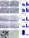
CDD-450 inhibits inflammatory responses in vivo. (A) Effects of CDD-450 and CDD-111 on LPS-induced TNF-α production. 8-wk-old WT female mice (six mice/group) were fed with normal chow, CDD-450 (1,000 ppm) chow, or CDD-111 (516 ppm) chow and challenged with LPS (0.025 mg/kg mouse) for 1 h at the indicated time (weeks). TNF-α serum levels were measured by ELISA. (B and C) Effects of CDD-450 on body weight and survival. 3-mo-old WT and NOMIDc littermate mice were fed with normal or CDD-450–formulated chow starting 3 d before sustained tamoxifen administration for 2 wk. Body weight (10–11 mice/group) and survival (12–13 mice/group) was scored every 2–3 d. (D) Analysis of IL-1β levels in bone marrow (four to eight mice/group). At the termination of the experiments, bone marrow was harvested and centrifuged, and IL-1β levels in the supernatants were measured by ELISA. (E) Spleen weights. (F–I) Complete blood counts (four to eight mice/group). Data are means ± SEM. *, P < 0.05; **, P < 0.01; ***, P < 0.001. (J–M) H&E staining of liver sections. Dotted circle indicates inflammatory cell infiltrates. Bars, 50 µm. RBC, red blood cell; WBC, white blood cell.
Figure 3.
Figure 3.
CDD-450 prevents bone destruction in NOMIDc mice. (A–F) 3-mo-old WT and NOMIDc littermate mice were fed with normal or CDD-450–formulated chow starting 3 d before sustained tamoxifen administration for 2 wk; the experiments were terminated 4 wk later. (A–D) Osteoclast (OC) formation in vivo. Bone sections (four to six mice/group) were stained for TRAP activity (red staining). (A and B) Osteoclasts (arrowheads) along trabecular bone surfaces were counted. (C and D) Osteoclasts along cortical bone surfaces were counted. BM, bone marrow; BS, bone surface; N, number; S, surface. (E and F) Effects of CDD-450 on osteoclast formation in vitro. WT BMMs were stimulated with 50 ng/ml RANKL for 4 d in the presence of DMSO (vehicle) or CDD-450. Cultures were stained for TRAP activity, and the number of osteoclasts was determined by counting TRAP+-multinucleated cells with at least three nuclei (arrowheads). In vivo data are means ± SEM; in vitro data are means ± SEM from experimental triplicates and are representative of at least three independent experiments. *, P < 0.05; **, P < 0.01; ***, P < 0.001. Bars, 100 µm.
Figure 4.
Figure 4.
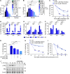
CDD-450 inhibits cytokine production in human cells. (A) Effects of CDD-450 on IL-1β production by PBMC isolated from CAPS patients or healthy controls. Cells were cultured for 16 h at 32°C or 37°C in the presence of DMSO or CDD-450. IL-1β levels in conditioned media were determined by ELISA. (B) Effects of CDD-450 on LPS-stimulated IL-1β production by normal human PBMC. Cells were stimulated with 100 ng/ml LPS for 16 h at 37°C in the presence of DMSO or CDD-450. (C) qPCR analysis of the effects of CDD-450 or IKK2 inhibitor on IL-1β mRNA stability in normal human PBMC. Cells were stimulated with 10 ng/ml LPS for 3 h and then simultaneously exposed to 1 µg/ml actinomycin D and CDD-450 or IKK2 inhibitor. (D–F) qPCR analysis of the effects of LPS (100 ng/ml), TNF-α (10 ng/ml), or IL-1β (10 ng/ml) on IL-1β, IL-18, and NLRP3 mRNA expression by synovial fibroblasts from an RA patient. (G) qPCR analysis of the effects of CDD-450 or IKK2 inhibitor on IL-1β mRNA expression by synovial fibroblasts from RA patient. Cells were preincubated with the inhibitors for 1 h before treatment with 10 ng/ml IL-1β for 3 h. (H and I) qPCR analysis of the effects of CDD-450 or IKK2 inhibitor on IL-1β and TNF-α mRNA stability in synovial fibroblasts from an RA patient. Cells were stimulated with 10 ng/ml IL-1β for 3 h and then simultaneously exposed to 1 µg/ml actinomycin D and CDD-450 or IKK2 inhibitor. Data are means ± SEM from experimental triplicates and are representative of at least two independent experiments. *, P < 0.05; **, P < 0.01; ***, P < 0.001. (J) Effect of CDD-450 on MK2 phosphorylation induced by IL-1β. Synovial fibroblasts were preincubated with 10 µM CDD-450 for 1 h and then treated with 10 ng/ml IL-1β. Samples were analyzed by Western blotting.
Figure 5.
Figure 5.
CDD-450 prevents joint destruction in inflammatory arthritis in rats. (A–C) Effects of CDD-450 or CDD-110 on paw swelling and bone destruction in SCW-induced arthritis. SCW preparations were injected into rats i.p. On day 9, rats displaying paw edema were sorted into groups (eight rats/group) and dosed daily with vehicle, 5, or 10 mg/kg CDD-450 2×/day (10 mg/kg/d or 20 mg/kg/d, respectively) or 1.5 mg/kg CDD-110 2×/day (3 mg/kg/d). (A) Paw volume measurements. (B) 3D µCT reconstruction of paws from rats treated with 5 mg/kg CDD-450 2×/day (10 mg/kg/d). (C) Bone mineral density (BMD) measurements corresponding with the area delineated by the dashed squares in B. Data are means ± SEM. *, P < 0.05; **, P < 0.01; ***, P < 0.001.

V体育官网 - Comment in

References

    1. Agostini L., Martinon F., Burns K., McDermott M.F., Hawkins P.N., and Tschopp J.. 2004. NALP3 forms an IL-1beta-processing inflammasome with increased activity in Muckle-Wells autoinflammatory disorder. Immunity. 20:319–325. 10.1016/S1074-7613(04)00046-9 - DOI - PubMed
    1. Alippe Y., Wang C., Ricci B., Xiao J., Qu C., Zou W., Novack D.V., Abu-Amer Y., Civitelli R., and Mbalaviele G.. 2017. Bone matrix components activate the NLRP3 inflammasome and promote osteoclast differentiation. Sci. Rep. 7:6630 10.1038/s41598-017-07014-0 - DOI - PMC - PubMed
    1. Bauernfeind F.G., Horvath G., Stutz A., Alnemri E.S., MacDonald K., Speert D., Fernandes-Alnemri T., Wu J., Monks B.G., Fitzgerald K.A., et al. . 2009. Cutting edge: NF-kappaB activating pattern recognition and cytokine receptors license NLRP3 inflammasome activation by regulating NLRP3 expression. J. Immunol. 183:787–791. 10.4049/jimmunol.0901363 - DOI - PMC - PubMed
    1. Bonar S.L., Brydges S.D., Mueller J.L., McGeough M.D., Pena C., Chen D., Grimston S.K., Hickman-Brecks C.L., Ravindran S., McAlinden A., et al. . 2012. Constitutively activated NLRP3 inflammasome causes inflammation and abnormal skeletal development in mice. PLoS One. 7:e35979 10.1371/journal.pone.0035979 - DOI - PMC - PubMed
    1. Bot F.J., Schipper P., Broeders L., Delwel R., Kaushansky K., and Löwenberg B.. 1990. Interleukin-1 alpha also induces granulocyte-macrophage colony-stimulating factor in immature normal bone marrow cells. Blood. 76:307–311. - PubMed

Publication types

MeSH terms

Substances