Skip to main page content
U.S. flag

An official website of the United States government

Dot gov

The . gov means it’s official. Federal government websites often end in . gov or . mil. Before sharing sensitive information, make sure you’re on a federal government site. VSports app下载.

Https

The site is secure V体育官网. The https:// ensures that you are connecting to the official website and that any information you provide is encrypted and transmitted securely. .

. 2018 Mar 1;69(5):729-743.e7.
doi: 10.1016/j.molcel.2018.02.005.

VSports app下载 - Dynamic Regulation of Long-Chain Fatty Acid Oxidation by a Noncanonical Interaction between the MCL-1 BH3 Helix and VLCAD

Affiliations

Dynamic Regulation of Long-Chain Fatty Acid Oxidation by a Noncanonical Interaction between the MCL-1 BH3 Helix and VLCAD

Silvia Escudero et al. Mol Cell. .

Abstract (V体育2025版)

MCL-1 is a BCL-2 family protein implicated in the development and chemoresistance of human cancer VSports手机版. Unlike its anti-apoptotic homologs, Mcl-1 deletion has profound physiologic consequences, indicative of a broader role in homeostasis. We report that the BCL-2 homology 3 (BH3) α helix of MCL-1 can directly engage very long-chain acyl-CoA dehydrogenase (VLCAD), a key enzyme of the mitochondrial fatty acid β-oxidation (FAO) pathway. Proteomic analysis confirmed that the mitochondrial matrix isoform of MCL-1 (MCL-1Matrix) interacts with VLCAD. Mcl-1 deletion, or eliminating MCL-1Matrix alone, selectively deregulated long-chain FAO, causing increased flux through the pathway in response to nutrient deprivation. Transient elevation in MCL-1 upon serum withdrawal, a striking increase in MCL-1 BH3/VLCAD interaction upon palmitic acid titration, and direct modulation of enzymatic activity by the MCL-1 BH3 α helix are consistent with dynamic regulation. Thus, the MCL-1 BH3 interaction with VLCAD revealed a separable, gain-of-function role for MCL-1 in the regulation of lipid metabolism. .

Keywords: BCL-2 family; MCL-1; VLCAD; apoptosis; fatty acid metabolism; mitochondria; mitochondrial matrix; stapled peptide; α helix; β-oxidation V体育安卓版. .


Conflict of interest statement

DECLARATION OF INTERESTS

L. D. W. is a scientific advisory board member and consultant for Aileron Therapeutics VSports最新版本. L. D. W. and S. E. have a patent filing related to this work.

Figures
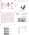
Figure 1
Figure 1. Identification of VLCAD as an MCL-1-Interacting Protein
(A) Proteomics workflow for streptavidin capture and identification of MEF lysate proteins that bound to a biotinylated and stapled MCL-1 BH3 helix. (B) Volcano plot demonstrating the distribution of identified proteins and high-confidence MCL-1 SAHBD interactors (blue), including VLCAD (red). Pull-down experiments and mass spectrometry analyses were performed in biological triplicate. (C) VLCAD was identified as a high-stringency hit with 49% sequence coverage (red) by mass spectrometry. (D) Interaction profiles of MCL-1, BIM, BID, and BAD BH3 stapled peptide helices with native MCL-1 and VLCAD, as demonstrated by streptavidin pull-down from MEF lysates, and MCL-1 and VLCAD western analyses. (E) Selective interaction of MCL-1 SAHBD with native VLCAD but not other members of the acyl-CoA dehydrogenase family, such as MCAD and SCAD, as assessed by streptavidin pull-down from MEF lysates, and VLCAD, MCAD, and SCAD western analyses. (F) Similar enrichment for native, full-length BIM, PUMA, and VLCAD proteins, but not MCAD or SCAD, in FLAG-MCL-1 immunoprecipitations from mouse liver mitochondria, isolated from Mcl-1fl/fl mice co-infected with scAAV-LP1-Cre and either scAAV-LP1-FLAG-MCL-1 or scAAV-LP1-GFP, and analyzed by mass spectrometry. Each immunoprecipitation represents the pooled mitochondria from 3 independent mice, and the mass spectrometry analysis was performed in duplicate. See also Figures S1–S2 and Tables S1–S2.
Figure 2
Figure 2. MCL-1 SAHBD Directly and Selectively Binds to VLCAD
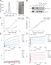
(A) Expression and purification of recombinant VLCAD, as demonstrated by size exclusion chromatography profile, Coomassie staining, and VLCAD western analysis. The collected peak corresponding to the obligate dimer is shaded in gray. (B) Interaction profiles of MCL-1, BIM, BID, and BAD BH3 stapled peptide helices with recombinant VLCAD and GST-MCL-1ΔNΔC, as demonstrated by streptavidin pull-down, and VLCAD and GST western analyses. (C–D) 19F NMR binding analyses demonstrated selective interaction of recombinant VLCAD with MCL-1 SAHBD (blue), but not MCL-1 SAHBB (purple) (C), whereas MCL-1ΔNΔC engaged with both SAHB constructs similarly (D). Data are mean ± S.D. for experiments performed in biological triplicate. (E–F) Comparative binding of MCL-1 SAHBD (blue) and MCL-1 SAHBB (purple) to recombinant VLCAD (E) and MCL-1ΔNΔC (F), as measured by biolayer interferometry, corroborated the 19F NMR findings. Representative traces are shown for BLI experiments performed in biological triplicate. See also Figures S2–S3, and Table S1.
Figure 3
Figure 3. Sequence Determinants for MCL-1 SAHBD Interaction with VLCAD
(A–B) Differential influence of alanine mutagenesis on the interactions between Btn-MCL-1 SAHBD and native VLCAD (A) and MCL-1 (B), as assessed by western blotting of streptavidin pull-downs from MEF lysates and densitometric analysis. Alanine mutations that reduced the wild-type interaction by more than 50% (red dotted line) are colored red on the helical wheels, whereas those constructs demonstrating less of a negative influence, no effect, or a binding enhancement are colored green. Native alanines and residues not mutated are colored gray. The dotted semicircles highlight the distinctive MCL-1 BH3 binding interfaces for VLCAD versus MCL-1 engagement, as defined by the differential sensitivities to alanine mutagenesis. The pull-downs were performed twice with similar results. See also Table S1.
Figure 4
Figure 4. Localization of the MCL-1 BH3 Binding Site on VLCAD
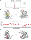
(A) Photoaffinity labeling of VLCAD by biotinylated MCL-1 pSAHBs that incorporate a benzophenone-containing residue (U). The plot represents the frequency and location of Btn-pSAHB/VLCAD crosslinks, as determined by LC-MS/MS analysis of biotinylated, streptavidin-captured, and trypsinized VLCAD protein. (B) Mapping of the pSAHB-crosslinked peptide fragments and specific residues (purple, green, and blue ribbons, and sticks, respectively) onto the VLCAD structure (PDB: 3B96) revealed their localization to a surface groove, where molecular docking calculations placed the MCL-1 BH3 helix (yellow cylinder). Intriguingly, fatty acid substrate (cyan) and the FAD co-factor (orange) lie adjacent to the putative helix-in-groove interaction. (C) The addition of MCL-SAHBD V220A to VLCAD (20 μM, 10:1 MCL-1 SAHB:VLCAD, 5 min incubation/10 sec deuteration) triggered a regiospecific decrease in deuterium incorporation compared to unliganded VLCAD, as measured by HXMS. The difference in deuterium uptake plot reflects the relative deuterium incorporation of MCL-1 SAHB/VLCAD minus the relative deuterium incorporation of VLCAD. Red arrows demarcate the boundary of the loop absent from the crystal structure of VLCAD (PDB: 3B96). Data are representative of two independent experiments. (D) The region of MCL-1 SAHB-induced protection encompasses peptide fragments corresponding to amino acids 431–489, which are highlighted in red on the ribbon diagram (PDB: 3B96) and map to the α-helical 2 region of VLCAD and the subjacent loop not found in the crystal structure (arrows). See also Table S1.
Figure 5
Figure 5. Elevated Long-Chain Fatty Acylcarnitine Levels upon Mcl-1 Deletion
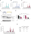
(A–B) Both chronic (Mcl-1−/−) (A) and acute (Mcl-1fl/flCreERT2, Tamoxifen) (B) deletion of Mcl-1 led to a similar profile of elevated long-chain fatty acylcarnitines compared to the corresponding control MEFs (wild-type and Mcl-1fl/flCreERT2, vehicle, respectively), as monitored by mass spectrometry of cellular lipid extracts. Error bars are mean ± S.D. for experiments performed in technical triplicate (A) and quadruplicate (B). (C) Murine livers with Mcl-1 deletion (Mcl-1fl/fl, scAAV-LP1-Cre/scAAV-LP1-GFP) demonstrated relative elevation of long-chain fatty acylcarnitines compared to those reconstituted with MCL-1 (Mcl-1fl/fl, scAAV-LP1-Cre/scAAV-LP1-MCL-1). Error bars are mean ± S.D. for experiments performed in technical triplicate. (D–E) MCL-1 western analysis of cellular lysates from tamoxifen-treated (d1–d3) Mcl-1fl/flCreERT2 + MCL-1OMM and Mcl-1fl/flCreERT2 + MCL-1Matrix MEFs at the indicated experimental time points (d1, d3, and d8; see experimental timeline, top) demonstrates the various levels of native and reconstituted MCL-1 protein isoforms (D), as also quantitated by densitometric analysis (E). Of note, reconstituted MCL-1Matrix protein is composed of the mitochondrial localization sequence of ATP synthase fused to an N-terminally truncated MCL-1 (Perciavalle et al., 2012), accounting for the lower molecular weight compared to native MCL-1Matrix. (F) Fatty acylcarnitines were quantified from the indicated MEFs on experimental day 8. The sequential elevation of long-chain fatty acylcarnitines inversely correlated with the level of MCL-1Matrix protein, rather than that of MCL-1OMM, as reflected by the color sequence (black>gray>magenta>pink) (E). Error bars are mean ± S.D. for experiments performed in technical quadruplicate. (G) Selective enrichment for BIM and PUMA by FLAG-MCL-1OMM, and VLCAD by FLAG-MCL-1Matrix, as evaluated by mass spectrometry of isoform-specific FLAG-MCL-1 immunoprecipitations from mouse liver mitochondria, which were isolated from Mcl-1fl/fl mice co-infected with scAAV-LP1-Cre and either scAAV-LP1-FLAG-MCL-1OMM or scAAV-LP1-FLAG-MCL-1Matrix. Each immunoprecipitation represents the pooled mitochondria from 3 independent mice, and the mass spectrometry analysis was performed in duplicate. See also Figures S4–S5.
Figure 6
Figure 6. Dynamic Regulation of Long-Chain Fatty Acid Oxidation by MCL-1Matrix
(A–B) Measurement of 3H-palmitic acid (A) and 3H-hexanoic acid (B) oxidation in wild-type and Mcl-1−/− MEFs in the presence and absence of serum. Error bars are mean ± S.D. for experiments performed in technical triplicate and repeated two (A) and four (B) times with similar results. **, p < 0.01. (C–D) Measurement of 3H-palmitic acid (C) and 3H-hexanoic acid (D) oxidation in vehicle- or tamoxifen-treated Mcl-1fl/flCreERT2 MEFs, in the presence and absence of serum. Error bars are mean ± S.D. for experiments performed in technical triplicate and repeated two times with similar results. **, p < 0.01. (E–F) Measurement of 3H-palmitic acid (E) and 3H-hexanoic acid (F) oxidation in tamoxifen-treated Mcl-1+/+CreERT2 control cells and tamoxifen-treated MCL1OMM or MCL-1Matrix-reconstituted Mcl-1fl/flCreERT2 MEFs, under serum-starved conditions. Error bars are mean ± S.D. for experiments performed in technical triplicate and repeated two times with similar results. ***, p < 0.001. (G) Measurement of 3H-palmitic acid in vehicle- or S63845-treated wild-type MEFs under serum-starved conditions. Error bars are mean ± S.D. for experiments performed in technical triplicate and repeated two times with similar results. (H) MCL-1 western analysis of lysates from wild-type MEFs subjected to the indicated duration of serum withdrawal. The experiment was performed twice with similar results. (I) Streptavidin pull-down of biotinylated MCL-1 SAHBD upon titration of palmitoyl-CoA or hexanoyl-CoA, followed by recombinant VLCAD detection by electrophoresis and Coomassie staining. (J) Effect of the indicated MCL-1 SAHBD constructs on the enzymatic activity of recombinant VLCAD. Error bars are mean ± S.E.M. for experiments performed in technical triplicate and repeated twice with similar results. (K) Initial velocity of recombinant VLCAD calculated based on the slope of the enzyme kinetic curves (J). Error bars are mean ± S.E.M for experiments performed in technical triplicate and repeated twice with similar results. *, p < 0.05; **, p < 0.01. See also Table S1.
Figure 7
Figure 7. Correlation Between MCL-1 Expression Level and Fatty Acid Metabolism in AML
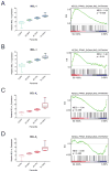
(A–D) Gene set enrichment analyses, performed on two distinct AML datasets, revealed a statistically significant correlation between MCL-1 expression level and a fatty acid β-oxidation signature (A–B). No such correlation was observed when the analysis was performed with an alternate anti-apoptotic BCL-2 family protein, BCL-XL (C–D). Error bars represent minimum and maximum relative expression level values.

References

    1. Aouacheria A, Combet C, Tompa P, Hardwick JM. Redefining the BH3 Death Domain as a ‘Short Linear Motif’. Trends Biochem Sci. 2015;40:736–748. - VSports最新版本 - PMC - PubMed
    1. Baeza J, Smallegan MJ, Denu JM. Mechanisms and Dynamics of Protein Acetylation in Mitochondria. Trends Biochem Sci. 2016;41:231–244. - PMC - PubMed
    1. Barclay LA, Wales TE, Garner TP, Wachter F, Lee S, Guerra RM, Stewart ML, Braun CR, Bird GH, Gavathiotis E, et al. Inhibition of Pro-apoptotic BAX by a noncanonical interaction mechanism. Mol Cell. 2015;57:873–886. - PMC - PubMed
    1. Beroukhim R, Mermel CH, Porter D, Wei G, Raychaudhuri S, Donovan J, Barretina J, Boehm JS, Dobson J, Urashima M, et al. The landscape of somatic copy-number alteration across human cancers. Nature. 2010;463:899–905. - PMC (VSports app下载) - PubMed
    1. Bird GH, Crannell WC, Walensky LD. Chemical synthesis of hydrocarbon-stapled peptides for protein interaction research and therapeutic targeting. Curr Protoc Chem Biol. 2011;3:99–117. - PMC - PubMed

Publication types

MeSH terms