Skip to main page content (VSports注册入口)
U.S. flag

An official website of the United States government

Dot gov

The VSports app下载. gov means it’s official. Federal government websites often end in . gov or . mil. Before sharing sensitive information, make sure you’re on a federal government site. .

Https

The site is secure V体育官网. The https:// ensures that you are connecting to the official website and that any information you provide is encrypted and transmitted securely. .

. 2018 Sep;25(9):1657-1670.
doi: 10.1038/s41418-018-0070-2. Epub 2018 Feb 19.

Lactobacillus accelerates ISCs regeneration to protect the integrity of intestinal mucosa through activation of STAT3 signaling pathway induced by LPLs secretion of IL-22

Affiliations

Lactobacillus accelerates ISCs regeneration to protect the integrity of intestinal mucosa through activation of STAT3 signaling pathway induced by LPLs secretion of IL-22

Qihang Hou et al. Cell Death Differ. 2018 Sep.

Erratum in

Abstract

The regeneration of intestinal epithelial are maintained by continuous differentiation and proliferation of intestinal stem cells (ISCs) under physiological and pathological conditions. However, little is known about the regulatory effect of intestinal microbiota on its recovery ability to repair damaged mucosal barrier. In this study, we established intestinal organoids and lamina propria lymphocytes (LPLs) co-cultured system, plus mice experiments, to explore the protective effect of Lactobacillus reuteri D8 on integrity of intestinal mucosa. We found that only live L. reuteri D8 was effective in protecting the morphology of intestinal organoids and normal proliferation of epithelial stained with EdU under TNF-α treatment, which was also further verified in mice experiments. L. reuteri D8 colonized in the intestinal mucosa and ameliorated intestinal mucosa damage caused by DSS treatment, including improvement of body weight, colon length, pathological change, and proliferation level VSports手机版. The repair process stimulated by L. reuteri D8 was also accompanied with increased numbers of Lgr5+ and lysozyme+ cells both in intestinal organoids and mice intestine. Furthermore, we demonstrated that D8 metabolite indole-3-aldehyde stimulated LPLs to secret IL-22 through aryl hydrocarbon receptor (AhR) and then induced phosphorylation of STAT3 to accelerate proliferation of intestinal epithelial, thus recovering damaged intestinal mucosa. Our findings indicate L. reuteri protects intestinal barrier and activates intestinal epithelial proliferation, which sheds light on treatment approaches for intestinal inflammation based on ISCs with probiotics Lactobacillus and daily probiotic consumption in heath foods. .


Conflict of interest statement

The authors declare that they have no conflict of interest.

Figures (V体育安卓版)

Fig. 1
Fig. 1
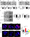
Building a co-cultured model with intestinal organoids and LPLs. a The co-culture model of LPLs and organoids. b Organoids cultured with LPLs were observed with a light microscope. Scale bar, 100 μm. c The growth status change of organoids from 1 to 6 days was observed with a light microscope. Scale bar, 50 μm
Fig. 2
Fig. 2
D8 increases the growth of intestinal organoids and the recovery of intestinal organoids after damage caused by TNF-α. a Size of organoids treated with/without D8 and HK-D8 (1 × 104 CFU per well); n = 50 organoids per group (day 3); total organoids number and budding organoids percentage of total organoids per well (day 3); n = 6 wells per group. Scale bars, 200 μm. b Organoids were treated with TNF-α (60 ng/ml) overnight, and the morphology of the organoids was observed with a light microscope. The number of total organoids and relative number of organoids with altered morphology per well were counted; n = 50 organoids per group. Scale bars, 200 μm. c Organoids were treated with TNF-α (60 ng/ml) overnight with/without D8 and HK-D8 (1 × 104 CFU per well). The number of total organoids and relative number of organoids with altered morphology per well were counted; n = 60 organoids per group. Scale bars, 200 μm. d Organoids were stained with EdU (red). Nuclei are stained blue. EdU-positive cells were found in the transit-amplifying region with obvious differences in the percentages among the four samples; n = 30 (control), n = 35 (D8), n = 28 (TNF-α), n = 33 (D8 + TNF-α) organoids per group. Scale bar, 50 μm. Data are the mean ± SD; comparisons performed with t-tests (two groups) or analysis of variance (ANOVA) (multiple groups). *P < 0.05, **P < 0.01, ***P < 0.001. Data combined from at least three independent experiments unless otherwise stated
Fig. 3
Fig. 3
D8 accelerates gut growth and ameliorates DSS-induced colitis in mice. a Mice were orally administrated with D8 (108 CFU), the number of D8 in mice feces at indicated time points were detected by MRS plates containing tetracycline (500 μg/ml). D8 labelled with Dylight 488 (108 CFU) was also administrated to mouse for 16 h, the distribution of D8 in intestine was detected by confocal microscope. D8 (green), DAPI (blue). b Changes in body weight were monitored daily starting form 21 day and presented relative to the initial body weight; n = 12 per group. c The colon lengths of mice treated with PBS, DSS, D8, or D8 + DSS. Treatment with DSS significantly reduced the colon length compared to the control group. D8 decreased the degree if reduction in colon length caused by DSS treatment. Scale bars, 200 μm. d Photomicrographs of the colons of mice treated with PBS, D8, DSS or DSS + D8. Note that treatment with D8 ameliorated DSS-induced colitis. Scale bars, 200 μm. e Confocal images (PCNA staining, red; and DAPI staining, blue) of colon of mice treated with PBS, D8, DSS or DSS + D8. The number of PCNA positive cells in each crypt was detected. Scale bars, 200 μm f Photomicrographs (×200) of mice treated with PBS, D8, DSS or DSS + D8. Administration of D8 decreased DSS-induced intestinal morphometric changes in mice. Segments of the jejunum were processed to measure the height of the villus and the crypt depth. Scale bars, 200 μm. g Confocal images (PCNA staining, red; and DAPI staining, blue) of jejunum of mice treated with PBS, D8, DSS or DSS + D8. The number of PCNA positive cells in each crypt was detected. Scale bars, 200 μm. h Administration of D8 reversed the increased levels of TNF-α and IL-1β in DSS-induced colitis. Note the increased levels of TNF-α and IL-1β due to exposure to DSS. Treatment with D8 decreased the levels of TNF-α and IL-1β; n = 6 per group. Data are the mean ± SD. The comparisons were performed with t-tests (two groups) or analysis of variance (ANOVA) (multiple groups). *P < 0.05, **P < 0.01, ***P < 0.001. Data combined from at least three independent experiments unless otherwise stated
Fig. 4
Fig. 4
D8 enhances the number of Paneth cells and stimulates ISCs regeneration. a Confocal images (Lgr5 staining, red; and DAPI staining, blue) of organoids cultured with/without D8 (104 CFU per well) and treated with/without TNF-α (60 ng/ml) for 24 h. The number of Lgr5-positive cells in each crypt were detected; n = 42 (control), n = 38 (D8), n = 45 (TNF-α), n = 53 (D8 + TNF-α) organoids per group. Scale bar, 10 μm. b Western blot results of Lgr5 expression in organoids cultured with/without D8 (104 CFU per well) and treated with/without TNF-α. n = 3 wells per group. c and f RT-qPCR was used to determine the relative mRNA expression of ISCs (Lgr5, Ascl2, and Olfm4) and Paneth cells (Lyz1 and Defa6) in organoids cultured with/without D8 and treated with/without TNF-α. n = 6 wells per group. d Confocal images (Lgr5 staining, red; and DAPI staining, blue) of the jejunum with different treatments. The number of Lgr5-positive cells in each crypt were detected (circled by dotted line). Scale bar, 100 μm. e Confocal images (lysozyme staining, green; and DAPI staining, blue) of organoids cultured with/without D8 (104 CFU per well) and treated with/without TNF-α (60 ng/ml). The number of lysozyme-positive cells in each crypt were detected; n = 50 organoids per group. Scale bar, 10 μm. g Confocal images (lysozyme staining, green; and DAPI staining, blue) of the jejunum with different treatments. The number of lysozyme-positive cells in each crypt were detected (circled by dotted line). Scale bar, 100 μm. Data are the mean ± SD. The comparisons were performed with t-tests (two groups) or analysis of variance (ANOVA) (multiple groups). *P < 0.05, **P < 0.01, ***P < 0.001. Data combined from at least three independent experiments unless otherwise stated
Fig. 5
Fig. 5
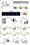
D8 up-regulates IL-22 expression ex vivo and in vivo. a Organoids were treated with TNF-α (60 ng/ml) or D8 (1 × 104 CFU per well). The expression of IL-22 in the four groups was detected using an ELISA kit; n = 6 well per group. b Size of organoids treated with/without D8 (104 CFU per well), IL-22 (5 ng/ml) or anti-IL-22 (0.1 μg/ml) for 24 h; n = 50 organoids per group; organoids number and budding organoids percentage of total organoids per well; n = 6 wells per group. Scale bars, 200 μm. c Organoids were stained with EdU (red). Nuclei were stained with DAPI (blue). EdU-positive cells were most distributed in the transit-amplifying region with obvious differences among the five groups; n = 30 organoids per group. Scale bar, 50 μm. d After the organoids were treated, the morphologies of the organoids with different treatments were assessed by light microscopy. The relative number of organoids with altered morphology was counted; n = 50 organoids per group. Scale bars, 200 μm. e Organoids were treated with HK-D8 or Lactobacillus ATCC 4356 (104 CFU per well) for 24 h. The expressions of IL-22 were detected using an ELISA kit; n = 6 well per group. f Organoids were treated with teichoic acid, exopolysaccharides, peptidoglycan (0.1, 1, 10 mM) for 24 h. The expression of IL-22 was detected using an ELISA kit; n = 6 well per group. g, Organoids were treated with acetate, propionate, butyrate, lactic acid, and indol-3-aldehyde (0.1, 1, 10 mM). The expression of IL-22 was detected using an ELISA kit; n = 6 well per group. h Organoids were cultured with/without D8 (1 × 104 CFU per well) or indole-3-aldehyde (1 mM) and treated with/without AHR inhibitor CH-223191 (1 mM). i Mice were treated with PBS, D8, DSS or DSS + D8. The expression of IL-22 in the four different groups was detected using an ELISA kit; n = 6 mice per group. Data are the mean ± SD; comparisons performed with t-tests (two groups) or analysis of variance (ANOVA) (multiple groups). *P < 0.05, **P < 0.01, ***P < 0.001. At least three independent experiments were performed
Fig. 6
Fig. 6
D8 activates pSTAT3 and the Reg3 signal pathway. a Confocal images of organoids of different treatments (pSTAT3 staining, red; UAE-1 staining, green; DAPI staining, blue). Obvious differences in the percentages of PSTAT3-positive and UAE-1-positive cells were observed among the four samples; n = 20 organoids per group. Scale bar, 50 μm. b Western blot results of pSTAT3 in organoids treated with IL-22 (5 ng/ml) or D8 (1 × 104 CFU per well) with/without anti-IL-22 (0.1 μg/ml); n = 3 wells per group. c The relative mRNA expressions of Reg3b and Reg3g in organoids treated with D8 (1 × 104 CFU per well) or TNF-α (60 ng/ml) for 24 h; n = 6 wells per group. d The relative mRNA expression of Reg3b and Reg3g in the jejunum and the colon treated with/without D8 and with/without DSS, n = 6 mice per group. Data are the mean ± SD. The comparisons were performed with t-tests or analysis of variance (ANOVA). *P < 0.05, **P < 0.01, ***P < 0.001. At least three independent experiments were performed
Fig. 7
Fig. 7
Our model depicts the protection conferred by Lactobacillus on the intestinal epithelial barrier via the modulation of ISCs. Lactobacillus stimulated LPLs to secret IL-22 via AhR and then activated phosphorylation of STAT3 to accelerate ISCs regeneration and thus maintain the epithelial barrier. Some factors, such as DSS, stimulated the intestinal mucosa to secret IL-1β and TNF-α, and then induced epithelial damage and intestinal inflammation. However, Lactobacillus stimulated ISCs proliferation through the pSTAT3 pathway activated by IL-22 and inhibited inflammatory cytokine secretion to protect the mucosa barrier

References

    1. Turner JR. Intestinal mucosal barrier function in health and disease. Nat Rev Immunol. 2009;9:799–809. doi: 10.1038/nri2653. - DOI - PubMed
    1. Tumanov AV, Koroleva EP, Guo XH, Wang YG, Kruglov A, Nedospasov S, et al. Lymphotoxin controls the IL-22 protection pathway in gut innate lymphoid cells during mucosal pathogen challenge. Cell Host Microbe. 2011;10:44–53. doi: 10.1016/j.chom.2011.06.002. - DOI - PMC - PubMed
    1. Bloemendaal AL, Buchs NC, George BD, Guy RJ. Intestinal stem cells and intestinal homeostasis in health and in inflammation: a review. Surgery. 2016;159:1237–48. doi: 10.1016/j.surg.2016.01.014. - DOI - PubMed
    1. Andersson-Rolf A, Zilbauer M, Koo BK, Clevers H. Stem cells in repair of gastrointestinal epithelia. Physiology. 2017;32:278–89. doi: 10.1152/physiol.00005.2017. - "V体育2025版" DOI - PMC - PubMed
    1. Matricon J, Barnich N, Ardid D. Immunopathogenesis of inflammatory bowel disease. Self Nonself. 2010;1:299–309. doi: 10.4161/self.1.4.13560. - DOI - PMC - PubMed

Publication types

MeSH terms (VSports手机版)