"V体育官网入口" Skip to main page content
U.S. flag

An official website of the United States government

Dot gov

The VSports app下载. gov means it’s official. Federal government websites often end in . gov or . mil. Before sharing sensitive information, make sure you’re on a federal government site. .

Https

The site is secure. The https:// ensures that you are connecting to the official website and that any information you provide is encrypted and transmitted securely V体育官网. .

. 2018 Jan 19;9(2):53.
doi: 10.1038/s41419-017-0081-9.

Epstein-Barr virus encoded latent membrane protein 1 suppresses necroptosis through targeting RIPK1/3 ubiquitination

Affiliations

Epstein-Barr virus encoded latent membrane protein 1 suppresses necroptosis through targeting RIPK1/3 ubiquitination

Xiaolan Liu et al. Cell Death Dis. .

"V体育ios版" Abstract

Necroptosis is an alternative programmed cell death pathway that is unleashed in the absence of apoptosis and mediated by signaling complexes containing receptor-interating protein kinase 1 (RIPK1) and RIPK3. This form of cell death has recently been implicated in host defense system to eliminate pathogen-infected cells VSports手机版. However, only a few viral species such as herpes simplex virus (HSV) and cytomegalovirus (CMV) have evolved mechanisms inhibiting necroptosis to overcome host antiviral defense, which is important for successful pathogenesis. Here, we show that the γ-herpesvirus Epstein-Barr virus (EBV) blocks necroptosis in EBV-infected human nasopharyngeal epithelial cells and nasopharyngeal carcinoma cells. Our findings indicate that EBV-encoded latent membrane protein 1 (LMP1), which lacks an RIP homotypic interaction motif (RHIM) domain, has mechanisms distinct from RHIM signaling competition to inhibit this necroptotic pathway. Intriguingly, LMP1 interacts directly with both RIPK1 and RIPK3 through its C-terminal activation region. More importantly, LMP1 can modulate the post-translational modification of the two receptor-interacting proteins. We then show that LMP1-mediated promotion of K63-polyubiquitinated RIPK1, suppression of RIPK1 protein expression and inhibition of K63-polyubiquitinated RIPK3 induced a switch in cell fate from necroptotic death to survival. These findings provide direct evidence for the suppression of necroptosis by EBV and define a mechanism of LMP1 to interrupt the initiation process of necroptosis before necrosome formation. .


Conflict of interest statement

All authors declare that they have no competing financial interests.

Figures

Fig. 1
Fig. 1
EBV infection prevents T/S/Z-induced necroptosis. a, b NP460hTERT and NP460hTERT-EBV cells were treated with T/S/Z, T/S/Z + Nec-1, T/S/Z + GSK’872, Nec-1, GSK’872, or DMSO solvent control for 24 h. At the end of treatment, cell viability and cell survival were determined by MTS and trypan blue exclusion assay. Data were presented as mean ± S.E.M. (n = 3). P values are by Student’s t test. T TNF-α, 100ng/ml; S Smac mimetic, 5 μM; Z z-VAD-fmk, 20 μM; Nec-1 Necrostatin-1, 40 μM; GSK’872: 5 μM. c Immunoblot analysis to detect phosphorylated RIPK3 (p-RIPK3), RIPK3, phosphorylated MLKL (p-MLKL), MLKL, cleaved Caspase-3 (Cl-Casp3), cleaved Caspase-8 (Cl-Casp8), and β-actin in lysates from NP460hTERT and NP460hTERT-EBV cells treated with T/S/Z, T/S/Z + Nec-1, T/S/Z + GSK’872, Nec-1, GSK’872, or DMSO solvent control and HT-29 cells treated with T/S or T/S/Z as indicated. d TEM photomicrographs of NP460hTERT and NP460hTERT-EBV cells treated with DMSO or T/S/Z. Black and white arrows denote plasma membrane rupture and cellular organelle swelling, respectively. Scale bars represent 5 μm
Fig. 2
Fig. 2
EBV-LMP1 plays a role in suppression of T/S/Z-induced necroptosis. a, b CNE1 and CNE1-LMP1 cells were treated with T/S/Z, T/S/Z + Nec-1, T/S/Z + GSK’872, Nec-1, GSK’872, or DMSO solvent control for 72 h. At the end of treatment, cell viability, and cell survival were determined by MTS and trypan blue exclusion assay. Data were presented as mean ± S.E.M. (n = 3). P values are by Student’s t test. T TNF-α, 100ng/ml; S Smac mimetic, 5 μM; Z z-VAD-fmk, 20 μM; Nec-1 Necrostatin-1, 40 μM; GSK’872: 5 μM. c CNE1 and CNE1-LMP1 cells were treated with T/S/Z or DMSO solvent control for 0, 24, 48, or 72 h. At the end of treatment, cell viability was determined by MTS assay. d Immunoblot analysis to detect phosphorylated RIPK3 (p-RIPK3), RIPK3, phosphorylated MLKL (p-MLKL), MLKL, and β-actin in lysates from CNE1 and CNE1-LMP1 cells treated with T/S/Z for 0, 24, 48, or 72 h. e TEM photomicrographs of CNE1 and CNE1-LMP1 cells treated with DMSO or T/S/Z. Black and white arrows denote plasma membrane rupture and cellular organelle swelling, respectively. Scale bars represent 5 μm. f CNE1 and CNE1-LMP1 cells were treated with T/S/Z, T/S/Z + Nec-1, T/S/Z + GSK’872, Nec-1, GSK’872, or DMSO solvent control for 72 h. Cells were then stained with Sytox Green and imaged by a fluorescent microscope. Scale bars represent 50 μm
Fig. 3
Fig. 3
LMP1 interacts directly with RIPK1 and RIPK3. a IP/IB was used to detect the interaction of LMP1 with RIPK1. Top panels show lysates of 293T cells transfected with Myc-RIPK1 and different amounts of pSG5-LMP1 subjected to IP with IgG or anti-Myc antibody followed by IB with anti-LMP1 or anti-Myc antibody. The lower panels depict IB of input cell lysates. Protein molecular size is shown to the right of the lanes. b IP/IB was used to detect the interaction of LMP1 with RIPK3. Top panels show lysates of 293T cells transfected with Flag-RIPK3 and different amounts of pSG5-LMP1 subjected to IP with IgG or anti-Flag antibody followed by IB with anti-LMP1 or anti-Flag antibody. The lower panels depict IB of input cell lysates. c, d CNE1-LMP1 cells were immunostained with anti-LMP1 (green) and anti-RIPK1 (red) or anti-RIPK3 (red) antibodies, and subjected to confocal microscopy. The nuclei were stained with DAPI. Scale bars represent 25 μm. e, f Proximity ligation assay was used to detect the LMP1–RIPK1 and LMP1–RIPK3 interactions in 293T cells transfected with the indicated expression plasmids (top of each panel). Red fluorescence corresponds to the PLA positive signal and blue fluorescence corresponds to nuclei (DAPI staining). Scale bars represent 10 μm. g Proximity ligation assay was used to detect the endogenous LMP1–RIPK1 and LMP–RIPK3 interactions in NP460hTERT-EBV cells treated with 10 μM MG132 for 16 h. Red fluorescence corresponds to the PLA positive signal and blue fluorescence corresponds to nuclei (DAPI staining). Scale bars represent 10 μm
Fig. 4
Fig. 4
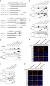
LMP1-CTAR2 interacts with RIPK1 and RIPK3 in an RHIM-independent manner. a LMP1, RIPK1, and RIPK3 domain architecture and schematic representation of their respective deletion or mutation constructs used in this study. LMP1 (ΔN), the N-terminal domain deletion mutant of LMP1; LMP1 (ΔC), the entire C-terminal domain deletion mutant of LMP1; LMP1 (ΔCTAR-2,3), the CTAR2 and CTAR3 deletion mutant of LMP1; LMP1 (ΔCTAR-1,3), the CTAR1 and CTAR3 deletion mutant of LMP1; LMP1 (ΔCTAR-1,2), the CTAR1 and CTAR2 deletion mutant of LMP1; RIPK1 (mutRHIM), the RHIM mutant RIPK1 with AAAA substitutions in IQIG(539-542); RIPK1 (ΔKD), the kinase domain deletion mutant of RIPK1; RIPK1 (ΔDD), the death domain deletion mutant of RIPK1; RIPK1 (ΔID), the entire intermediate domain deletion mutant of RIPK1; RIPK3 (mutRHIM), the RHIM mutant RIPK3 with AAAA substitutions in VQVG(458-461); RIPK3 (ΔCD), the C-terminal domain deletion mutant of RIPK3. b, c IP/IB was used to detect the interaction between Flag-tagged LMP1 mutants and Myc-RIPK1 or Myc-RIPK3. IP of cell lysates employed anti-Flag antibody followed by IB with anti-Myc or anti-Flag-HRP antibody. d, f IP/IB was used to detect the interaction between Myc-tagged RIPK1 mutants or RIPK3 mutants and Flag-LMP1. IP of cell lysates employed anti-Myc antibody followed by IB with anti-Flag-HRP or anti-Myc antibody. e, g Proximity ligation assay was used to detect the interaction between Myc-tagged RIPK1 mutants or RIPK3 mutants and Flag-LMP1
Fig. 5
Fig. 5
LMP1 promotes K63-linked polyubiquitination of RIPK1 through TRAF2 and inhibits RIPK1 protein level. a The top panel shows a schematic diagram of ubiquitin mutants. The bottom panel is an in vivo ubiquitination assay performed in 293T cells transfected with vectors encoding Myc-RIPK1, pSG5-LMP1, and HA-UB mutants. Ubiquitinated RIPK1 was detected by IP using anti-Myc antibody followed by IB with anti-HA antibody. The relative levels of RIPK1 ubiquitination were quantified and normalized to vehicle controls. b In vivo ubiquitination assay was performed in 293T cells transfected with vectors encoding Myc-RIPK1, HA-Ub-K63, pSG5-LMP1, and V5-TRAF2 or Flag-TRAF4 or Flag-TRAF6 as indicated. K63 ubiquitinated RIPK1 was detected by IP using anti-Myc antibody followed by IB with anti-HA antibody. c IB analysis was performed to examine the expression of TRAF2 in CNE1/CNE1-LMP1, NP69/NP69-LMP1 cells as well as LMP1 transiently transfected CNE1 cells. d IB analysis was performed to examine p-IκBα expression in NP460hTERT/NP460 hTERT-EBV, CNE1/CNE1-LMP1, and NP69/NP69-LMP1 cells treated with DMSO or T/S/Z. e The effect of LMP1 on stability of RIPK1 was examined by cycloheximide chase assay. HNE3 cells transfected with LMP1 or empty vector were treated with cycloheximide (20 μg/ml) for the indicated times. RIPK1 expression levels were determined by immunoblot analysis. f IB analysis was performed to examine the expression of RIPK1 in CNE1/ CNE1-LMP1, HNE2/HNE2-LMP1, SUNE1/SUNE1-LMP1 cells as well as LMP1 transiently transfected CNE1 cells
Fig. 6
Fig. 6
LMP1 inhibits K63-linked polyubiquitination of RIPK3 and blocks the RIPK1–RIPK3 necrosome formation. a An in vivo ubiquitination assay was performed in 293T cells transfected with vectors encoding GFP-RIPK3, Flag-LMP1, and HA-UB mutants. Ubiquitinated RIPK3 was detected by IP using anti-GFP antibody followed by IB with anti-HA antibody. The relative levels of RIPK3 ubiquitination were quantified and normalized to vehicle controls. b Proximity ligation assay was used to detect the RIPK1–RIPK3 interactions in NP460hTERT and NP460hTERT-EBV cells treated with DMSO or T/S/Z. Red fluorescence corresponds to the PLA positive signal and blue fluorescence corresponds to nuclei (DAPI staining). Scale bars represent 25 μm. c IP/IB demonstrating LMP1 disruption of the RIPK1 and RIPK3 interaction. Myc-RIPK1, Flag-RIPK3, and pSG5-LMP1 were transfected into 293T cells as indicated. The RIPK1–RIPK3 immunocomplex was analyzed by IP with anti-Flag antibody followed by IB with anti-Myc or anti-Flag-HRP antibody. d CNE1 and CNE1-LMP1 cells were treated with DMSO or T/S/Z for 72 h. The RIPK1–RIPK3 immunocomplex was analyzed by IP with anti-RIPK3 antibody followed by IB with anti-RIPK1 or anti-RIPK3 antibody
Fig. 7
Fig. 7
EBV-LMP1 regulates T/S/Z-induced necroptosis. EBV-LMP1 (−) cells stimulated with T/S/Z undergo necroptosis through RIPK1–RIPK3 signaling. However, EBV-LMP1 (+) cells can survive under this stimulation. On the one hand, LMP1 interacts directly with both RIPK1 and RIPK3 through its C-terminal activation region. On the one hand, LMP1 promotes K63-linked polyubiquitination of RIPK1 and suppresses the protein expression while inhibiting K63-linked polyubiquitination of RIPK3. These effects contribute to the activation of NF-κB and disruption of necrosome formation, collectively switching cell fate from death to survival

References (VSports手机版)

    1. Vandenabeele P, Galluzzi L, Vanden Berghe T, Kroemer G. Molecular mechanisms of necroptosis: an ordered cellular explosion. Nat. Rev. Mol. Cell Biol. 2010;11:700–714. doi: 10.1038/nrm2970. - DOI - PubMed
    1. Li J, et al. The RIP1/RIP3 necrosome forms a functional amyloid signaling complex required for programmed necrosis. Cell. 2012;150:339–350. doi: 10.1016/j.cell.2012.06.019. - DOI - PMC - PubMed
    1. Sun L, et al. Mixed lineage kinase domain-like protein mediates necrosis signaling downstream of RIP3 kinase. Cell. 2012;148:213–227. doi: 10.1016/j.cell.2011.11.031. - DOI - PubMed
    1. Wang H, et al. Mixed lineage kinase domain-like protein MLKL causes necrotic membrane disruption upon phosphorylation by RIP3. Mol. Cell. 2014;54:133–146. doi: 10.1016/j.molcel.2014.03.003. - DOI (VSports手机版) - PubMed
    1. Kaiser WJ, et al. Toll-like receptor 3-mediated necrosis via TRIF, RIP3, and MLKL. J. Biol. Chem. 2013;288:31268–31279. doi: 10.1074/jbc.M113.462341. - DOI - PMC - PubMed

Publication types

MeSH terms

Substances