Skip to main page content
U.S. flag

An official website of the United States government

Dot gov

The . gov means it’s official. Federal government websites often end in . gov or VSports app下载. mil. Before sharing sensitive information, make sure you’re on a federal government site. .

Https

The site is secure V体育官网. The https:// ensures that you are connecting to the official website and that any information you provide is encrypted and transmitted securely. .

. 2017 Nov 7;8(68):112530-112549.
doi: 10.18632/oncotarget.22486. eCollection 2017 Dec 22.

Decreased expression of the long non-coding RNA SLC7A11-AS1 predicts poor prognosis and promotes tumor growth in gastric cancer

Affiliations

Decreased expression of the long non-coding RNA SLC7A11-AS1 predicts poor prognosis and promotes tumor growth in gastric cancer

Yajun Luo et al. Oncotarget. .

Abstract

Many lncRNA and mRNA sense-antisense transcripts have been systematically identified in malignant cells. However, the molecular mechanisms of most lncRNA-mRNA pairs in gastric cancer remain largely unknown. We found the gastric cancer-associated lncRNA SLC7A11-AS1 and coding transcript mRNA SLC7A11 in human gastric cancer specimens by microarray. SLC7A11-AS1, antisense to SLC7A11, is significantly down-regulated in gastric cancer and could promote tumor growth in vitro and in vivo. The effects of SLC7A11-AS1 depend on the regulation of SLC7A11 via the ASK1-p38MAPK/JNK signaling pathway. These findings suggest that decreased expression of SLC7A11-AS1 contributes to the progression of gastric cancer and may be a novel diagnostic biomarker and effective therapeutic target in gastric cancer patients VSports手机版. .

Keywords: SLC7A11; SLC7A11-AS1; gastric cancer; long non-coding RNA; proliferation V体育安卓版. .


Conflict of interest statement

CONFLICTS OF INTEREST There are no conflicts of interest.

"VSports在线直播" Figures
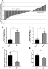
Figure 1
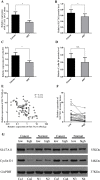
Figure 1. SLC7A11-AS1 expression levels were assessed in human gastric cancer tissues and PBMCs
(A) SLC7A11-AS1 expression was assessed in human GC tissues and paired adjacent noncancerous tissues (ANTs). Bars represent the ratio between expression in GC tissues and ANT (C/N, log scale) from 100 patients was presented. GC tissues express significantly lower levels of SLC7A11-AS1 compared with paired ANTs in the majority of patients (64%). (B) The relative expression levels of SLC7A11-AS1 in GC tissues was significantly lower than those in ANTs (P < 0.001, N = 100). (C) The relative expression levels of SLC7A11-AS1 in 33 GC patients’ PBMCs was significantly lower than those in 20 healthy controls (P = 0.011). (D) SLC7A11-AS1 expression level was down-regulated in the stage III/IV GC tissues compared with the stage I/II GC tissues (P = 0.003). (E) Ki-67 positive rate was higher in the SLC7A11-AS1 low expression group (N = 48) than that in the SLC7A11-AS1 high expression group (N = 22) (P < 0.001). Expression levels were normalized to β-actin levels. Results are shown as mean ± SEM. (*P < 0.05, **P < 0.01, ***P < 0.001, two-tailed Student's t-test).
Figure 2
Figure 2. Immunohistochemical staining for Ki-67 in pairs of representative gastric cancer tissues with adjacent non-tumorous tissues (ANTs)
Ki-67 labeling tumor cells were clearly identified by their brown nuclear staining. Ki-67 was mainly found in the nuclei of the carcinoma cells. High expression of Ki-67 in GC tissues compare with ANTs (200×). Scale bars, 100 mm.
Figure 3
Figure 3. The ROC curves
ROC curves analyses SLC7A11-AS1 for detecting gastric cancer patients in the tissues(AUC = 0.632, N = 100, P = 0.001,) and PBMCs (AUC = 0.791, N = 33, P < 0.001).
Figure 4
Figure 4. SLC7A11 and cyclin D1 expression level were assessed in human GC tissues
(A) Levels of SLC7A11 in GC tissues was significantly higher than those in paired ANTs (P = 0.037, N = 80), expression levels were normalized to β-actin levels. (B). Western Blot analysis protein level of SLC7A11 between GC tissues and paired ANTs (P = 0.025, N = 75), expression levels were normalized to GAPDH levels. (C) Levels of cyclin D1 in GC tissues was significantly higher than those in paired ANTs (P = 0.040, N = 32), and expression levels were normalized to β-actin levels. (D) Western Blot analysis of protein level of cyclin D1 between GC tissues and paired ANTs (P = 0.385, N = 32), expression levels were normalized to GAPDH levels. (E) Bivariate correlation analysis of the relationship between SLC7A11-AS1 and SLC7A11 expression level, and the resulting Spearman correlation was calculated as 0.252 where P = 0.024 (N = 80). (F) SLC7A11 expression level was higher in the SLC7A11-AS1low GC tissues than that SLC7A11-AS1high GC tissues (P = 0.045), expression levels were normalized to β-actin levels. (G) SLC7A11 and cyclin D1 protein levels were increased in SLC7A11-AS1low GC tissues compare with SLC7A11-AS1high GC tissues. Results are shown as mean ± SEM. (*P < 0.05, NS: No significantly, two-tailed Student's t-test).
Figure 5
Figure 5. Immunohistochemical staining of SLC7A11 in pairs of representative gastric cancer tissues with adjacent non-tumorous tissues (ANT)
Marked SLC7A11 expression was observed in GC. SLC7A11 was mainly found in the membrane of the carcinoma cells. High expression of SLC7A11 in GC tissues compare with ANTs (200×). Scale bars, 100 mm.
Figure 6
Figure 6. The expression of SLC7A11-AS1 and SLC7A11 were changed after transfection of SLC7A11-AS1 specific shRNA or negative control shRNA
(A) The relative expression of SLC7A11-AS1 was measured in various cell lines were detected by qPCR, including 4 GC cell lines (SGC-7901, BGC-823, MGC-803 and HGC-27). (B) Schematic diagram was for exhibiting the location between SLC7A11-AS1 and SLC7A11. (C) SLC7A11-AS1 knocked down in SGC-7901 (P=0.001), MGC-803 (P = 0.023) and HGC-27 (P = 0.008) cells infected adenovirus shRNA by sh-NC and sh-SLC7A11-AS1 vector. (D) The SLC7A11 expression level was up-regulated in SGC-7901 (P = 0.034), MGC-803 (P = 0.025) and HGC-27 (P = 0.005) cells after knocked down SLC7A11-AS1. Expression levels were normalized to GAPDH levels. (E) The SLC7A11 protein level was up-regulated in SGC-7901, MGC-803 and HGC-27 cells after knocked down SLC7A11-AS1. Expression levels were normalized to GAPDH levels. Results are shown as mean ± SEM. (*P < 0.05, **P < 0.01, two-tailed Student's t-test)
Figure 7
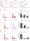
Figure 7. SLC7A11-AS1 low expression promotes gastric cancer cells proliferation and causes G1/S progress
(A) The function of SLC7A11-AS1 on cell proliferation was determined by CCK8 assay. Cell proliferation was increased after knocked down SLC7A11-AS1 in SGC-7901 (P < 0.001), MGC-803(P = 0.046) and HGC-27(P < 0.001) cells. (BD) The function of SLC7A11-AS1 on cell cycle was measured by flowcytometry after PI staining in SGC-7901, MGC-803 and HGC-27 cells. Error bars suggest mean ± SEM. (*P < 0.05, **P < 0.01, ***P < 0.001, NS: No significantly).
Figure 8
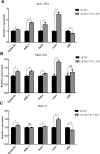
Figure 8. The expression of cyclin D1, ASK1, Gclm, p38 and c-Jun were changed after knocking down SLC7A11-AS1 with specific shRNA or negative control shRNA
(A) The cyclin D1 (P = 0.035), ASK1 (P < 0.001), Gclm (P = 0.037), p38 (P = 0.032) and c-Jun (P = 0.001) expressed level in SGC-7901 cells when SLC7A11-AS1 was knocked down. (B) The cyclin D1 (P=0.018), ASK1 (P = 0.021), Gclm (P = 0.015), p38 (P = 0.415) and c-Jun (P=0.003) expressed level in MGC-803 cells when SLC7A11-AS1 was knocked down. (C) The cyclin D1 (P = 0.025), ASK1 (P = 0.826), Gclm (P = 0.008), p38 (P < 0.001) and c-Jun (P = 0.008) expressed level in HGC-27 cells when SLC7A11-AS1 was knocked down. Expression levels were normalized to GAPDH levels. Results are shown as mean ± SEM. (*P < 0.05, **P < 0.01, ***P < 0.001, NS: No significantly, two-tailed Student's t-test).
Figure 9
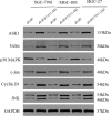
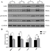
Figure 9. The protein of cyclin D1, ASK1, Gclm, p38, c-Jun and JNK were changed after knocking down SLC7A11-AS1 with specific shRNA or negative control shRNA
The ASK1, Gclm,, c-Jun, cyclin D1 and JNK protein levels were up-regulated and p38 protein level was down-regulated in SGC-7901, MGC-803 and HGC-27 cells after knocking down SLC7A11-AS1, expression levels were normalized to GAPDH levels.
Figure 10
Figure 10. Expression changes of ASK1, Gclm, c-Jun and p38 were assessed in human SLC7A11-AS1low gastric cancer tissues
(A) The protein level of ASK1, Gclm and c-Jun were increased and p38MAPK was decreased in 30 paired SLC7A11-AS1low GC tissues compared with ANTs, expression levels were normalized to GAPDH levels. (B) The expression of ASK1 (P = 0.027) and Gclm (P = 0.217) mRNA level were up-regulated and p38 (P = 0.638) and c-Jun (P = 0.193) mRNA levels were down-regulated in 30 paired SLC7A11-AS1low group GC tissues and respective ANTs. Expression levels were normalized to β-actin levels. Results are shown as mean ± SEM. (*P < 0.05, NS: No significantly, Ca: cancer tissue, N: respective ANTs, two-tailed Student's t-test)
Figure 11
Figure 11. The role of the SLC7A11-AS1–SC7A11 model interacts in tumor development
Decreased expression of SLC7A11-AS1 is increases GSH levels within cells by up-regulating SLC7A11 expression. The resultant increase in cellular GSH levels activates ASK1-p38MAPK/JNK signaling in cancer cells, thereby promoting tumor development. ASCT, system ASC glutamine transporter.

"V体育官网" References

    1. Siegel RL, Miller KD, Jemal A. Cancer statistics, 2015. CA Cancer J Clin. 2015;65:5–29. - PubMed
    1. GBD Mortality and Causes of Death Collaborators. Global, regional, and national age-sex specific all-cause and cause-specific mortality for 240 causes of death, 1990–2013: a systematic analysis for the Global Burden of Disease Study 2013. Lancet. 2013;2015;385:117–171. - PMC - PubMed
    1. Ponting CP, Oliver PL, Reik W. Evolution and functions of long noncoding RNAs. Cell. 2009;136:629–641. - PubMed
    1. Cheetham SW, Gruhl F, Mattick JS, Dinger ME. Long noncoding RNAs and the genetics of cancer. Br J Cancer. 2013;108:2419–2425. - PMC (VSports手机版) - PubMed
    1. Spizzo R, Almeida MI, Colombatti A, Calin GA. Long non-coding RNAs and cancer: a new frontier of translational research? Oncogene. 2012;31:4577–4587. - V体育ios版 - PMC - PubMed