"VSports app下载" Skip to main page content
U.S. flag

An official website of the United States government

Dot gov

The . gov means it’s official. Federal government websites often end in . gov or . mil. Before sharing sensitive information, make sure you’re on a federal government site. VSports app下载.

Https

The site is secure. The https:// ensures that you are connecting to the official website and that any information you provide is encrypted and transmitted securely. V体育官网.

. 2018 Feb 5;215(2):699-718.
doi: 10.1084/jem.20171116. Epub 2018 Jan 16.

Targeting IRF3 as a YAP agonist therapy against gastric cancer

Affiliations

Targeting IRF3 as a YAP agonist therapy against gastric cancer

"VSports手机版" Shi Jiao et al. J Exp Med. .

Abstract

The Hippo pathway plays a vital role in tissue homeostasis and tumorigenesis. The transcription factor IRF3 is essential for innate antiviral immunity. In this study, we discovered IRF3 as an agonist of Yes-associated protein (YAP). The expression of IRF3 is positively correlated with that of YAP and its target genes in gastric cancer; the expression of both IRF3 and YAP is up-regulated and prognosticates patient survival. IRF3 interacts with both YAP and TEAD4 in the nucleus to enhance their interaction, promoting nuclear translocation and activation of YAP. IRF3 and YAP-TEAD4 are associated genome-wide to cobind and coregulate many target genes of the Hippo pathway. Overexpression of active IRF3 increased, but depletion of IRF3 reduced, the occupancy of YAP on the target genes. Knockdown or pharmacological targeting of IRF3 by Amlexanox, a drug used clinically for antiinflammatory treatment, inhibits gastric tumor growth in a YAP-dependent manner VSports手机版. Collectively, our study identifies IRF3 as a positive regulator for YAP, highlighting a new therapeutic target against YAP-driven cancers. .


Figures

None
Graphical abstract
Figure 1.
Figure 1.
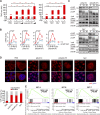
Viral infection induces YAP activation. (A) Luciferase activity of TEAD promoter in YAP-overexpressing cells after poly(I:C)/poly(dA:dT) stimulation or virus infection. HEK293FT cells were transfected with empty vector or Flag-YAP plasmid, together with TEAD-luciferase reporter, and renilla luciferase reporter for 24 h. Then, cells at 80% confluence was transfected with 0.5 or 1 µg/ml poly(I:C)/poly(dA:dT) or infected with SeV (MOI 1), VSV (MOI 0.1), or HCV (MOI 0.1) for 0 to ∼48 h, and subsequently the luciferase assay was performed. (B) Transcriptional levels of CTGF and CYR61 in YAP/TAZ-depleted cells after transfection with poly(I:C)/poly(dA:dT). Cells (70% confluence) were transfected with 1 µg/ml poly(I:C)/poly(dA:dT) for the indicated times (∼0–72 h), and then real-time PCR was performed. (C) Immunoblotting analysis of the protein levels of YAP(S127), pYAP, YAP, pIRF3(S396), and IRF3 in HEK293FT cells after nucleic acids/virus treatment. When HEK293FT cells reached ∼70% confluence, they were transfected with 1 µg/ml poly(I:C)/poly(dA:dT) or infected with SeV (MOI 1)/VSV (MOI 0.1) for the indicated duration of time (0, 24, or 48 h). Cell lysates were prepared and subjected to immunoblotting for the indicated proteins and phosphorylation. Phos-tag denotes phos-tag gel used to resolve phosphorylated YAP based on mobility shift. Molecular mass is indicated in kilodaltons. (D) Localization of YAP in cells with or without nucleic acids/virus treatment. After nucleic acids/virus treatment for 48 h, cells were cultured sparsely or to confluence. YAP was then stained with anti-YAP antibody. Bars, 10 µm. (E) GSEA analysis showing significant positive enrichment of three sets of YAP targets genes in SeV-infected HGC-27 cells by RNA-seq. HGC-27 cells (∼80% confluence) were infected with SeV (MOI 1) for 48 h. Total RNA was extracted, and RNA-seq was subsequently performed. At least two independent experiments were performed for all data. Two biological replicates were used for RNA-seq. For bar figures and curve figures, data are presented as means ± SD. Unpaired Student’s t tests were used for comparing two variables. One-way ANOVA was used for multiple variables comparison. *, P < 0.05; **, P < 0.01; ***, P < 0.001; n.s., no significance in comparison with control group.
Figure 2.
Figure 2.
IRF3-mediated antiviral signaling regulates YAP activity. (A) Transactivity of TEAD4 promoter and transcription of CTGF in YAP-overexpressing HEK293FT cells after transfection with the CARD domain of RIG-I, MAVS, STING, TBK1, or IRF3(5D). (B) mRNA levels of CTGF in YAP-overexpressing cells after transfection with the indicated plasmids. (C) YAP staining in cells overexpressing the indicated plasmids. (D) Nuclear localization of YAP in HGC-27 cells after transfection with siMAVS, siSTING, or siIRF3, respectively. (E) Immunoblotting analysis of the protein levels of p-YAP(S127) in HEK293FT cells transfected with the indicated plasmids. (F) Immunoblotting of the protein levels of pYAP(S127) in HGC-27 cells after transfection with specific IRF3 siRNAs. Molecular mass is indicated in kilodaltons. (G) YAP staining in IRF3-depletion cells. After transfection with IRF3 siRNAs (a mixture of siIRF3-1 and siIRF3-2) for 48 h, HGC-27 cells were seeded in 33-mm dishes to ∼20–30% confluence. YAP was then stained with anti-YAP antibody. Bars, 10 µm. (H) CTGF mRNA in cells after transfection with indicated siRNAs. At least two independent experiments were performed for all data. For bar figures, data are presented as means ± SD. Unpaired Student’s t tests were used for comparing two variables. One-way ANOVA was used for multiple variables comparison. **, P < 0.01; ***, P < 0.001; 5D, IRF3(S396D/S398D/S402D/S405D/T404D); e.v., empty vector; n.c., negative control siRNA.
Figure 3.
Figure 3.
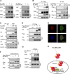
IRF3 binds both YAP and TEAD4 to form a complex in the nucleus. (A) CoIP of endogenous YAP and IRF3 in HGC-27 cells after treatment with poly(I:C) and poly(dA:dT). (B) CoIP analysis of WT or mutant YAP with WT or mutant IRF3. (C) CoIP of YAP with TEAD4 in HEK293FT cells after transfection with the indicated plasmids. (D) Exogenous coIP of Flag-TEAD4 with HA-IRF3 or HA-IRF3(5D). (E) Colocalization of Flag-TEAD4 with HA-IRF3. (F) Flag pull-down assay to assess the interaction of TEAD4 and IRF3. (G) CoIP of endogenous TEAD4 and IRF3 in SeV-infected cells with or without 10 µg/ml DNase. Molecular mass is indicated in kilodaltons. (H) Schematic model showing that IRF3 binds both YAP and TEAD4 to form a complex, thus retaining YAP in the nucleus. For all data, experiments were repeated two times.
Figure 4.
Figure 4.
Genome-wide association of IRF3 with YAP and TEAD4. (A) Heat map representing YAP, TEAD4, and IRF3 binding sites located on promoters (top) and enhancers (bottom). YAP, TEAD4, and IRF3 peaks are ranked from the strongest to weakest signal. (B) Absolute distance of YAP peaks (n = 7,606), TEAD4 peaks (n = 8,325), IRF3 peaks (n = 8,715), or overlapping YAP/TEAD4/IRF3 peaks (n = 6,275) to the nearest TSS. (C) Overlap of peaks identified with YAP, TEAD4, and IRF3 antibodies. (D) Linear correlation between the signal of YAP or TEAD4 and IRF3 peaks in the 6,275 shared binding sites. r is the coefficient of determination of the two correlations. (E) Representative examples of YAP/TEAD4/IRF3 binding profiles in the genome of HGC-27 cells. (F) ChIP assay showing IRF3 bound to the indicated genes’ promoters in 293FT cells transfected with HA-IRF3 or HA-IRF3(5D). Chromatin was immunoprecipitated with the HA antibody followed by qPCR using primer pairs spanning the human CTGF or CYR61 locus. (G) ChIP-qPCR showing YAP binding to the promoter of indicate genes in 293FT cells transfected with WT IRF3 and its mutant. (H) ChIP experiment performed with YAP antibody or IRF3 antibody in cells after transfection with poly(I:C). (I) ChIP experiment performed with YAP antibody in IRF3-depleted HGC-27 cells. (J) Primary component analysis for each siRNA. n.c. (gray), siYAP (blue), and siIRF3 (red) are indicated in the 3D scatter plot. A mixture of the two siRNAs for each gene was used. (K) GSEA analysis showing significant negative enrichment of three sets of YAP targets genes in siIRF3 group. (L) Heat map indicating the YAP targets genes that are significantly down-regulated in IRF3 knockdown state. (M) RNA-seq reads for YAP target genes (CTGF, CYR61, and AXL) by using IGV browser. For all ChIP assays, results are presented as percentage immunoprecipitated over input (0.5%) and are representative of three independent experiments. *, P < 0.05; **, P < 0.01; ***, P < 0.001; n.s., not significant relative to control group.
Figure 5.
Figure 5.
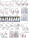
Targeting IRF3 inhibits GC growth. (A) Cell proliferation of HGC27, BGC-823, and MKN45 cells after transfection with IRF3 siRNAs. (B) Colony formation of IRF3-depleted cells. (C) Cell proliferation of HGC27 and BGC-823 cells after transfection with IRF3 siRNAs (a mixture of two siRNAs) together with YAP(S127A). (D) Colony formation of IRF3-depleted cells after transfection with YAP(S127A). (E) Knockdown of endogenous IRF3 inhibited xenograft tumor growth. Mice were photographed after being killed. BALB/cA nu/nu mice (aged 4 wk) were injected with the GC cell lines (HGC-27, BGC-823, and MKN-45). Once palpable tumors were detected, pairs of mice were randomized and treated with lentivirus-delivered shIRF3 or scramble shRNA by subcutaneous injection (n = 10). (F) Tumor volumes for the mice from E. (G) Tumor numbers in WT and IRF3−/− mice after administration of H. pylori intragastrically with alkylating agent MNNG in drinking water. 4-wk-old IRF3−/− mice and their WT littermates were orally gavaged with 50 µl of bacterial suspension (∼106 CFU) every day, which persisted for at least 6 mo before sacrifice. 100 mg/ml MNNG was added to the drinking water for a period of up to 2 mo. A total of 40 mice were reared, including 20 normal controls. (H) Ki67 staining of adenomas from G. Bar, 50 µm. (I) Relative mRNA levels of YAP target genes in gastric tissue from G. (J) Tumor numbers in WT and IRF3−/− mice after administration of YAP lentivirus intragastrically with alkylating agent MNNG in drinking water. (K) Ki67 staining of adenomas from J. Bar, 50 µm. (L) Relative mRNA levels of YAP target genes in gastric tissue from J. At least two independent experiments were performed for all data. For curve figures and bar figures, data are presented as means ± SD. Unpaired Student’s t tests were used for comparing two variables. One-way ANOVA was used for multiple variables comparison. *, P < 0.05; **, P < 0.01; ***, P < 0.001; n.s., no significance in comparison with control group.
Figure 6.
Figure 6.
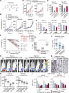
Pharmacological inhibition of IRF3 suppresses GC growth. (A) Chemical structures of Amlexanox. (B) Cell proliferation of HGC27, BGC-823, and MKN45 cells after treatment with different doses of Amlexanox. (C) Colony formation of Amlexanox-treated cells. (D) Cell proliferation of YAP(S127)-overexpressing cells after treatment with Amlexanox. (E) Colony formation of YAP(S127A)-overexpressing cells after treatment with Amlexanox. (F) Cell viability of various cancer cells after treatment with different doses of Amlexanox. (G) Xenograft tumor growth of GC cell lines after treatment with Amlexanox. (H) Tumor volumes for the mice from G. (I) mRNA levels of YAP target genes in samples from G. (J) Tumor numbers in MNNG/HP-induced GC model after treatment with different doses of Amlexanox. (K) Ki67 staining of adenomas from J. Bar, 50 µm. At least two independent experiments were performed for all data. For curve figures and bar figures, data are presented as means ± SD. Unpaired Student’s t tests were used for comparing two variables. One-way ANOVA was used for multiple variables comparison. *, P < 0.05; **, P < 0.01; ***, P < 0.001; n.s., no significance in comparison with control group.
Figure 7.
Figure 7.
Pathological association of IRF3 with YAP in GC. (A) Scatter plot of positive correlation between IRF3 and YAP/CTGF at the transcriptional level in different cancer cell lines. mRNA levels of IRF3 were compared with those of YAP (left) and CTGF (right) by Spearman’s correlation. (B) Western blotting of YAP, CTGF, and IRF3 in GC cell lines. (C) CoIP assay for detecting the association of YAP/IRF3 with TEAD4 in GC cells. (D) Box plots for mRNA levels of IRF3 and YAP in MNNG/HP samples. (E) mRNA levels of IRF3, YAP, CTGF, and AXL in GC. (F) Protein levels of IRF3, YAP, CTGF, CYR61, and CDX2 were elevated in GC samples. Molecular mass is indicated in kilodaltons. (G) Representative cores of YAP and IRF3 staining on tissue microarray. Bar, 100 μm. (H) Staining levels of YAP and IRF3 in normal and cancerous colon tissue indicating negative (–), weak (+), moderate (++), and strong (+++) expression levels. (I) Kaplan–Meier survival analysis of patients with YAP/IRF3 at high or low levels from tissue microarray. At least two independent experiments were performed for all data. For box figures, data are presented as means ± SD. Unpaired Student’s t tests were used for comparing two variables. One-way ANOVA was used for multiple variables comparison. For correlation, the Spearman rank correlation was used for continuous variables. Survival curves were calculated according to the Kaplan–Meier method; survival analysis was performed using the log-rank test. **, P < 0.01; ***, P < 0.001 in comparison with control group.

References

    1. Akira S., Uematsu S., and Takeuchi O.. 2006. Pathogen recognition and innate immunity. Cell. 124:783–801. 10.1016/j.cell.2006.02.015 - DOI - PubMed
    1. Akram N., Imran M., Noreen M., Ahmed F., Atif M., Fatima Z., and Bilal Waqar A.. 2017. Oncogenic role of tumor viruses in humans. Viral Immunol. 30:20–27. 10.1089/vim.2016.0109 - DOI - PubMed
    1. Bahrami A., Amerizadeh F., ShahidSales S., Khazaei M., Ghayour-Mobarhan M., Sadeghnia H.R., Maftouh M., Hassanian S.M., and Avan A.. 2017. Therapeutic potential of targeting Wnt/β-catenin pathway in treatment of colorectal cancer: Rational and progress. J. Cell. Biochem. 118:1979–1983. 10.1002/jcb.25903 - VSports最新版本 - DOI - PubMed
    1. Cai J., Zhang N., Zheng Y., de Wilde R.F., Maitra A., and Pan D.. 2010. The Hippo signaling pathway restricts the oncogenic potential of an intestinal regeneration program. Genes Dev. 24:2383–2388. 10.1101/gad.1978810 - DOI (V体育安卓版) - PMC - PubMed
    1. Chan S.W., Lim C.J., Chen L., Chong Y.F., Huang C., Song H., and Hong W.. 2011. The Hippo pathway in biological control and cancer development. J. Cell. Physiol. 226:928–939. 10.1002/jcp.22435 - V体育ios版 - DOI - PubMed

Publication types

MeSH terms