Skip to main page content
U.S. flag

An official website of the United States government

Dot gov

The . gov means it’s official. Federal government websites often end in . gov or . mil VSports app下载. Before sharing sensitive information, make sure you’re on a federal government site. .

Https

The site is secure V体育官网. The https:// ensures that you are connecting to the official website and that any information you provide is encrypted and transmitted securely. .

. 2018 Jan 11;553(7687):208-211.
doi: 10.1038/nature25172. Epub 2018 Jan 3.

Precision editing of the gut microbiota ameliorates colitis

Affiliations

Precision editing of the gut microbiota ameliorates colitis

"VSports app下载" Wenhan Zhu et al. Nature. .

Abstract

Inflammatory diseases of the gastrointestinal tract are frequently associated with dysbiosis, characterized by changes in gut microbial communities that include an expansion of facultative anaerobic bacteria of the Enterobacteriaceae family (phylum Proteobacteria). Here we show that a dysbiotic expansion of Enterobacteriaceae during gut inflammation could be prevented by tungstate treatment, which selectively inhibited molybdenum-cofactor-dependent microbial respiratory pathways that are operational only during episodes of inflammation VSports手机版. By contrast, we found that tungstate treatment caused minimal changes in the microbiota composition under homeostatic conditions. Notably, tungstate-mediated microbiota editing reduced the severity of intestinal inflammation in mouse models of colitis. We conclude that precision editing of the microbiota composition by tungstate treatment ameliorates the adverse effects of dysbiosis in the inflamed gut. .


Conflict of interest statement

The authors declare competing financial interests: details are available in the online version of the paper.

Figures

Extended Data Figure 1
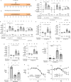
Extended Data Figure 1. Effect of tungstate on anaerobic respiration in vitro
a, Nitrate reductase activity of E. coli Nissle 1917 measured in media supplemented with sodium nitrate and the indicated concentrations of sodium tungstate. b, Competitive growth of the E. coli Nissle 1917 wild-type strain and the isogenic molybdenum cofactor-deficient mutant (moaA) in the presence of the indicated electron acceptors under anaerobic conditions. c, Competitive growth of E. coli NRG857c wild-type strain and the isogenic molybdenum cofactor-deficient mutant (moaA) in the presence of the indicated electron acceptors under anaerobic conditions. a–c, n=3 replicates per condition. n denotes the number of biological replicates. Bars represent the geometric mean of three experiments ± geometric standard deviation. **, P < 0.01; ***, P < 0.001; ns, not statistically significant compared to media containing no sodium tungstate.
Extended Data Figure 2
Extended Data Figure 2. Tungstate treatment of mice colonized with E. coli K-12
Conventionally-raised C57BL/6 mice were treated with 0.2 % sodium tungstate (W), dextran sulfate sodium (DSS), DSS+W, or left untreated (mock). After 4 days, animals were orally inoculated with the E. coli K-12 wild-type strain and the isogenic moaA mutant. C57BL/6 mice with naïve microbiota (endogenous Enterobacteriaceae only) or germ-free C57BL/6 mice were similarly treated but were not inoculated with E. coli indicator strains. Samples were analyzed after a total of 9 days of treatment. a–b, Schematic representation of the colitis models used in this figure. c–h Transcription of Nos2 (c), Tnf (d), Il6 (e), Lcn2 (f), Cxcl2 (g) and Ifng (h) in the cecal mucosa was determined by RT-qPCR, E. coli K-12: n=6 per group; Endogenous Enterobacteriaceae, Mock (n=14), W (n=6), DSS (n=19), DSS+W (n=19); Germ-free: Mock (n=5), DSS (n=4), DSS+W (n=5). i, Cumulative histopathology score for the colon tissue, Mock (n=14), W (n=6), DSS (n=19), DSS+W (n=19); bars represent means ± standard error and each dot represents one animal. j, Colon length, n=8 per group. k, Body weight of mice harboring endogenous Enterobacteriaceae, n=8 per group. l, Body weight of mice experimentally colonized with E. coli K-12, n=6 per group. Unless noted otherwise, bars represent the geometric mean ± geometric standard deviation. *, P < 0.05; **, P < 0.01; ***, P < 0.001. n.s., not significant.
Extended Data Figure 3
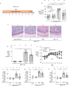
Extended Data Figure 3. Impact of tungstate treatment on mice experimentally colonized with E. coli Nissle 1917
Groups of conventionally-raised C57BL/6 mice were orally inoculated with the E. coli Nissle 1917 wild-type strain and treated with 0.2 % sodium tungstate (W), dextran sulfate sodium (DSS), DSS and sodium tungstate, or left untreated (mock) for 9 days. a, Schematic representation of colitis model used in this figure. b, Bacterial load in the cecum (white bars) and colon content (black bars). c–d Formalin-fixed, hematoxylin and eosin-stained sections of the cecum were scored for the presence of inflammatory lesions. c, Representative images of stained cecal sections. d, Cumulative histopathology score for the cecum tissue; bars represent means ± standard error and each dot represents one animal. b–d, Mock and W (n=11 per group), DSS and DSS+W (n=15 per group). e, Animal body weight, n=8 per group. f–h The transcription of the inflammatory marker genes Cxcl1 (f), Nos2, (g) and Tnf (h) in the cecal mucosa was determined by RT-qPCR, n=11 per group. Unless noted otherwise, bars represent the geometric mean ± geometric standard deviation. *, P < 0.05; **, P < 0.01; ***, P < 0.001. n.s, not significant.
Extended Data Figure 4
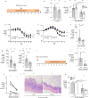
Extended Data Figure 4. Effect of tungstate treatment on mice experimentally colonized with Enterobacter cloacae and Adherent Invasive E. coli
a–h, Conventionally-raised C57BL/6 mice were treated with DSS or DSS+W. After 4 days, animals were intragastrically inoculated with the indicated bacterial strains. Samples were collected 5 days after inoculation. a, Schematic representation of the experiments. b and c, The total population of E. cloacae (b) and NRG857c (c) in the large intestinal content was determined by plating on selective media. d and e, Animal body weight. b and d, DSS (n=8), DSS+W (n=10). c, DSS (n=12), DSS+W (n=10). e, n=5 per group. f–h The transcription of the inflammatory marker genes Cxcl1 (f) Nos2 (g), and Tnf (h) in the cecal mucosa was determined by RT-qPCR, DSS (n=12), DSS+W (n=10). i–m, Paired germ-free Swiss-Webster mice received human fecal transplant and were subjected to DSS or DSS + 0.2 % sodium tungstate (W) treatment for 7 days, DSS (n=4), DSS+W (n=4) i, Schematic representation of the experiments. j, The abundance of Enterobacteriaceae in the cecal content was determined by plating on selective media (MacConkey agar). k, Formalin-fixed, hematoxylin and eosin-stained sections of the murine cecum were scored for the presence of inflammatory lesions. l, Representative images of stained murine cecal sections. m, The transcription of the inflammatory marker genes Nos2 and Il17 in the murine cecal mucosa. Bars represent the geometric mean ± geometric standard deviation. *, P < 0.05; **, P < 0.01; ***, P < 0.001. n.s, not significant.
Extended Data Figure 5
Extended Data Figure 5. Impact of tungstate on the naïve gut microbiome
Groups of C57BL/6 mice naturally harboring Enterobacteriaceae were treated with 0.2 % sodium tungstate (W), dextran sulfate sodium (DSS), DSS+W, or left untreated (mock) for 9 days (see also Extended Data Fig. 3b). a, Relative abundance of genes involved in formate and nitrate utilization in the cecal content revealed by shotgun metagenomic sequencing (MEGAN5). Each section of the pie chart is representative of the number of reads mapped obtained for the individual animals (n=6 per group). b, Box-and-whisker plot (Min to Max) of Chao1 alpha diversity of the cecal microbiota community based on 16S profiling (n=6 per group). c, Abundance of endogenous Enterobacteriaceae family members determined by plating on selective media (MacConkey agar), Mock (n=14), W (n=6), DSS (n=19), DSS+W (n=19). Bars represent the geometric mean ± geometric standard deviation. *, P < 0.05; ***, P < 0.001. n.s., not significant.
Extended Data Figure 6
Extended Data Figure 6. Effect of tungstate treatment on obligate anaerobic commensal bacteria
a–c Metagenomic analysis of the cecal content of mice described in Extended Data Fig. 3b. a, Principal coordinates (PC) analysis of global metabolic pathway and quantification of reads involved in fumarate respiration (b) and butyrate production (c). Ellipses in a denotes 95% confident interval. Bars represent the mean of ± standard deviation. a–c, n=6 per group. d–f B. thetaiotaomicron or B. fragilis were cultured anaerobically in mucin broth at 37 °C for 48 hours. Media was supplemented with sodium tungstate or metronidazole as indicated. Succinate production by B. thetaiotaomicron (d) and B. fragilis (f) was assessed by GC-MS. The growth of B. thetaiotaomicron was determined by plating serial dilution of bacterial culture on blood agar (e). g, C. symbiosum was inoculated into chopped meat broth and incubated anaerobically at 37 °C for 36 hours. Butyrate concentration in the media was measured using GC-MS. h, C. symbiosum was anaerobically cultured in chopped meat broth at 37 °C for 48 hours. The growth of C. symbiosum was determined by plating serial dilution of bacterial culture on thioglycolate plates. d–h, n=3 replicates per condition. Bars represent the geometric mean of three experiments ± geometric standard deviation. **, P < 0.01; ***, P < 0.001. n.s., not significant.
Extended Data Figure 7
Extended Data Figure 7. Assessment of overall health of mice orally treated with tungstate
Groups of seven mice were either mock treated or treated with 0.2 % sodium tungstate (W) in the drinking water for 9 days. a, Complete blood count. b, Serum alanine amino-transferase concentration. a–b, n=7 animals per group. Bars represent the geometric mean ± geometric standard deviation. n.s., not statistically significant.
Extended Data Figure 8
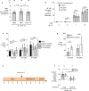
Extended Data Figure 8. Exposure of tissue culture cells to sodium tungstate
a, Daily water consumption of the E. coli K-12 inoculated mice. Each dot represents the average daily water consumption (ml/day) of 3 mice, obtained during 8 time points, with 2 cages per treatment group, n=16. b, HeLa57A cells, expressing luciferase under control of a NF-κB-dependent promoter, were treated with Phorbol 12-myristate 13-acetate (PMA) and sodium tungstate (W) at the indicated concentrations. Relative luciferase activity was determined after 5 hours. c–d, MODE-K or bone marrow derived macrophage (BMDMs) cells were treated with DSS or DSS+sodium tungstate at the indicated concentration for 24 hours. The release of lactate dehydrogenases into the culture supernatant by MODE-K cells (c) or BMDMs (d) was measured. e–f, Groups of conventionally-raised C57BL/6 mice were treated with DSS for 4 days. Animals were intragastrically inoculated with an equal mixture of the indicated E. coli Nissle 1917 wild-type strain and an isogenic moaA mutant. On the day of inoculation, a subset of mice was switched to DSS+sodium tungstate (W) for 4 days while a control group remained on DSS treatment. b–d, n=3 replicates per condition. e, Schematic representation of experiment. f, The competitive index in the cecal (white bars) and colon content (black bars) was analyzed 5 days after inoculation, DSS (n=5), DSS+W (n=6). Bars represent the geometric mean ± geometric standard deviation. *, P < 0.05; ***, P < 0.001. n.s., not significant.
Figure 1
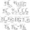
Figure 1. Effect of tungstate on molybdenum cofactor-dependent anaerobic respiration
a–c Nitrate reductase activity in E. coli K-12 (a), commensal Enterobacteriaceae strains (b) and an Enterobacter cloacae strain (c). d–e Competitive anaerobic growth of the E. coli K-12 wild type and a moaA mutant in the presence of electron acceptors (d) or microaerobic growth with the electron donor formate (e). a–e, n=3 replicates for each condition. f–j C57BL/6 mice received tungstate (W), dextran sulfate sodium (DSS), and DSS+W in the drinking water for 4 days. Animals were intragastrically inoculated with an equal mixture of the indicated E. coli wild-type strains and isogenic moaA mutants. E. coli populations in the cecal and colonic content were analyzed 5 days after inoculation: competitive index (f) and total population (g) of E. coli K-12; competitive index for Nissle 1917 (h), E. cloacae (i) and NRG857c (j). f–g, n=6 per group. h, Mock (n=4), DSS (n=8), DSS+W (n=8). i, DSS (n=8), DSS+W (n=10). j, DSS (n=12), DSS+W (n=10). k, Il10−/− mice on piroxicam-fortified diet were intragastrically inoculated with the murine E. coli strain NC101 and received W in the drinking water or were mock-treated. Abundance of NC101 was assessed after 14 days, Mock (n=4), W (n=5). Unless stated otherwise, n indicates the number of animals per group. Bars represent geometric means ± geometric standard deviation. *, P < 0.05; **, P < 0.01; ***, P < 0.001; ns, not statistically significant.
Figure 2
Figure 2. Impact of tungstate treatment on gut bacterial community composition and metabolic landscape
DNA extracted from the cecal contents of C57BL/6 mice (n=6/group) receiving indicated treatment was analyzed by metagenomic sequencing and 16S profiling. a, Principal coordinates (PC) analysis plots and analysis of similarity (ANOSIM) of the predicted coding capacity. Ellipses denote 95% confident interval. The number of animals per group (N) is indicated above each graph. b, Tallied metagenomic reads mapped to anaerobic respiration and formate utilization pathways. c, PC analysis of the microbiota composition (weighted UniFrac distances). d, Box-and-whisker plot (Min to Max) of intercommunity β-diversity determined by weighted 16S UniFrac distances. e, Phylum-level microbiota composition. f, Enterobacteriaceae abundance quantified by qPCR. g, Changes in the population size of the 25 most abundant operational taxonomic units as the result of tungstate administration in the DSS colitis model. Unless noted otherwise, bars represent geometric means ± geometric standard deviation. *, P < 0.05; **, P < 0.01; ***, P < 0.001; ns, not statistically significant.
Figure 3
Figure 3. Influence of tungstate treatment on mucosal inflammation
a–c Conventionally raised C57BL/6 mice, treated with DSS or DSS+tungstate (W) for 4 days, were inoculated with E. coli K-12 and samples analyzed after 5 days. C57BL/6 mice with a naïve microbiota (including endogenous Enterobacteriaceae) or germ-free C57BL/6 mice were similarly treated with W, DSS, and DSS+W. E. coli K-12: n=6 for all groups; Endogenous Enterobacteriaceae: Mock (n=14), W (n=6), DSS (n=19), DSS+W (n=19); Germ-free: n=5 for all groups (for one DSS-treated mouse, no mRNA was obtained). a, Representative images of H+E stained cecal sections. b, Cumulative histopathology score for the cecum; Bars represent means ± standard error and each dot represents one animal. c, Transcription of Cxcl1 (Kc) in the cecal mucosa by RT-qPCR. d–g Groups of Il10−/− mice were orally inoculated with NC101. Animals received piroxicam-fortified diet or piroxicam-fortified diet plus tungstate in the drinking water for 2 weeks, Mock (n=4), W (n=5). d, Representative images of H+E stained colonic sections. e, Cumulative histopathology score for the colon; Bars represent means ± standard error and each dot represents one animal. Abundance of Cxcl1 (f) and Cxcl2 (g) mRNA in the colonic mucosa by RT-qPCR. Unless noted otherwise, bars represent geometric means ± geometric standard deviation. *, P < 0.05; **, P < 0.01; ***, P < 0.001; ns, not statistically significant.

Comment in

References

    1. Frank DN, et al. Molecular-phylogenetic characterization of microbial community imbalances in human inflammatory bowel diseases. Proc Natl Acad Sci U S A. 2007;104:13780–13785. doi: 10.1073/pnas.0706625104. - DOI - PMC - PubMed
    1. Lupp C, et al. Host-mediated inflammation disrupts the intestinal microbiota and promotes the overgrowth of Enterobacteriaceae. Cell Host Microbe. 2007;2:204. - PubMed (V体育安卓版)
    1. Garrett WS, et al. Enterobacteriaceae act in concert with the gut microbiota to induce spontaneous and maternally transmitted colitis. Cell Host Microbe. 2010;8:292–300. doi: 10.1016/j.chom.2010.08.004. - DOI - PMC - PubMed
    1. Mshvildadze M, et al. Intestinal microbial ecology in premature infants assessed with non-culture-based techniques. J Pediatr. 2010;156:20–25. doi: 10.1016/j.jpeds.2009.06.063. - DOI - PMC - PubMed
    1. Vujkovic-Cvijin I, et al. Dysbiosis of the gut microbiota is associated with HIV disease progression and tryptophan catabolism. Sci Transl Med. 2013;5:193ra191. doi: 10.1126/scitranslmed.3006438. - DOI - PMC - PubMed

Publication types

MeSH terms