Skip to main page content
U.S. flag

An official website of the United States government

Dot gov

The . gov means it’s official VSports app下载. Federal government websites often end in . gov or . mil. Before sharing sensitive information, make sure you’re on a federal government site. .

Https

The site is secure V体育官网. The https:// ensures that you are connecting to the official website and that any information you provide is encrypted and transmitted securely. .

. 2018 Mar;8(3):320-335.
doi: 10.1158/2159-8290.CD-17-0993. Epub 2017 Dec 28.

MYC Drives a Subset of High-Risk Pediatric Neuroblastomas and Is Activated through Mechanisms Including Enhancer Hijacking and Focal Enhancer Amplification (V体育官网)

Affiliations

MYC Drives a Subset of High-Risk Pediatric Neuroblastomas and Is Activated through Mechanisms Including Enhancer Hijacking and Focal Enhancer Amplification (V体育平台登录)

Mark W Zimmerman et al. Cancer Discov. 2018 Mar.

Abstract

The amplified MYCN gene serves as an oncogenic driver in approximately 20% of high-risk pediatric neuroblastomas. Here, we show that the family member MYC is a potent transforming gene in a separate subset of high-risk neuroblastoma cases (∼10%), based on (i) its upregulation by focal enhancer amplification or genomic rearrangements leading to enhancer hijacking, and (ii) its ability to transform neuroblastoma precursor cells in a transgenic animal model. The aberrant regulatory elements associated with oncogenic MYC activation include focally amplified distal enhancers and translocation of highly active enhancers from other genes to within topologically associating domains containing the MYC gene locus. The clinical outcome for patients with high levels of MYC expression is virtually identical to that of patients with amplification of the MYCN gene, a known high-risk feature of this disease. Together, these findings establish MYC as a bona fide oncogene in a clinically significant group of high-risk childhood neuroblastomas. Significance: Amplification of the MYCN oncogene is a recognized hallmark of high-risk pediatric neuroblastoma. Here, we demonstrate that MYC is also activated as a potent oncogene in a distinct subset of neuroblastoma cases through either focal amplification of distal enhancers or enhancer hijacking mediated by chromosomal translocation. Cancer Discov; 8(3); 320-35. ©2017 AACR VSports手机版. This article is highlighted in the In This Issue feature, p. 253. .


Conflict of interest statement

Disclosure of potential conflicts of interest

The authors do have any conflicts of interest to disclose.

"V体育ios版" Figures

Figure 1
Figure 1. Expression of c-MYC and MYCN in neuroblastoma tumors and cell lines
A) Gene expression analysis of RNAs from a set of primary neuroblastomas from patients with disseminated disease (n = 123) demonstrates an inverse correlation between the expression levels of c-MYC and MYCN (r = −0.495, p < 0.0001). MYCN-amplified tumors are shown in red, MYCN non-amplified stage 4 tumors in blue, and MYCN non-amplified stage 4S tumors in yellow. Correlation coefficients (r) and statistical significance levels were determined by linear regression analysis. B) Overall survival is shown for subsets of neuroblastoma patients; i) MYCN-amplified (n=30), ii) c-MYC high (upper 10%, n=12), iii) MYCN non-amplified and low c-MYC expression stage 4 (n=61) or iv) MYCN non-amplified and non-high c-MYC stage 4S tumors (n=19). Patients with stage 4S tumors had an overall survival rate approaching 90%, in marked contrast to the uniformly low survival probability of <50% for patients with stage 4 tumors (p<0.005). The unfavorable outcomes for patients whose tumors had MYCN gene amplification or high levels of c-MYC expression were not significantly different (p=0.346). C) Gene expression profiling of human neuroblastoma cell lines (n = 25; R2 database) demonstrates an inverse correlation between the expression levels of c-MYC and MYCN (r = −0.826, p < 0.0001). Correlation coefficients (r) and statistical significance were determined by linear regression analysis. D) Western blot analysis of MYCN-amplified (n = 5) and MYCN non-amplified (n = 7) cell lines demonstrates exclusively high c-MYC or MYCN protein levels in neuroblastoma cell lines.
Figure 2
Figure 2. Transgenic overexpression of human c-MYC and MYCN in zebrafish results in hyperplasia of the peripheral sympathetic nervous system progressing to neuroblastoma
A) Transgenic strategy illustrating the three transgenic lines used in the experiments described in this study. The zebrafish dβh (5.2kb) promoter was used to drive expression of EGFP, mCherry, c-MYC and MYCN. The dβh:EGFP and dβh:MYCN lines were previously established (13). The dβh:c-MYC construct was coinjected with dβh:mCherry to make the dβh:c-MYC;dβh:mCherry line. The dβh:EGFP line (top) was crossed to the dβh:MYCN (middle) and dβh:c-MYC;dβh:mCherry (bottom) lines. B) Weekly quantification of IRG size by EGFP fluorescence microscopy in the indicated transgenic zebrafish from 4 to 7 wpf. EGFP-expressing regions were normalized to the surface area of the head of the fish being analyzed, as fish size was variable. Dotted line indicates the threshold of normal IRG fluorescent coverage and red bars the average value for each group. C) Representative fluorescent images showing EGFP-expressing sympathoadrenal cells in the indicated transgenic lines at 5 and 9 wpf. D) Tumor onset curves generated after biweekly monitoring of the indicated transgenic lines by EGFP fluorescence microscopy starting at 5 wpf. dβh:c-MYC transgenic lines reach nearly complete tumor penetrance by 7 wpf, while dβh:MYCN-induced latent tumors show lower penetrance. E) H&E staining, as well as immunohistochemical analysis, to detect expression of tyrosine hydroxylase (Th) and the pan-neuronal marker Hu-c in tumor sections derived from the indicated transgenic lines (black bar = 1 mm, white bar = 10 μm).
Figure 3
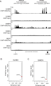
Figure 3. Focal amplification of noncoding, transcriptional enhancer regions occurs downstream of the c-MYC gene in neuroblastoma patients
A) ChIP-seq for H3K27ac in SJNB3, GIMEN and BE2C cells reveal a super-enhancer with a high H3K27ac signal downstream of the c-MYC gene in SJNB3 cells, corresponding to the sequence amplified in primary patient tumor PATEPF. GIMEN shows a super-enhancer immediately downstream of the c-MYC coding region, in the region showing enhancer amplification in the PASUCB primary sample. B) Ranking of H3K27ac signals across the SJNB3 genome demonstrates the high H3K27ac signal of the downstream super-enhancer regulating c-MYC gene expression. C) Ranking of H3K27ac signals across the GIMEN genome demonstrates a relatively lower H3K27ac signal regulating c-MYC gene expression.
Figure 4
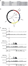
Figure 4. Elevated c-MYC expression is concomitant with segmental 8q chromosomal translocations
A) List of c-MYC-expressing primary tumors and cell lines with the genomic coordinates of each chromosomal translocation. B) Circos plot illustrating the observed chromosomal translocations associated with the c-MYC gene on chromosome 8q24 in patient tumors and c-MYC expressing neuroblastoma cell lines. Gray lines indicate the translocation of separate chromosomes observed in the indicated patient tumors and cell lines. C) ChIP-seq for H3K27ac shown with input control data for the chromosomal regions harboring the c-MYC and HAND2 gene loci in NB69 cells. H3K27ac modifications are indicative of super-enhancers that mediate high levels of gene expression. Dotted lines indicate the breakpoints where translocation was detected by WGS. Merged tracks for H3K27ac ChIP-seq with input control demonstrate that the super-enhancer formerly driving expression of HAND2 and FBXO8, is repositioned by the t(4;8) proximal to the c-MYC gene locus in NB69 cells.
Figure 5
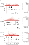
Figure 5. Delineation of topologically associated domains (TADs) and chromatin interactions by Hi-C demonstrates c-MYC interaction with translocated super-enhancers
A) ChIP-seq for H3K27ac and CTCF overlaid with the Hi-C chromatin contact maps in NB69 cells, demonstrating that on the translocated allele the c-MYC gene locus resides with an insulated chromatin neighborhood also containing the translocated HAND2 super-enhancer. Blue parallelogram indicates the apex and blue arrows the boundaries of the TAD; vertical red line denoting the breakpoint where chromosomes 4 and 8 (NB69 and SKNAS) and chromosomes 7 and 8 (SH-SY5Y) are joined together. B) Ranking of H3K27ac signals across the genome in NB69 cells demonstrating elevated super-enhancer signal of the enhancer associated with HAND2/FBXO8 (normal allele) and c-MYC (translocated allele). C) H3K27ac and CTFC ChIP-seq and Hi-C chromatin contact maps in SKNAS cells, demonstrating interaction of the c-MYC gene locus with the HAND2 super-enhancer (shaded signal on diagonal ending at the red arrow). D) Ranking of H3K27ac signals across the genome in SKNAS cells. E) H3K27ac and CTCF ChIP-seq and Hi-C chromatin contact maps in SH-SY5Y cells, demonstrating interaction of the c-MYC gene locus with the super-enhancer within the EXOC4 gene (shaded signal on diagonal ending at the red arrow). Additionally, this super-enhancer is also focally amplified in the region of high H3K27ac modifications (indicated by the bar). F) Ranking of H3K27ac signals across the genome in SH-SY5Y cells.
Figure 6
Figure 6. Global expression array for 123 human primary neuroblastoma tumors, including those driven my MYCN or MYC
Relative RNA expression for 123 high risk and stage 4S human primary neuroblastoma tumors (see red to blue scale, left of the figure). Groups 1- 6 (vertical white bars) are defined by unbiased hierarchical clustering. Primary samples are annotated by c-MYC expression in the top 10th percentile (orange bars); c-MYC expression biallelic verified based on expressed SNPs (blue bars); c-MYC locus alutered by chromosomal translocation (purple bars) or focal amplification (green bars); MYCN-amplification status (brown bars); INSS stage (stage 4S orange, stage 4 red and stage 3 blue); whether WES or WGS results were obtained (yes, black bars).

References (VSports在线直播)

    1. Matthay KK, Maris JM, Schleiermacher G, Nakagawara A, Mackall CL, Diller L, et al. Neuroblastoma. Nat Rev Dis Primers. 2016;16078:2. - PubMed (VSports手机版)
    1. Brodeur GM, RC S, M S, HE V, JM B. Amplification of N-myc in untreated human neuroblastomas correlates with advanced disease stage. Science. 1984;224:1121–4. - PubMed
    1. Wang LL, Suganuma R, Ikegaki N, Tang X, Naranjo A, McGrady P, et al. Neuroblastoma of undifferentiated subtype, prognostic significance of prominent nucleolar formation, and MYC/MYCN protein expression: a report from the Children’s Oncology Group. Cancer. 2013;119:3718–26. - PMC - PubMed
    1. Wang LL, Teshiba R, Ikegaki N, Tang XX, Naranjo A, London WB, et al. Augmented expression of MYC and/or MYCN protein defines highly aggressive MYC-driven neuroblastoma: a Children’s Oncology Group study. Br J Cancer. 2015;113:57–63. - "V体育安卓版" PMC - PubMed
    1. Westermann F, Muth D, Benner A, Bauer T, Henrich KO, Oberthuer A, et al. Distinct transcriptional MYCN/c-MYC activities are associated with spontaneous regression or malignant progression in neuroblastomas. Genome Biol. 2008;9:R150. - PMC (V体育2025版) - PubMed

Publication types

MeSH terms