VSports最新版本 - Skip to main page content
U.S. flag

An official website of the United States government

Dot gov

The . gov means it’s official. Federal government websites often end in . gov or . mil. Before sharing sensitive information, make sure you’re on a federal government site VSports app下载. .

Https

The site is secure. The https:// ensures that you are connecting to the official website and that any information you provide is encrypted and transmitted securely. V体育官网.

. 2018 Feb 16;122(4):606-615.
doi: 10.1161/CIRCRESAHA.117.312380. Epub 2017 Dec 13.

Pannexin 1 Channels as an Unexpected New Target of the Anti-Hypertensive Drug Spironolactone

Affiliations

"VSports" Pannexin 1 Channels as an Unexpected New Target of the Anti-Hypertensive Drug Spironolactone

"VSports app下载" Miranda E Good et al. Circ Res. .

"VSports最新版本" Abstract

Rationale: Resistant hypertension is a major health concern with unknown cause. Spironolactone is an effective antihypertensive drug, especially for patients with resistant hypertension, and is considered by the World Health Organization as an essential medication VSports手机版. Although spironolactone can act at the mineralocorticoid receptor (MR; NR3C2), there is increasing evidence of MR-independent effects of spironolactone. .

Objective: Here, we detail the unexpected discovery that Panx1 (pannexin 1) channels could be a relevant in vivo target of spironolactone V体育安卓版. .

Methods and results: First, we identified spironolactone as a potent inhibitor of Panx1 in an unbiased small molecule screen, which was confirmed by electrophysiological analysis V体育ios版. Next, spironolactone inhibited α-adrenergic vasoconstriction in arterioles from mice and hypertensive humans, an effect dependent on smooth muscle Panx1, but independent of the MR NR3C2. Last, spironolactone acutely lowered blood pressure, which was dependent on smooth muscle cell expression of Panx1 and independent of NR3C2. This effect, however, was restricted to steroidal MR antagonists as a nonsteroidal MR antagonist failed to reduced blood pressure. .

Conclusions: These data suggest new therapeutic modalities for resistant hypertension based on Panx1 inhibition VSports最新版本. .

Keywords: hypertension; mice; mineralocorticoid; pannexin 1; spironolactone; vasoconstriction. V体育平台登录.


Figures

Figure 1
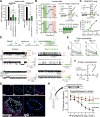
Figure 1. Spironolactone inhibits Pannexin 1 channels
(A) Inhibition of ATP release or TO-PRO-3 uptake (median fluorescence intensity, MFI) by spironolactone or positive control CBX in apoptotic Jurkat T cells that were treated with anti-Fas (n= 4, mean ± s.e.m.). TO-PRO-3 uptake was specifically scored on Annexin V+ apoptotic cells via flow cytometry. ** p<0.01 and **** p<0.0001 by one-way ANOVA, with Bonferroni’s test for multiple comparisons; n.s.: not statistically significant. (B) Inhibition of whole-cell pannexin currents in apoptotic Jurkat cells by spironolactone. (left) Whole-cell current amplitude from membrane potentials (Em) of +80 mV and −50 mV were assessed from apoptotic Jurkat cells after spironolactone treatment. Washing off spironolactone reverses the inhibition and treatment with CBX (positive control for PANX1 inhibition) again inhibits the PANX1 currents. (top right) The current-voltage relationship graphs indicate that spironolactone inhibits PANX1 across the voltage range, without affecting ionic selectivity. (bottom right) Grouped results show that spironolactone (20 µmol/L) inhibited 70.3 ± 4.5 % (at +80 mV) and 58.6 ± 8.1 % (at −50 mV) CBX-sensitive currents (n=7, mean ± s.e.m.). (C) Dose-dependent inhibition of PANX1 whole-cell currents in HEK293T cells co-expressing PANX1(TEV) and Tobacco Etch Virus (TEV) protease, a system in which TEVp cleaves and activates PANX1(TEV) in an apoptosis-independent manner. (top) Representative current-voltage relationship graph is shown. (bottom) Grouped results (n=8) show an IC50 of spironolactone as 8.7 ± 1.5 µmol/L at +80 mV and 8.0 ± 1.3 µmol/L at −50 mV. (D) Spironolactone addition directly affects single channel currents in inside-out patches of HEK293T cells expressing an active form of PANX1. Schematic of the inside out patch measurements is shown at the top. Representative inside-out patch recording from HEK293T cell co-expressing PANX1(TEV) and TEV protease before (left) and after (right) spironolactone (20 µmol/L) application, at patch potentials of +60 mV (upper) or −60 mV (lower). C: closed state; O1: level for one open channel; O2: level for two open channels. The traces on the right are shown to depict data indicating that majority of the channels are in the ‘closed state’ (C) after spironolactone addition compared to control conditions. (E) Open probability (NPo) of PANX1 channels was reduced by spironolactone (20 µmol/L, n=7). ** p<0.01 by two-tailed paired t-test. (F) Unitary conductance of PANX1 channels was unaffected by spironolactone (20 µmol/L, n=7). Unitary conductance (mean ± s.e.m.) are 91.0 ± 1.5 pS (control) and 89.5 ± 1.7 pS (spironolactone) at patch potentials between +50 mV and +80 mV, and 13.5 ± 1.9 pS (control) or 11.5 ± 1.5 pS (spironolactone) at patch potentials between −70 mV and −50 mV, respectively. (G) Arterioles from human patients with resistant hypertension showed expression for PANX1 (magenta) that were not seen when secondary antibodies alone (IgG) were used. The internal elastic lamina autofluorescence (green), α-smooth muscle actin (cyan) and DAPI (blue) are visualized in each image. Bottom images are higher magnification images. Arrowheads indicate endothelial Panx1, and arrows point to smooth muscle Panx1. Star (*) indicates lumen of arteriole. Scale bar: 30 µm. (H) Phenylephrine induced vasoconstriction was impaired in human arterioles from patients with treatment resistant hypertension, pretreated with either 5 µmol/L PxIL2P (n=3) or 80 µmol/L spironolactone (n=7 vessels). Mean ± s.e.m. * p<0.05 by two-way ANOVA (PxIL2P vs. untreated).
Figure 2
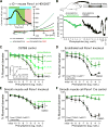
Figure 2. Spironolactone inhibition of Pannexin 1 channels on smooth muscle cells regulates α-adrenergic vasoconstriction
(A) Exemplar whole-cell recording from a HEK293T cell co-expressing mouse Panx1 channels and α1D adrenergic receptors. Phenylephrine-induced currents (cyan shading) are inhibited by spironolactone (20 µmol/L; green shading) and further by CBX (50 µmol/L; red shading). Inset: whole-cell current at +80 mV during sequential application of phenylephrine, spironolactone and CBX (in the continued presence of phenylephrine). (B) Representative trace of phenylephrine-induced vasoconstriction of a C57Bl/6 thoracodorsal artery. Vasoconstriction was blunted when 80 µmol/L spironolactone was present; after washing off the spironolactone, the thoracodorsal artery regained the ability to contract in response to phenylephrine. To confirm vessel integrity was not affected the spironolactone treatment, 10 µmol/L acetylcholine (ACh) was used for vasodilation, and 40 mmol/L KCl was used for inducing vasoconstriction, followed by maximal dilation achieved via a Ca2+ free solution. (C) Control vessels from C57Bl/6 mice treated with DMSO vehicle control (N=6) or 10 (N=4), 20 (N=5), 40 (N=5), or 80 (N=5) µmol/L spironolactone show dose-dependent inhibition of PE-induced vasoconstriction. Repeated measures two-way ANOVA showed significant differences between DMSO vehicle control vs. 20, 40, or 80 µmol/L spironolactone at 10−6 – 10−3 mol/L phenylephrine. Inset: IC50 for spironolactone was determined to be ~ 18.9 µmol/L at 1 µmol/L phenylephrine (10−6 mol/L). (D) Endothelial cell deletion of Panx1 retained significant reduction of phenylephrine-induced vasoconstriction upon treatment with 80 µmol/L spironolactone. DMSO vehicle control: N=4 mice; n=6 vessels; spironolactone: N=4; n=7. Repeated measures two-way ANOVA showed significant differences between DMSO vehicle control vs. 80 µmol/L spironolactone at 10−6 – 10−3 mol/L phenylephrine. (E and F) Smooth muscle cell deletion of Panx1 after tamoxifen injection (E), prevented the inhibitory effect of 80 µmol/L spironolactone on phenylephrine-induced vasoconstriction, while this was retained in in Panx1fl/fl/SMC-CreERT2+ mice injected with the control vehicle (peanut oil) (F). Tamoxifen injected mice: DMSO vehicle control N=6, n=11; spironolactone treated N=5, n=10. Vehicle injected: DMSO vehicle control N=6, n=11; spironolactone treated N=6, n=9. Two-way ANOVA and significant differences at any dose of phenylephrine are shown. For all graphs: mean ± s.e.m. and * p<0.05.
Figure 3
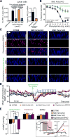
Figure 3. Spironolactone acutely lowers blood pressure by inhibiting Pannexin 1 channels in a mineralocorticoid receptor Nr3c2-independent manner
(A) Jurkat cells transfected with control siRNA or NR3C2 siRNA were induced to undergo apoptosis via anti-Fas treatment (250 ng/ml) in the presence or absence of spironolactone, and TO-PRO-3. Dye uptake was measured using flow cytometry (left). Knockdown efficiency of NR3C2 is shown on the right. Data are presented as mean ± s.e.m. (n=4). ** p<0.01 and **** p<0.0001 by one-way ANOVA, with Bonferroni’s multiple comparison test (left) or by two-tailed unpaired t-test (right). (B) Spironolactone significantly blunted phenylephrine-induced vasoconstriction in mice with smooth muscle cell deletion of Nr3c2 by injection with tamoxifen. DMSO vehicle control: N=5 mice; n=9 vessels; spironolactone (80 µmol/L): N=5; n=9. Repeated measures two-way ANOVA showed significant differences between untreated vs. spironolactone at 10−7 – 10−3 mol/L phenylephrine. * p<0.05. (C) Phenylephrine-induced uptake of TO-PRO-3 (red) in thoracodorsal arteries from C57Bl6 and SMC Nr3c2-KO mice was prevented by pretreatment with 80 µmol/L spironolactone (blue, DAPI counterstained to mark nuclei from smooth muscle cells). However, SMC Panx1-KO mice failed to take up TO-PRO-3, and this is not affected by vehicle or spironolactone treatment. (D) Continuous recording of blood pressure (5 min averages) reveals a significant drop in mean arterial pressure (MAP) following intraperitoneal (i.p.) injection of 40 mg/kg spironolactone in C57Bl6 control (N=5 mice), SMC Nr3c2-KO (N=5 mice), and hypertensive BPH/2 mice (N=5 mice). In contrast, spironolactone did not decrease MAP in SMC Panx1-KO mice (N=5). Data points are presented as averaged MAP taken every 5 min (mean ± s.e.m.). Dashed green line indicates time of injection. Blood pressure returned to pre-injection values 24 hr after injection. (E) Mean arterial pressure (MAP) is significantly reduced from baseline in C57Bl6 control, SMC Nr3c2-KO, and hypertensive BPH/2 mice following injection of 40 mg/kg spironolactone. (Δ MAP = 2-hr baseline MAP, 30-min post-injection MAP). However, injection of 40 mg/kg finerenone, a non-steroidal mineralocorticoid antagonist, or vehicle (DMSO) failed to reduce the blood pressure in all genotypes. Notably, spironolactone, did not reduce MAP in SMC Panx1-KO mice. N=5 mice per group (N=4 for each group for BPH/2 mice); mean ± s.e.m. (one-way ANOVA; * p<0.05; *** p<0.001). Boxed: Representative whole-cell recording from a HEK293T cell co-expressing human PANX1(TEV) and TEV protease shows that finerenone (50 µmol/L) does NOT inhibit PANX1 currents. Inset shows whole-cell currents at +80 mV upon sequential application of finerenone and CBX (% inhibition is 15.3 ± 5.3 for currents at +80 mV and 30.1 ± 15.6 for −50 mV; n=6).
Figure 4
Figure 4. Metabolites and analogs of spironolactone inhibit Panx1 channel function, but not other nucleotide-release channels
(A) Jurkat cells transfected with control siRNA or NR3C2 siRNA were induced to undergo apoptosis via anti-Fas treatment (250 ng/ml) in the presence or absence of spironolactone or its metabolites and analogs. Representative histograms of TO-PRO-3 uptake are shown on the left. TO-PRO-3 uptake was measured using flow cytometry (right). Data are presented as mean ± s.e.m. (n=4). TO-PRO-3 uptake by apoptotic Jurkat cells was significantly (p<0.0001) reduced by spironolactone (green), canrenone (blue), and eplerenone (purple), compared to vehicle controls (black); no statistical significance was found between scramble and NR3C2 siRNA groups by the same treatment of spironolactone and derivatives (two-way ANOVA, with Bonferroni’s test). (B) Inhibition of CBX-sensitive whole-cell currents by spironolactone and its derivatives (mean ± s.e.m.). Whole-cell currents were obtained from HEK293T cells co-expressing human PANX1(TEV) and TEV protease. All compounds caused voltage-independent inhibition of PANX1 currents. (C) Spironolactone and its derivatives show negligible effect on mouse pannexin 2 (Panx2) channel currents. Whole-cell currents were recorded from HEK293T cells exogenously expressing mouse Panx2. Cells were treated with spironolactone (20 µmol/L), canrenone (20 µmol/L), or eplerenone (20 µmol/L), and followed by CBX (50 µmol/L) exposure. Exemplar I-V curves and grouped percent inhibition (mean ± s.e.m.) are presented. (D) Spironolactone and its derivatives display negligible effect on Cx43 hemichannel currents. Whole-cell currents were recorded from HEK293T cells exogenously expressing Cx43. Cells were treated with spironolactone (20 µmol/L), canrenone (20 µmol/L), or eplerenone (20 µmol/L), and followed by gadolinium (Gd3+, 100 µmol/L) inhibition. Exemplar I-V curves and grouped percent inhibition (Gd3+-sensitive, mean ± s.e.m.) are presented.

Comment in (V体育平台登录)

References

    1. Cekic C, Linden J. Purinergic regulation of the immune system. Nat Rev Immunol. 2016;16:177–192. - PubMed
    1. Chekeni FB, Elliott MR, Sandilos JK, Walk SF, Kinchen JM, Lazarowski ER, Armstrong AJ, Penuela S, Laird DW, Salvesen GS, Isakson BE, Bayliss DA, Ravichandran KS. Pannexin 1 channels mediate 'find-me' signal release and membrane permeability during apoptosis. Nature. 2010;467:863–867. - "V体育平台登录" PMC - PubMed
    1. Yang D, He Y, Munoz-Planillo R, Liu Q, Nunez G. Caspase-11 requires the pannexin-1 channel and the purinergic p2×7 pore to mediate pyroptosis and endotoxic shock. Immunity. 2015;43:923–932. - V体育ios版 - PMC - PubMed
    1. Furlow PW, Zhang S, Soong TD, Halberg N, Goodarzi H, Mangrum C, Wu YG, Elemento O, Tavazoie SF. Mechanosensitive pannexin-1 channels mediate microvascular metastatic cell survival. Nat Cell Biol. 2015;17:943–952. - PMC - PubMed
    1. Billaud M, Chiu YH, Lohman AW, Parpaite T, Butcher JT, Mutchler SM, DeLalio LJ, Artamonov MV, Sandilos JK, Best AK, Somlyo AV, Thompson RJ, Le TH, Ravichandran KS, Bayliss DA, Isakson BE. A molecular signature in the pannexin1 intracellular loop confers channel activation by the alpha1 adrenoreceptor in smooth muscle cells. Sci Signal. 2015;8:ra17. - PMC - PubMed

V体育官网入口 - Publication types

V体育官网 - MeSH terms