Skip to main page content
U.S. flag

An official website of the United States government

Dot gov

The . gov means it’s official. Federal government websites often end in . gov or . mil. Before sharing sensitive information, make sure you’re on a federal government site. VSports app下载.

Https

The site is secure V体育官网. The https:// ensures that you are connecting to the official website and that any information you provide is encrypted and transmitted securely. .

. 2018 Feb 1;24(3):696-707.
doi: 10.1158/1078-0432.CCR-17-1872. Epub 2017 Nov 14.

Targeting the MYCN-PARP-DNA Damage Response Pathway in Neuroendocrine Prostate Cancer (V体育平台登录)

Affiliations

Targeting the MYCN-PARP-DNA Damage Response Pathway in Neuroendocrine Prostate Cancer

Wei Zhang et al. Clin Cancer Res. .

Abstract

Purpose: We investigated MYCN-regulated molecular pathways in castration-resistant prostate cancer (CRPC) classified by morphologic criteria as adenocarcinoma or neuroendocrine to extend the molecular phenotype, establish driver pathways, and identify novel approaches to combination therapy for neuroendocrine prostate cancer (NEPC). Experimental Design and Results: Using comparative bioinformatics analyses of CRPC-Adeno and CRPC-Neuro RNA sequence data from public data sets and a panel of 28 PDX models, we identified a MYCN-PARP-DNA damage response (DDR) pathway that is enriched in CRPC with neuroendocrine differentiation (NED) and CRPC-Neuro. ChIP-PCR assay revealed that N-MYC transcriptionally activates PARP1, PARP2, BRCA1, RMI2, and TOPBP1 through binding to the promoters of these genes. MYCN or PARP1 gene knockdown significantly reduced the expression of MYCN-PARP-DDR pathway genes and NED markers, and inhibition with MYCNsi and/or PARPsi, BRCA1si, or RMI2si significantly suppressed malignant activities, including cell viability, colony formation, and cell migration, in C4-2b4 and NCI-H660 cells. Targeting this pathway with AURKA inhibitor PHA739358 and PARP inhibitor olaparib generated therapeutic effects similar to those of gene knockdown in vitro and significantly suppressed tumor growth in both C4-2b4 and MDACC PDX144-13C subcutaneous models in vivoConclusions: Our results identify a novel MYCN-PARP-DDR pathway that is driven by N-MYC in a subset of CRPC-Adeno and in NEPC. Targeting this pathway using in vitro and in vivo CRPC-Adeno and CRPC-Neuro models demonstrated a novel therapeutic strategy for NEPC. Further investigation of N-MYC-regulated DDR gene targets and the biological and clinical significance of MYCN-PARP-DDR signaling will more fully elucidate the importance of the MYCN-PARP-DDR signaling pathway in the development and maintenance of NEPC. Clin Cancer Res; 24(3); 696-707 VSports手机版. ©2017 AACR. .


V体育官网入口 - Conflict of interest statement

Disclosure of Potential Conflicts of Interest: No potential conflicts of interest are to be disclosed by authors.

"VSports注册入口" Figures

Figure 1
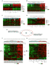
Figure 1. Increased DNA damage response and mitotic gene expression in CRPC-Neuro samples
(A) Unsupervised clustering of Beltran 70 classifier genes in PDX RNA-seq dataset. Samples were classified into CRPC-Neuro and CRPC-Adeno using NE scores. The pathology and NE score classifications are shown in top covariate bars. The over (in red) and under (in green) expression status of these genes in CRPC-Neuro samples (see also Supplementary Table 2) is shown on the right covariate bar. (B) Unsupervised clustering of significant DDR-M genes in Beltran et al. RNA-seq dataset. The DDR-M significant genes (FDR < or = 0.05) are derived from differential analysis comparing NE score classified CRPC-Neuro to CRPC-Adeno Beltran et al. samples. The pathology and NE score classifications are shown in top covariate bars. (C) Unsupervised clustering of significant DDR-M genes in Grasso et al. microarray dataset. The DDR-M significant genes (FDR < or = 0.05) are derived from differential analysis comparing NE score classified CRPC-Neuro to CRPC-Adeno samples. The pathology and NE score classifications are shown in top covariate bars. (D) Unsupervised clustering of significant DDR-M genes in PDX RNA-seq dataset. The DDR-M significant genes (FDR < or = 0.05) are derived from differential analysis comparing NE score classified CRPC-Neuro to CRPC-Adeno PDX samples. The pathology and NE score classifications are shown in top covariate bars. (E) Significant DDR-M genes in Beltran and PDX datasets at FDR 0.15 were compared and 149 common genes were identified. (F) Unsupervised clustering of AR, MYCN and common significant DDR-M genes (FDR < or = 0.15) in Beltran et al. RNA-seq dataset. The NE score classifications (CRPC-Neuro and CRPC-Adeno) are shown in top covariate bar. The NE score for each samples are shown in covariate scatter plot with 0.4 as cutoff. (G) Unsupervised clustering of AR, MYCN and common genes (FDR < or = 0.15) in PDX dataset. The NE score classifications (CRPC-Neuro and CRPC-Adeno) are shown in top covariate bar. The NE score for each samples are shown in covariate scatter plot with 0.35 as cutoff.
Figure 2
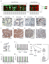
Figure 2. Analysis of proposed MYCN-PARP-DDR signaling pathway
(A) The expression pattern of MYCN pathway genes in Beltran CRPC-Adeno (left panel heat map) and CRPC-Neuro (right panel heat map) are shown. The samples are ordered based on MYCN expression level. The top covariate bars indicate neuroendocrine scores. The Pearson Correlation Coefficients of pathway genes with MYCN expression are shown in the box plot next to heat map. (B) MYCN ISH in untreated adenocarcinoma. (C) MYCN ISH in ADT-treated adenocarcinoma or in SCPC (D and E). In one SCPC sample MYCN expression in some cancer cells with adenocarcinoma morphology was lower than that in adjacent SCPC cells (right in D). (F–I) positive PARP1, PARP2, BRCA1, and TOPBP1, respectively in SCPC. (J) Western blotting analysis of N-MYC and NE markers in C4-2b, C4-2b4 and NCI-H660. (K–M) Effect of MYCNsi on mRNA expression of MYCN-PARP-DDR pathway genes in C4-2b4 and NCI-H660. (N) Proposed MYCN-PARP-DDR signaling pathway.
Figure 3
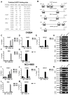
Figure 3. ChIP-PCR analysis of N-MYC binding to PARP1/2 and selected DDR-M genes
(A, B) Predicted N-MYC binding sites on PARP1, PARP2, BRCA1, RMI2 and TOPBP1 promoters according to JASPAR database. (C–G) ChIP-PCR assay testing the direct binding of N-MYC to PARP1, PARP2, BRCA1, RMI2 and TOPBP1promoter in C4-2b4. (I–M) ChIP-PCR assay testing the direct binding of N-MYC to PARP1, PARP2, BRCA1, RMI2 and TOPBP1 promoter in H660. (H, N) DNA gel electrophoresis were conducted to confirm N-MYC occupancy on PARP1, PARP2, BRCA1, RMI2 and TOPBP1 promoter C4-2b4 and NCI-H660. P1, PARP1; P2, PARP2; B1, BRCA1; R2, RMI2; T1, TOPBP1; BS, binding site.
Figure 4
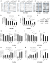
Figure 4. Effect of MYCN and/or PARP knockdown on MYCN-PARP-DDR pathway and CRPC oncogenic activities
(A, B) Effect of MYCNsi, or PARP1/2si on protein expression of MYCN-PARP-DDR pathway genes in C4-2b4 and NCI-H660. (C) Effect of MYCN or PARP1 overexpression on protein levels of MYCN-PARP-DDR pathway genes in C4-2b4 and NCI-H660. (D, E) Colony growth and cell migration after cooperative oncogenic effects of N-MYC and PARP by genetic knockdown in C4-2b4 cell. (F) Effect of MYCNsi on protein expression of γ-H2AX. (G, I) Cell proliferation/viability, (K, M) Sub-G1 cell distribution in C4-2b4 and H660 cells transfected with MYCNsi, PARP1/2si, or MYCNsi+PARP1/2si. (H, J) Cell proliferation/viability, (L, N) Sub-G1 cell distribution in C4-2b4 and H660 cells transfected with BRCA1si and RMI2si. In Figs D–M, Msi, MYCNsi; P1si, PARP1si; P2si, PARP2si. *, p < 0.05; **, p< 0.01; ***, p< 0.001 (t-test).
Figure 5
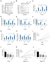
Figure 5. AURKA and PARP inhibitors cooperatively suppress prostate cancer oncogenic activities in vitro and xenograft tumor growth in vivo
(A, B) Effect of PHA, OLA and combination of PHA and OLA on protein expression of MYCN-PARP-DDR pathway genes in C4-2b4 and NCI-H660 cells. PHA1.5, PHA 1.5 μM; OLA5, OLA 5 μM; OLA10, OLA 10 μM. (C–H) Demonstration of the cooperative oncogenic effects of N-MYC and PARP by pharmacological inhibition of AURKA and PARP in C4-2b4 and NCI-H660 cells. PHA1.5, PHA 1.5 μM; OLA5, OLA 5 μM; OLA10, OLA 10 μM. Sub-G1 cell distribution (C and G), cell proliferation/viability (D, H), colony growth (E), and cell migration (F) in C4-2b4 and/or H660 cells treated with AURKA inhibitor PHA, PARP inhibitor OLA, or PHA+OLA. (I–L) Results from C4-2b4 and MDA 144-13C subcutaneous model. (I) Tumor growth curve for C4-2b4. (J) Tumor wet weight for C4-2b4. (K) Tumor wet weight for MDA PCa 144-13C. (L) Tumor growth curve for MDA PCa 144-13C. *, p < 0.05; **, p< 0.01; ***, p< 0.001 (t-test).

V体育安卓版 - Comment in

References

    1. Wang HT, Yao YH, Li BG, Tang Y, Chang JW, Zhang J. Neuroendocrine Prostate Cancer (NEPC) progressing from conventional prostatic adenocarcinoma: factors associated with time to development of NEPC and survival from NEPC diagnosis-a systematic review and pooled analysis. J Clin Oncol. 2014;32:3383–90. - PubMed
    1. Helpap B, Kollermann J, Oehler U. Neuroendocrine differentiation in prostatic carcinomas: histogenesis, biology, clinical relevance, and future therapeutical perspectives. Urol Int. 1999;62:133–8. - PubMed
    1. Palmgren JS, Karavadia SS, Wakefield MR. Unusual and underappreciated: small cell carcinoma of the prostate. Semin Oncol. 2007;34:22–9. - PubMed
    1. Beltran H, Rickman DS, Park K, Chae SS, Sboner A, MacDonald TY, et al. Molecular characterization of neuroendocrine prostate cancer and identification of new drug targets. Cancer Discov. 2011;1:487–95. - PMC - PubMed
    1. Spiess PE, Pettaway CA, Vakar-Lopez F, Kassouf W, Wang X, Busby JE, et al. Treatment outcomes of small cell carcinoma of the prostate: a single-center study. Cancer. 2007;110:1729–37. - PubMed (V体育官网)

"V体育安卓版" Publication types

MeSH terms (V体育ios版)

Substances