Skip to main page content
U.S. flag

An official website of the United States government

Dot gov

The . gov means it’s official. Federal government websites often end in VSports app下载. gov or . mil. Before sharing sensitive information, make sure you’re on a federal government site. .

Https

The site is secure. The https:// ensures that you are connecting to the official website and that any information you provide is encrypted and transmitted securely V体育官网. .

. 2017 Nov 28;114(48):E10399-E10408.
doi: 10.1073/pnas.1716015114. Epub 2017 Nov 13.

Human resistin protects against endotoxic shock by blocking LPS-TLR4 interaction

Affiliations

V体育2025版 - Human resistin protects against endotoxic shock by blocking LPS-TLR4 interaction

Jessica C Jang et al. Proc Natl Acad Sci U S A. .

Abstract

Helminths trigger multiple immunomodulatory pathways that can protect from sepsis. Human resistin (hRetn) is an immune cell-derived protein that is highly elevated in helminth infection and sepsis. However, the function of hRetn in sepsis, or whether hRetn influences helminth protection against sepsis, is unknown. Employing hRetn-expressing transgenic mice (hRETNTg+) and recombinant hRetn, we identify a therapeutic function for hRetn in lipopolysaccharide (LPS)-induced septic shock. hRetn promoted helminth-induced immunomodulation, with increased survival of Nippostrongylus brasiliensis (Nb)-infected hRETNTg+ mice after a fatal LPS dose compared with naive mice or Nb-infected hRETNTg- mice VSports手机版. Employing immunoprecipitation assays, hRETNTg+Tlr4-/- mice, and human immune cell culture, we demonstrate that hRetn binds the LPS receptor Toll-like receptor 4 (TLR4) through its N terminal and modulates STAT3 and TBK1 signaling, triggering a switch from proinflammatory to anti-inflammatory responses. Further, we generate hRetn N-terminal peptides that are able to block LPS proinflammatory function. Together, our studies identify a critical role for hRetn in blocking LPS function with important clinical significance in helminth-induced immunomodulation and sepsis. .

Keywords: LPS; TLR4; inflammation; resistin; sepsis. V体育安卓版.


Conflict of interest statement

The authors declare no conflict of interest.

Figures
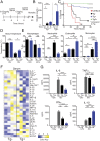
Fig. 1.
Fig. 1.
hRetn protects against endotoxic shock. (A) Experimental design of LPS-induced sepsis model. (B) hRetn serum levels in hRETNTg+ and Tg2+ mice were measured by ELISA. (C) Survival rate was evaluated after high-dose LPS (12 mg/kg) injection. (D) Rectal body temperature was measured at 0 and 6 h after high-dose LPS injection. (E) hRETNTg and hRETNTg+ mice were left naive or challenged with LPS, followed by PEC recovery and flow cytometric analysis at 24 h after high-dose LPS. (F and G) Serum was assayed for cytokines by Luminex. (F) Cytokines induced 6 h after LPS treatment of hRETNTg+ and hRETNTg mice were plotted as a heat map. (G) Significantly changed proinflammatory and anti-inflammatory cytokines are represented in bar graphs. Data are presented as mean ± SEM (n = 7–12 for survival, n = 3–5 for other parameters) and representative of three separate experiments. ns, not significant; *P ≤ 0.05; **P ≤ 0.01; ***P ≤ 0.001; ****P ≤ 0.0001.
Fig. 2.
Fig. 2.
Therapeutic administration of hRetn during septic shock. (A) Experimental design to test the role of hRetn in LPS-induced sepsis. (B) Survival rate of C57BL/6 mice treated with PBS or 500 ng/mouse hRetn before LPS injection. (C) Rectal body temperature at 6 h after LPS injection. (D) Lung vascular permeability was measured with Evan’s blue dye at 24 h after LPS injection. (E) Serum cytokines at 6 h after LPS injection were measured by Luminex. (F) PECs were recovered at 24 h after LPS injection and cells analyzed by flow cytometry. Data are presented as mean ± SEM (n = 6 for survival, n = 3–5 for other parameters) and are representative of two separate experiments. ns, not significant; *P ≤ 0.05; **P ≤ 0.01; ***P ≤ 0.001; ****P ≤ 0.0001.
Fig. 3.
Fig. 3.
hRetn enhances helminth-mediated protection during endotoxic shock. (A) Experimental design of hRETNTg and hRETNTg+ mice infected with Nb for 14 d followed by LPS challenge. Serum hRetn was measured in naive (n) or Nb-infected mice at −48 h (Left) or 6 h (Right) after LPS injection. (B) Survival rate after LPS injection. (C) Serum cytokines from Nb-infected and LPS-challenged hRETNTg and hRETNTg+ mice were evaluated by Luminex. (D and E) PECs from Nb+LPS mice were recovered at 48 h after LPS challenge and assayed directly ex vivo. (D) Flow cytometric analysis of PEC populations. (E) Representative Western blot (Left) and band density of phosphorylated STAT3, phosphorylated TBK1, and IκBα over endogenous β-actin was quantified for three mice per group (Right). Data are presented as mean ± SEM (n = 6–10 for survival, n = 3–4 for other parameters) and are representative of two separate experiments. ns, not significant; *P ≤ 0.05; **P ≤ 0.01; ***P ≤ 0.001.
Fig. 4.
Fig. 4.
TLR4 is a receptor for hRetn. (A) Cap1 and Tlr4 mRNA expression in naive or day 7 Nb-infected lungs from hRETNTg and hRETNTg+ lungs, as measured by RNA-seq. (BD) hRetn binding of day 7 Nb-infected Tlr4+/+ and Tlr4−/− lung cells was measured as frequency of hRetn-bound cells and Δmean fluorescence intensity. (E) Lung population frequency. (F) Pie chart of proportion of hRetn-bound cells. (G) Anti-His or anti-hRetn Western blot of pull-down assay with His-tagged TLR4, His-CAP1 or His-MBP with E.coli or 293T cell-derived hRetn. Mouse data are presented as mean ± SEM (n = 3–4 per group), and all data are representative of two to three separate experiments. ns, not significant; *P ≤ 0.05; **P ≤ 0.01; ***P ≤ 0.001.
Fig. 5.
Fig. 5.
hRetn outcompetes LPS for binding to TLR4. (A) Prediction of the hRetn (green) structure based on the structure of mRetn (cyan) was performed with ClusPro web server. (B) Structural modeling of hRetn (blue, N-terminal; green, C-terminal) and TLR4 (red) reveals that hRetn binds in the same binding pocket of MD2 (white), the adaptor protein for LPS. (C) Predicted molecular interactions between the N-terminal helical trimer (cyan, red, yellow) and the TLR4 monomer (blue). (D) Pie chart of proportion of hRetn-bound cells in human PBMC. (E and F) LPS binding assay in human PBMC with or without prior incubation with hRetn (Left), followed by flow cytometric analysis of LPS-bound CD14+CD11b+ monocytes (E) and statistical analysis (Right) (F). (G) TNFα secretion measured in PBMC treated with PBS or hRetn followed LPS stimulation. (H) Primary sequence of the synthesized hRetn N-terminal peptide and CD spectrum of the agent measured at 100 μM. (I) His-tagged TLR4 was incubated with control buffer or hRetn N-terminal peptide (N-pep), followed by incubation with 293T cell-derived Flag-tagged hRetn, His-pulldown, and Western blot with anti-His and anti-Flag. (J and K) LPS binding assay in human PBMC with or without prior incubation with hRetn or N-pep followed by flow cytometric analysis of monocytes for LPS binding (J) and MD2/TLR4 surface expression (K). (L) TNFα secretion was measured in PBMC treated with PBS, hRetn, or N-pep, followed by LPS stimulation. PBMC data are presented as mean ± SEM (n = 3–4 replicates), and all are representative of two to three separate experiments. *P ≤ 0.05; **P ≤ 0.01; ***P ≤ 0.001.
Fig. 6.
Fig. 6.
hRetn downstream signaling is TLR4-dependent. PECs were collected from 6- to 8-wk-old naive hRETNTg+ or hRETNTg mice on the Tlr4+/+ or Tlr4−/− background and analyzed directly ex vivo. (A) Representative Western blot (Left) and band density of phosphorylated STAT3, phosphorylated TBK1, and IκBα over endogenous β-actin was quantified for three to four mice per group (Right). (B) Flow cytometric analysis of PEC populations. (C) The ability of hRetn to inhibit LPS-induced TNFα and IL-10 secretion was measured in human PBMC pretreated with control DMSO, TBK1, and STAT3 inhibitors, followed by hRetn and LPS treatment. Percentage TNFα inhibition or IL-10 induction by hRetn over PBS-treatment was calculated. Data are presented as mean ± SEM (n = 3–4) and representative of two separate experiments. ns, not significant; *P ≤ 0.05; **P ≤ 0.01; ***P ≤ 0.001.

References

    1. Angus DC, et al. Epidemiology of severe sepsis in the United States: Analysis of incidence, outcome, and associated costs of care. Crit Care Med. 2001;29:1303–1310. - PubMed
    1. Cauwels A, Brouckaert P. Survival of TNF toxicity: Dependence on caspases and NO. Arch Biochem Biophys. 2007;462:132–139. - PubMed
    1. Roger T, et al. Protection from lethal gram-negative bacterial sepsis by targeting Toll-like receptor 4. Proc Natl Acad Sci USA. 2009;106:2348–2352. - PMC - PubMed
    1. Marshall JC. Why have clinical trials in sepsis failed? Trends Mol Med. 2014;20:195–203. - PubMed
    1. Szkudlapski D, et al. The emering role of helminths in treatment of the inflammatory bowel disorders. J Physiol Pharmacol. 2014;65:741–751. - PubMed

MeSH terms

Substances