Skip to main page content
U.S. flag

An official website of the United States government

Dot gov

The . gov means it’s official. Federal government websites often end in . gov or . mil. Before sharing sensitive information, make sure you’re on a federal government site VSports app下载. .

Https

The site is secure V体育官网. The https:// ensures that you are connecting to the official website and that any information you provide is encrypted and transmitted securely. .

. 2018 Jan;41(1):107-118.
doi: 10.3892/ijmm.2017.3244. Epub 2017 Nov 7.

Detection of calprotectin in inflammatory bowel disease: Fecal and serum levels and immunohistochemical localization

Affiliations

Detection of calprotectin in inflammatory bowel disease: Fecal and serum levels and immunohistochemical localization

Shuhei Fukunaga et al. Int J Mol Med. 2018 Jan.

Abstract

The aim of the present study was to quantify calprotectin levels using an enzyme-linked immunosorbent assay (ELISA) and a point-of-care test (POCT) in patients with inflammatory bowel disease. Overall, 113 patients with ulcerative colitis (UC; 51 men and 62 women) and 42 patients with Crohn's disease (CD; 29 men and 13 women), who were scheduled to undergo a colonoscopy, were prospectively enrolled and scored endoscopically and clinically. An additional 96 healthy, age-matched subjects served as the normal controls. Feces and blood samples from the patients with UC and CD, and the normal controls were analyzed. These patients had received adequate medical treatment. The tissue distribution of calprotectin was investigated using immunohistochemistry. The fecal calprotectin levels, as measured using an ELISA, were correlated with the endoscopic and clinical disease activities and laboratory parameters, including serum levels of hemoglobin (Hb), albumin and C-reactive protein, and erythrocyte sedimentation rate, particularly among the patients with UC. The fecal Hb level was close to that of the fecal calprotectin level (r=0 VSports手机版. 57; P<0. 0001). The fecal calprotectin level measured using an ELISA was well-correlated with the fecal calprotectin level measured using the POCT (r=0. 81; P<0. 0001), but was not correlated with the serum calprotectin level (r=0. 1013; P=0. 47). An immunohistochemical investigation revealed that patients with both UC and CD had higher neutrophil and monocyte/macrophage calprotectin-positive cell expression levels, compared with those in the normal controls. Fecal calprotectin was considered a reliable marker for disease activity, and the assessment of fecal calprotectin via POCT showed potential as a rapid and simple measurement in clinical settings. .


Figures

Figure 1
Figure 1
Fecal calprotectin levels in patients with ulcerative colitis. Fecal calprotectin levels according to (A) endoscopic disease activity as assessed using the Rachmilewitz score and (B) clinical disease activity as assessed using the Mayo score.
Figure 2
Figure 2
Fecal calprotectin levels in patients with CD. Fecal calprotectin levels according to the (A) endoscopic disease activity as assessed using the simple endoscopic score for CD and (B) clinical disease activity as assessed using the CD activity index. CD, Crohn's disease.
Figure 3
Figure 3
Fecal hemoglobin levels in patients with ulcerative colitis. Fecal hemoglobin levels according to (A) endoscopic disease activity as assessed using the Rachmilewitz score and (B) clinical disease activity as assessed using the Mayo score.
Figure 4
Figure 4
Fecal hemoglobin levels in patients with CD. Fecal hemoglobin levels according to (A) endoscopic disease activity as assessed using the simple endoscopic score for CD and (B) clinical disease activity as assessed using the CD activity index. CD, Crohn's disease.
Figure 5
Figure 5
Correlation between the fecal calprotectin and fecal hemoglobin levels in patients with (A) UC, (B) CD and (C) inflammatory bowel disease (UC+CD). UC, ulcerative colitis; CD, Crohn's disease.
Figure 6
Figure 6
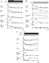
Time-courses for fecal calprotectin levels and other inflammatory parameters in three patients with inflammatory bowel disease. (A) Twenty-four-year male patient with UC involving the entire colon; (B) 26-year male patient with UC involving the entire colon; (C) 21-year female patient with CD involving the small and large bowel. 5ASA, 5-aminosalicylic acid; PSL, prednisolone; anti-TNF-α, antitumor necrosis factor-α antibody; TPN, total parenteral nutrition; CRP, C-reactive protein; CDAI, CD activity index; SES-CD, Simple Endoscopic Score for CD; UC, ulcerative colitis; CD, Crohn's disease.
Figure 7
Figure 7
Correlations between fecal calprotectin levels. ELISA and a POCT were used to measure correlations between fecal calprotectin levels in patients with (A) UC, (B) CD and (C) inflammatory bowel disease (UC+CD). ELISA, enzyme-linked immunosorbent assay; POCT, point-of-care test; UC, ulcerative colitis; CD, Crohn's disease.
Figure 8
Figure 8
Mean differences in fecal calprotectin levels between ELISA and POCT values in 80 patients with inflammatory bowel disease, including 56 patients with ulcerative colitis and 24 patients with Crohn's disease. The differences between the ELISA and POCT values for each of the patients are plotted against the mean of the two measurements; the LoA, defined as the mean ± 2 standard deviations of the difference, are shown. ELISA, enzyme-linked immunosorbent assay; POCT, point-of-care test; LoA, limit of agreement.
Figure 9
Figure 9
Correlation between fecal and serum calprotectin levels. Correlation between fecal and serum calprotectin levels were measured using an enzyme-linked immunosorbent assay in patients with (A) UC, (B) CD and (C) inflammatory bowel disease (UC+CD). UC, ulcerative colitis; CD, Crohn's disease.
Figure 10
Figure 10
Correlation between serum concentrations of calprotectin and CRP in patients with (A) UC, (B) CD and (C) inflammatory bowel disease (UC+CD). UC, ulcerative colitis; CD, Crohn's disease.
Figure 11
Figure 11
Immunohistochemistry for the detection of calprotectin-stained cells in colonic samples. Samples from a patient with (A) UC, a patient with (B) Crohn's disease and a (C) normal subject. (D) Double staining of colonic tissue specimens from a patient with UC showed calprotectin-positive cells (brown), anti-neutrophil elastase stain-positive neutrophils (red) and double-positive cells (purple). (E) Double staining of colonic tissue specimens from a patient with UC showed calprotectin-positive cells (brown), anti-CD68-staining-positive macrophages (brown), and double-positive cells (purple). These tissue sections revealed that neutrophils and certain macrophage populations expressed calprotectin. UC, ulcerative colitis. Original magnification, ×20.

References

    1. Strober W, Fuss I, Mannon P. The fundamental basis of inflammatory bowel disease. J Clin Invest. 2007;117:514–521. doi: 10.1172/JCI30587. - DOI - PMC - PubMed
    1. Pineton de Chambrun G, Peyrin-Biroulet L, Lémann M, Colombel JF. Clinical implications of mucosal healing for the management of IBD. Nat Rev Gastroenterol Hepatol. 2010;7:15–29. doi: 10.1038/nrgastro.2009.203. - DOI - PubMed
    1. Baert F, Moortgat L, Van Assche G, Caenepeel P, Vergauwe P, De Vos M, Stokkers P, Hommes D, Rutgeerts P, Vermeire S, et al. Mucosal healing predicts sustained clinical remission in patients with early-stage Crohn's disease. Gastroenterology. 138:463–468. - "V体育平台登录" PubMed
    1. Frøslie KF, Jahnsen J, Moum BA, Vatn MH, Group I, IBSEN Group. Mucosal healing in inflammatory bowel disease: Results from a Norwegian population-based cohort. Gastroenterology. 2007;133:412–422. doi: 10.1053/j.gastro.2007.05.051. - DOI - PubMed
    1. Sands BE. Biomarkers of inflammation in inflammatory bowel disease. Gastroenterology. 2015;149:1275–1285.e1272. doi: 10.1053/j.gastro.2015.07.003. - DOI - PubMed

MeSH terms