VSports注册入口 - Skip to main page content
U.S. flag

An official website of the United States government

Dot gov

The . gov means it’s official VSports app下载. Federal government websites often end in . gov or . mil. Before sharing sensitive information, make sure you’re on a federal government site. .

Https

The site is secure V体育官网. The https:// ensures that you are connecting to the official website and that any information you provide is encrypted and transmitted securely. .

. 2017 Dec;48(12):3336-3346.
doi: 10.1161/STROKEAHA.117.018505. Epub 2017 Nov 7.

"V体育安卓版" Fingolimod Protects Against Ischemic White Matter Damage by Modulating Microglia Toward M2 Polarization via STAT3 Pathway

Affiliations

Fingolimod Protects Against Ischemic White Matter Damage by Modulating Microglia Toward M2 Polarization via STAT3 Pathway

Chuan Qin et al. Stroke. 2017 Dec.

Abstract (V体育官网入口)

Background and purpose: White matter (WM) ischemic injury, a major neuropathological feature of cerebral small vessel diseases, is an important cause of vascular cognitive impairment in later life. The pathogenesis of demyelination after WM ischemic damage are often accompanied by microglial activation VSports手机版. Fingolimod (FTY720) was approved for the treatment of multiple sclerosis for its immunosuppression property. In this study, we evaluated the neuroprotective potential of FTY720 in a WM ischemia model. .

Methods: Chronic WM ischemic injury model was induced by bilateral carotid artery stenosis. Cognitive function, WM integrity, microglial activation, and potential pathway involved in microglial polarization were assessed after bilateral carotid artery stenosis V体育安卓版. .

Results: Disruption of WM integrity was characterized by demyelination in the corpus callosum and disorganization of Ranvier nodes using Luxol fast blue staining, immunofluorescence staining, and electron microscopy V体育ios版. In addition, radial maze test demonstrated that working memory performance was decreased at 1-month post-bilateral carotid artery stenosis-induced injury. Interestingly, FTY720 could reduce cognitive decline and ameliorate the disruption of WM integrity. Mechanistically, cerebral hypoperfusion induced microglial activation, production of associated proinflammatory cytokines, and priming of microglial polarization toward the M1 phenotype, whereas FTY720 attenuated microglia-mediated neuroinflammation after WM ischemia and promoted oligodendrocytogenesis by shifting microglia toward M2 polarization. FTY720's effect on microglial M2 polarization was largely suppressed by selective signal transducer and activator of transcription 3 (STAT3) blockade in vitro, revealing that FTY720-enabled shift of microglia from M1 to M2 polarization state was possibly mediated by STAT3 signaling. .

Conclusions: Our study suggested that FTY720 might be a potential therapeutic drug targeting brain inflammation by skewing microglia toward M2 polarization after chronic cerebral hypoperfusion VSports最新版本. .

Keywords: cognitive dysfunction; corpus callosum; microglia; multiple sclerosis; white matter V体育平台登录. .


Conflict of interest statement

Disclosures

The authors declare that they have no conflict of interest.

Figures
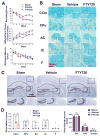
Figure 1
Figure 1. Working memory impairment and white matter lesions were partially reduced by FTY720 treatment
(A) The working memory and reference memory of mice were assessed by the 8-arm maze test at 1 month post injury (n=8 for Sham, n=9 for Vehicle and FTY720). Vehicle mice made much more revisiting errors (P<0.001) and less different arm choices (P<0.001) comparing to the sham-operated mice. FTY720-treated group made much less revisiting errors and more different arm choices (P=0.016) comparing to Vehicle group. No impairment in spatial reference memory was revealed between different groups (P>0.05). (B) White matter lesions were detected by LFB staining in different WM regions. Scale bar, 200μm. (C) Representative images of Nissl stained coronal sections depicting the morphology of hippocampus and cerebral cortex. Scale bar, 1mm. (D) Dot-plot with median and interquartile range of the severity of white matter lesions in CCm, CPu, AC, IC, and F in histogram. Quantitative analysis of Nissl stained neuron numbers in DG, CA3 region in hipocampus and cerebral cortex was performed. n.s. no significant changes between different groups. * P<0.05 ** P<0.01 versus Sham, # P<0.05 ## P<0.01 versus Vehicle. n=6 per group.
Figure 2
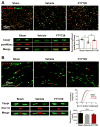
Figure 2. Disruption of Ranvier’s nodes due to hypoperfusion was reversed by FTY720 treatment
(A) Representative 3 dimensional confocal images labeled with panNfasc/Caspr. Scale bar, 10μm. A higher magnification of representative single Ranvier’s node was shown below. Quantitative analysis of panNfasc/Caspr colocalization was shown in the histogram. **P<0.01 versus Sham, #P<0.05 versus Vehicle. n=5 per group. (B) Representative confocal images labeled with Nav1.6/Caspr. Scale bar, 10μm. A higher magnification of representative single Ranvier’s node was shown below. Summary of the length of Nav1.6 domain was shown in curve. P<0.0001 FTY720 versus Vehicle, Vehicle versus Sham. n=5 mice per group and 100 Nav1.6 domains for each mouse. Quantitative analysis of Ranvier’s nodes number is shown in the histogram. n.s. no significant changes between different groups. n=5 per group.
Figure 3
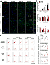
Figure 3. Activation of microglia due to hypoperfusion injury was retained by FTY720 treatment
(A) Representative confocal images of coronal sections labeled with Iba-1 and CD68 at different time points post BCAS. Scale bar, 50μm. (B, C) Quantitative analysis of Iba-1+ and CD68+ cells was shown in the histogram. **P<0.01 versus Sham, #P<0.05 ##P<0.01 versus Vehicle. n=6 per group.
Figure 4
Figure 4. Hypoperfusion induced microglia M1 polarization was partially retained by FTY720 treatment
(A) Representative confocal images of coronal sections labeled with Iba-1, CD16/32 and CD206 at different time points post BCAS. Scale bar, 50μm. (B) Quantitative analysis of Iba-1, CD16/32 double positive cells and Iba-1, CD206 double positive cells was shown in the histogram. Ratio of CD206 positive cells to CD16/32 positive cells was performed. *P<0.05 **P<0.01 versus Sham, #P<0.05 ##P<0.01 versus Vehicle. n=6 per group. (C) FACS analysis of microglia in WM after BCAS. Dot plots represent CD16/32 staining (upper panel), CD86 staining (second panel), CD206 staining (third panel), CD23 staining (lower panel) of CD11b positive microglia. (D) Percentage of CD16/32/CD86/CD206/CD23 positive cells in total CD11b positive microglia. *P<0.05 versus Sham, #P<0.05 ##P<0.01 versus Vehicle. n=5–6 mice for each group.
Figure 5
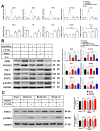
Figure 5. FTY720 modulated microglia toward M2 polarization via STAT3 pathway
(A) Primary microglia were exposed to vehicle as Control, LPS plus IFN-γ stimulation, LPS plus IFN-γ stimulation and FTY720 treatment, and IL-4 stimulation. The mRNA expression of pro-inflammatory markers (CD86, TNF-α, iNOS, CD32 and IL-1β) and anti-inflammatory markers (TGF-β, YM, Arg-1, CD206 and IL-10) in response to different stimulation and treatment was detected by RT-PCR. n.s. no significant changes between different groups. *P<0.05 **P<0.01 versus Control, #P<0.05 ##P<0.01 versus LPS plus IFN-γ stimulation. n=6 per group. (B) The protein expression of M1 markers (iNOS, CD16/32), M2 markers (Arg-1, CD206), STAT3 and p-STAT3 in primary microglia were detected by western-blots. Quantitative analysis was performed. **P<0.01 versus Control, #P<0.05 ##P<0.01 versus LPS plus IFN-γ stimulation, ΦP<0.05 ΦΦP<0.01 versus LPS plus IFN-γ stimulation and FTY720 treatment. n=6 per group. (C) Total protein expression and phosphorylation level of STAT3 were detected by western-blots in mice from Sham, Vehicle and FTY720 groups at different time points after BCAS. Quantitative analysis was performed. #P<0.05 ##P<0.01 versus Vehicle. n=8 per group.
Figure 6
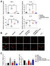
Figure 6. FTY720 modulated OPCs apoptosis and maturation via M1 to M2 switch of microglia
(A) Microglia supernatants under different stimulation and treatment were analyzed by ELISA. Quantitative analysis of expression level of cytokines TNF-α, IL-1β, TGF-β and IL-13 was performed. **P<0.01 *P<0.05 versus Control, #P<0.05 ##P<0.01 versus LPS plus IFN-γ stimulation. n=8 per group. (B) Representative images of oligodendrocytes labeled with Olig2 and TUNEL (upper label), Olig2 and MBP (lower label) in response to different stimulation and treatment. Scale bar, 200 μm. (C) Quantitative analysis was performed as percentage of TUNEL+ and MBP+ in Olig2+ cells. **P<0.01 versus Control, ##P<0.01 versus LPS plus IFN-γ stimulation, ΦΦP<0.01 ΦP<0.05 versus LPS plus IFN-γ stimulation and FTY720 treatment. n=8 per group.

References

    1. Miyamoto N, Maki T, Pham LD, Hayakawa K, Seo JH, Mandeville ET, et al. Oxidative stress interferes with white matter renewal after prolonged cerebral hypoperfusion in mice. Stroke. 2013;44:3516–3521. doi: 10.1161/STROKEAHA.113.002813. - DOI - PMC - PubMed
    1. Verden D, Macklin WB. Neuroprotection by central nervous system remyelination: Molecular, cellular, and functional considerations. Journal of neuroscience research. 2016;94:1411–1420. doi: 10.1002/jnr.23923. - DOI - PMC - PubMed
    1. Choi BR, Kim DH, Back DB, Kang CH, Moon WJ, Han JS, et al. Characterization of white matter injury in a rat model of chronic cerebral hypoperfusion. Stroke. 2016;47:542–547. doi: 10.1161/STROKEAHA.115.011679. - DOI (VSports在线直播) - PubMed
    1. Fu Y, Liu Q, Anrather J, Shi FD. Immune interventions in stroke. Nature reviews. Neurology. 2015;11:524–535. doi: 10.1038/nrneurol.2015.144. - DOI (VSports app下载) - PMC - PubMed
    1. Hu X, Li P, Guo Y, Wang H, Leak RK, Chen S, et al. Microglia/macrophage polarization dynamics reveal novel mechanism of injury expansion after focal cerebral ischemia. Stroke. 2012;43:3063–3070. doi: 10.1161/STROKEAHA.112.659656. - DOI - PubMed

Publication types

VSports app下载 - MeSH terms

VSports手机版 - Substances