Skip to main page content
U.S. flag

An official website of the United States government

Dot gov

The . gov means it’s official. Federal government websites often end in . gov or VSports app下载. mil. Before sharing sensitive information, make sure you’re on a federal government site. .

Https

The site is secure. The https:// ensures that you are connecting to the official website and that any information you provide is encrypted and transmitted securely. V体育官网.

. 2017 Oct 17;8(1):978.
doi: 10.1038/s41467-017-00880-2.

"VSports手机版" Extrafollicular CD4+ T-B interactions are sufficient for inducing autoimmune-like chronic graft-versus-host disease

Affiliations

Extrafollicular CD4+ T-B interactions are sufficient for inducing autoimmune-like chronic graft-versus-host disease

"V体育官网入口" Ruishu Deng et al. Nat Commun. .

VSports app下载 - Abstract

Chronic graft-versus-host disease (cGVHD) is an autoimmune-like syndrome mediated by pathogenic CD4+ T and B cells, but the function of extrafollicular and germinal center CD4+ T and B interactions in cGVHD pathogenesis remains largely unknown. Here we show that extrafollicular CD4+ T and B interactions are sufficient for inducing cGVHD, while germinal center formation is dispensable. The pathogenesis of cGVHD is associated with the expansion of extrafollicular CD44hiCD62loPSGL-1loCD4+ (PSGL-1loCD4+) T cells. These cells express high levels of ICOS, and the blockade of ICOS/ICOSL interaction prevents their expansion and ameliorates cGVHD. Expansion of PSGL-1loCD4+ T cells is also prevented by BCL6 or Stat3 deficiency in donor CD4+ T cells, with the induction of cGVHD ameliorated by BCL6 deficiency and completely suppressed by Stat3 deficiency in donor CD4+ T cells. These results support that Stat3- and BCL6-dependent extrafollicular CD4+ T and B interactions play critical functions in the pathogenesis of cGVHD. Chronic graft-versus-host disease (cGVHD) is mediated by specific CD4 and B cells, but the relative contribution of extrafollicular and germinal centre (GC) T-B interaction is unclear. Here the authors show that the extrafollicular expansion of a specific CD4 T subset is sufficient for inducing cGVHD while GC is dispensable. VSports手机版.


Conflict of interest statement

The authors declare no competing financial interests.

Figures
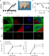
Fig. 1
Fig. 1
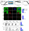
No germinal center formation is observed in cGVHD recipients. BALB/c mice were irradiated (850 cGy) and given 2.5 × 106 TCD-BM alone (n = 6) or 2.5 × 106 TCD-BM plus 1 × 106 (n = 8) or 0.01 × 106 (n = 8) splenocytes from C57BL/6 donors. Mice were monitored for cGVHD. a Cutaneous cGVHD score (severe cGVHD group versus no-GVHD group: P < 0.001, severe cGVHD group versus mild cGVHD group: P < 0.001, two-way ANOVA). b Picture taken on day 60 after transplantation (1–no GVHD, 2–mild cGVHD, 3–severe cGVHD). c Survival curve (severe cGVHD group versus no GVHD group: P < 0.001, severe cGVHD group versus mild cGVHD group: P < 0.001, log-rank test). d Immunofluorescent staining of B220, CD3, and PNA on cryosections of spleen harvested on day 60 after HCT. e Germinal center number and area were measured and are shown as mean ± SE (n = 6). Scale bar, 50 µm
Fig. 2
Fig. 2
Chronic GVHD was induced in recipients without germinal center formation. BALB/c recipients were irradiated (850 cGy) and given 2.5 × 106 TCD-BM alone (n = 12) or 2.5 × 106 TCD-BM plus 1 × 106 (n = 12) splenocytes from either WT C57BL/6 or BCL6fl/fl Mb1-Cre C57BL/6 donors. cGVHD development was monitored. a Cutaneous cGVHD score. b Picture taken at day 60 after HCT (1 and 3–B-BCL6+/+ no GVHD and cGVHD, 2 and 4–B-BCL6−/− no GVHD and cGVHD). c Percent body weight changes. d Survival curve. e Sixty days after transplantation, spleens were harvested and germinal centers were identified by immunofluorescent staining of B220, CD3, and PNA, and GC area and numbers were measured and are shown as mean ± SE (n = 6). f Donor splenocytes were stained for CD4, CD19, CXCR5, and PD-1. Tfh were gated as CD4+CD19and are shown as CXCR5hiPD-1hi. Percentages of CXCR5hiPD-1hi cells among CD4+CD19 cells were shown as mean ± SE (n = 6). g Donor splenocytes were stained for CD19, GL7, and Fas. Germinal center B cells were gated on CD19+ and are shown as GL7+Fas+. Percentages of GL7+Fas+ among CD19+ cells are shown as mean ± SE (n = 6). **P < 0.01, ***P < 0.001, unpaired two-tailed Student’s t test. B-BCL6+/+ no GVHD = B-BCL6+/+ TCD-BM; B-BCL6−/− no GVHD = B-BCL6−/−TCD-BM; B-BCL6+/+ cGVHD = B-BCL6+/+ TCD-BM + B-BCL6+/+ splenocytes; B-BCL6−/− cGVHD = B-BCL6−/− TCD-BM + B-BCL6−/− splenocytes. Scale bar, 50 µm
Fig. 3
Fig. 3
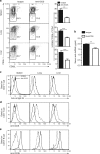
cGVHD is associated with expansion of PSGL-1loCD4+ T cells. BALB/c recipients were irradiated (850 cGy) and given 2.5 × 106 TCD-BM or 2.5 × 106 TCD-BM plus 1 × 106 splenocytes from C57BL/6 donors. a, b Twenty-one, 30, and 45 days after HCT, spleen and lung were harvested. Splenocytes and mononuclear cells isolated from lung were stained for CD4, CD44, PSGL-1, and CD62L. Gated CD4+CD44hi are shown as PSGL-1 versus CD62L. PSGL-1 low and CD62L low cells were gated as extrafollicular CD4+ T cells. Percentages of PSGL-1loCD62Llo cells among CD4+CD44hi cells are shown as mean ± SE (n = 8). c Twenty-one days after HCT, splenocytes from no-GVHD or cGVHD recipients given wild-type C57BL/6 transplants were harvested and stained for CD4, CD44, PSGL-1, and CD62L. CD44hiCD62LloPSGL-1loCD4+ T cells were sorted and used for RNA isolation and RNA-Seq microarray analysis. Heat maps of RNA expression of CXCR4, CXCR5, and CCR7 are shown as mean centered log2 expression. RNA-Seq microarray measurements were performed on duplicate samples from no-GVHD group and cGVHD group. Each sample represents splenocytes from eight recipients. d Twenty-one days after HCT, sorted CD4+CD44hiPSGL-1loCD62Llo cells were stimulated with PMA and ionomycin for 24 h. Stimulated cells were stained and are shown as CD4 versus IFN-γ, IL-13, IL-17, or IL-21. Percentages of CD4+IFN-γ+, CD4+IL-13+, CD4+IL-17+, or CD4+IL-21+ cells among CD4+ T cells are shown as mean ± SE (n = 9). *P < 0.05, ***P < 0.001, unpaired two-tailed Student’s t test
Fig. 4
Fig. 4
Anti-ICOS treatment ameliorates cGVHD in recipients without GC formation. a As described in Fig. 3, 21 days after HCT, splenocytes from no-GVHD or cGVHD recipients given wild-type C57BL/6 donor cells were harvested and stained for CD4, CD44, PSGL-1, and CD62L. CD44hiCD62LloPSGL-1loCD4+ T cells were sorted and used for RNA isolation and RNA-Seq microarray analysis. Heat maps of RNA expression levels of costimulatory and coinhibitory markers are shown as mean centered log2 expression. RNA-Seq microarray measurements were performed on duplicate samples from no-GVHD group and cGVHD group. Each sample represents splenocytes from eight recipients. bf BALB/c recipients were irradiated (850 cGy) and given either 2.5 × 106 TCD-BM (n = 8) or 2.5 × 106 TCD-BM plus 1 × 106 splenocytes (n = 12) from B-BCL6−/− C57BL/6 donors. Recipients given 2.5 × 106 TCD-BM plus 1 × 106 splenocytes were treated with anti-ICOS or isotype control of rat IgG2b, 200 µg/mouse i.p., starting on day 0 and repeated every other day until day 45 after HCT. Chronic GVHD was monitored. b Cutaneous cGVHD score (Group 3 versus Group 2: P < 0.001 two-way ANOVA). c Picture taken on day 60 after HCT(1–no-GVHD, 2–isotype, 3–anti-ICOS). d Survival curve (Group 3 versus Group 2: P < 0.05, log-rank test). e H&E staining of salivary gland, skin, lung, and liver. f Pathology scores of cGVHD for salivary gland, skin, lung, and liver are shown as mean ± SE (n = 6). **P < 0.01, unpaired two-tailed Student’s t test. Scale bar, 50 µm
Fig. 5
Fig. 5
Anti-ICOS treatment reduces PSGL-1loCD4+ T-cell expansion. As described in Fig. 4, BALB/c recipients were irradiated (850 cGy) and given 2.5 × 106 TCD-BM plus 1 × 106 splenocytes. Recipients were treated with anti-ICOS or control rat IgG2b (200 µg/mouse i.p.) every other day from day 0 to day 45 after HCT. a Twenty-one days after HCT, mononuclear cells from spleen, lung, and liver were stained for CD4, CD44, PSGL-1, and CD62L. Gated CD4+CD44hi cells are shown as PSGL-1 versus CD62L (n = 8). b Serum anti-dsDNA was measured at 45 days after HCT. ce Twenty-one days after HCT, mononuclear cells from spleen, lung, and liver were stained with CD4, anti-rat IgG2b, or anti-ICOS, or stained with anti-CD19 and anti-ICOSL. Gated CD4+ T cells are shown as anti-rat IgG2b (c) or anti-ICOS (d) staining. e Gated CD19+ B cells are shown as ICOSL staining. One representative of four experiments is shown. **P < 0.01, ***P < 0.001, unpaired two-tailed Student’s t test
Fig. 6
Fig. 6
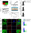
BCL6 deficiency in donor CD4+ T cells prevents expansion of PSGL-1loCD4+ T cells and cutaneous cGVHD. BALB/c recipients were irradiated (850 cGy) and given 2.5 × 106 TCD-BM alone or 2.5 × 106 TCD-BM plus 1 × 106 splenocytes from either WT or B-BCL6−/− C57BL/6 donors (n = 12). cGVHD was monitored. a Percent body weight changes. b Cutaneous cGVHD scores (Group 3 versus Group 2: P < 0.001 two-way ANOVA). c Picture taken at day 60 after HCT (1 and 2–CD4-BCL6+/+ no GVHD and GVHD, 3–CD4-BCL6−/− GVHD). d Survival curve. e Sixty days after transplantation, spleens were harvested, and germinal centers were identified by immunofluorescent staining of B220, CD3, and PNA. GC area and numbers were measured and are shown as mean ± SE (n = 4). f Twenty-one days after HCT, mononuclear cells from spleen, lung, and liver were stained for CD4, CD44, PSGL-1, and CD62L. Gated CD4+CD44hi cells are shown as PSGL-1 versus CD62L. Percentages of CD62LloPSGL-1lo cells among CD4+CD44hi are shown as mean ± SE (n = 8). ***P < 0.001, unpaired two-tailed Student’s t test. CD4-BCL6+/+ no-GVHD = CD4-BCL6+/+ TCD-BM; CD4-BCL6−/− no-GVHD = CD4-BCL6−/− TCD-BM; CD4-BCL6+/+ cGVHD = CD4-BCL6+/+ TCD-BM + CD4-BCL6+/+ splenocytes; CD4-BCL6−/− cGVHD = CD4-BCL6−/− TCD-BM + CD4-BCL6−/− splenocytes. Scale bar, 50 µm
Fig. 7
Fig. 7
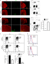
Stat3 deficiency in donor CD4+ T cells prevents expansion of PSGL-1loCD4+ T cells and systemic cGVHD. a Twenty-one days after HCT, splenocytes from no-GVHD or cGVHD recipients given wild-type C57BL/6 donors were harvested and stained for CD4, CD44, PSGL-1, and CD62L. CD44hiCD62LloPSGL-1loCD4+ T cells were sorted and used for RNA isolation and RNA-Seq microarray analysis. Heat maps of RNA expression of transcription factor in extrafollicular T cells are shown as mean centered log2 expression. RNA-Seq microarray measurements were performed on duplicate samples from no-GVHD and cGVHD groups. Each sample represents splenocytes from eight recipients. bg BALB/c recipients were irradiated (850 cGy) and given 2.5 × 106 TCD-BM alone or 2.5 × 106 TCD-BM plus 1 × 106 splenocytes (n = 12) from either WT or CD4-STAT3−/− C57BL/6 donors. b Percent body weight changes (Group 3 versus Group 4: P < 0.001, two-way ANOVA). c Cutaneous cGVHD scores (Group 3 versus Group 4: P < 0.001, two-way ANOVA). d Picture taken on day 60 after HCT (1 and 3–Stat3+/+–no GVHD and cGVHD, 2 and 4–Stat3−/−–no GVHD and cGVHD). e Survival curve (Group 3 versus Group 4: P < 0.05, log-rank test). f Twenty-one days after HCT, mononuclear cells from spleen, lung, and liver were stained with CD4, CD44, PSGL-1, and CD62L. Percentages of CD62LloPSGL-1lo cells among CD4+CD44hi are shown as mean ± SE (n = 6). g Sixty days after transplantation, spleens were harvested and germinal centers were identified by immunofluorescent staining of B220, CD3, and PNA, and GC area and numbers were measured and are shown as mean ± SE (n = 4). ** P < 0.01*** P < 0.001, unpaired two-tailed Student’s t test. CD4-Stat3+/+ no GVHD = CD4-Stat3+/+ TCD-BM; CD4-Stat3−/− no GVHD = CD4-Stat3−/−TCD-BM; CD4-Stat3+/+ cGVHD = CD4-Stat3+/+ TCD-BM + CD4-Stat3+/+ splenocytes; CD4-Stat3−/−cGVHD = CD4-Stat3−/− TCD-BM + CD4-Stat3−/− splenocytes. Scale bar, 50 µm
Fig. 8
Fig. 8
Thymus recovery in recipients given Stat3−/− transplants is associated with reduced PSGL-1loCD4+ T-cell infiltration in the thymus. BALB/c recipients were conditioned with 850 cGy TBI and given 2.5 × 106 TCD-BM plus 1 × 106 splenocytes from either WT or CD4-Stat3−/− C57BL/6 donors. a, b 10 days (a) and 30 days (b) after HCT, thymus specimens were harvested and stained with CK8 for the cortex and UEA-1 for the medulla epithelial cells. Percentage of CD4+CD8+ thymocytes was measured with flow cytometry. c Ten days after HCT, spleen and thymus were harvested and stained for CD4, CD44, PSGL-1, and CD62L. Gated CD4+CD44hi are shown as PSGL-1 versus CD62L. PSGL-1loCD62LloCD4+CD44hi cells were identified as extrafollicular PSGL1lo CD4+ T cells. Percentages of PSGL-1lo CD4+ T cells among CD4+CD44hi cells are shown as mean ± SE (n = 6). d CCR9 and FasL expression on splenic PSGL-11oCD4+ T cells were measured by flow cytometry and one representative histogram is shown (n = 6). Scale bar, 50 µm
Fig. 9
Fig. 9
Hypothesis on the potential function of PSGL-1loCD4+ T and B interactions in the pathogenesis of chronic GVHD

"V体育安卓版" References

    1. M. Horowitz, M. in Thomas’ Hematopoietic Cell Transplantation 5th edn (eds Forman, S. J., Negrin, R. S., Antin J. H. & Appelbaum F. R.) (Wiley-Blackwell, 2016).
    1. Cooke K. R., et al. The biology of chronic graft-versus-host disease: a task force report from the national institutes of health consensus development project on criteria for clinical trials in chronic graft-versus-host disease. Biol. Blood Marrow Transplant23, 211–234 (2016). - PMC - PubMed
    1. Flowers ME, Martin PJ. How we treat chronic graft-versus-host disease. Blood. 2015;125:606–615. doi: 10.1182/blood-2014-08-551994. - DOI - PMC - PubMed
    1. Filipovich AH, et al. National Institutes of Health consensus development project on criteria for clinical trials in chronic graft-versus-host disease: I. Diagnosis and staging working group report. Biol. Blood Marrow Transplant. 2005;11:945–956. doi: 10.1016/j.bbmt.2005.09.004. - DOI - PubMed
    1. Zhao D, et al. Alloimmune response results in expansion of autoreactive donor CD4+ T cells in transplants that can mediate chronic graft-versus-host disease. J. Immunol. 2011;186:856–868. doi: 10.4049/jimmunol.1002195. - DOI - PMC - PubMed

Publication types

MeSH terms