Skip to main page content
U.S. flag

An official website of the United States government

Dot gov

The . gov means it’s official. Federal government websites often end in VSports app下载. gov or . mil. Before sharing sensitive information, make sure you’re on a federal government site. .

Https

The site is secure. The https:// ensures that you are connecting to the official website and that any information you provide is encrypted and transmitted securely. V体育官网.

. 2017 Oct 17;114(42):11027-11033.
doi: 10.1073/pnas.1711395114. Epub 2017 Sep 25.

Resistin-like molecule β is a bactericidal protein that promotes spatial segregation of the microbiota and the colonic epithelium

Affiliations

Resistin-like molecule β is a bactericidal protein that promotes spatial segregation of the microbiota and the colonic epithelium

Daniel C Propheter et al. Proc Natl Acad Sci U S A. .

Abstract

The mammalian intestine is colonized by trillions of bacteria that perform essential metabolic functions for their hosts. The mutualistic nature of this relationship depends on maintaining spatial segregation between these bacteria and the intestinal epithelial surface. This segregation is achieved in part by the presence of a dense mucus layer at the epithelial surface and by the production of antimicrobial proteins that are secreted by epithelial cells into the mucus layer VSports手机版. Here, we show that resistin-like molecule β (RELMβ) is a bactericidal protein that limits contact between Gram-negative bacteria and the colonic epithelial surface. Mouse and human RELMβ selectively killed Gram-negative bacteria by forming size-selective pores that permeabilized bacterial membranes. In mice lacking RELMβ, Proteobacteria were present in the inner mucus layer and invaded mucosal tissues. Another RELM family member, human resistin, was also bactericidal, suggesting that bactericidal activity is a conserved function of the RELM family. Our findings thus identify the RELM family as a unique family of bactericidal proteins and show that RELMβ promotes host-bacterial mutualism by regulating the spatial segregation between the microbiota and the intestinal epithelium. .

Keywords: antibacterial protein; innate immunity; intestinal epithelium; microbiota. V体育安卓版.


Conflict of interest statement

The authors declare no conflict of interest.

Figures (V体育2025版)

Fig. 1.
Fig. 1.
RELMβ is a bactericidal protein that targets Gram-negative bacteria. (A) Crystal structure of mRELMβ (PDB ID code 1RH7) (14) showing the N-terminal α-helix and the C-terminal β-sheet with its aromatic residues. (B and C) RELMβ bactericidal activity. Purified recombinant mouse RELMβ (mRELMβ) (B) or human RELMβ (hRELMβ) (C) were added to midlogarithmic phase bacteria for 2 h, and numbers of surviving bacteria were quantified by dilution plating. Means ± SD are plotted. (D) Transmission electron microscopy of P. aeruginosa following a 2-h exposure to purified recombinant mRELMβ. (Scale bar: 0.5 μm.) (E) RELMβ permeabilizes bacterial membranes. C. rodentium was treated with 5 μM mRELMβ, hRELMβ, or BSA, and PI uptake was measured over 2 h. (F) PI uptake by C. rodentium in the presence of increasing concentrations of mRELMβ or hRELMβ. Assays were performed at least twice and repeated in triplicate within each experiment.
Fig. S1.
Fig. S1.
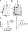
Expression and purification of recombinant RELM family proteins. (A and B) SDS/PAGE analysis of mRELMβ, mRELMβ C terminus, hRELMβ, and hRETN under reducing (A, +DTT) and nonreducing (B, −DTT) conditions. (C) Size exclusion chromatography of recombinant RELMs on a Superdex 75/300 column. (D) Circular dichroism (CD) spectroscopy of mRELMβ and hRELMβ. The CD spectra for both proteins exhibit maximal negative ellipticity in the range of 205–215 nm, indicating a predominantly β-sheet structure that is consistent with previously published spectra of members of this family (36) and with the RELMβ crystal structure (14). These results indicate that the proteins have acquired their expected secondary structures and are thus correctly refolded.
Fig. S2.
Fig. S2.
Dimeric hRELMβ has antibacterial activity. (A) SDS/PAGE analysis of hRELMβ produced in-house and commercially available hRELMβ (Shenandoah Biotechnology) under reducing (−DTT) and nonreducing (+DTT) conditions. Under nonreducing conditions, hRELMβ produced in-house migrates as a monomer while commercially available hRELMβ migrates as a dimer. (B) Antibacterial activity of monomeric hRELMβ and commercially available, dimeric hRELMβ against P. aeruginosa. Bactericidal assays were performed at least twice and repeated in triplicate within each experiment.
Fig. S3.
Fig. S3.
Characterization of mRELMβ antibacterial activity. (A) mRELMβ antibacterial activity is dependent on bacterial growth phase. C. rodentium was grown to either midlogarithmic phase or stationary phase before the addition of mRELMβ with incubation for 2 h at 37 °C, followed by dilution plating. (B) mRELMβ antibacterial activity is pH dependent, with higher activity at acidic pH (5.5) than at slightly basic pH (7.5). Bacteria were incubated with 5 μM mRELMβ. (C) Low pH (5.5) and a relatively low salt concentration (25 mM NaCl) are optimal for bacterial membrane permeabilization by mRELMβ. C. rodentium was treated with 5 μM mRELMβ, and PI uptake was measured over 2 h. Assays were performed at least twice and repeated in triplicate within each experiment. Means ± SD are plotted. Statistics were performed with Student’s t test; **P < 0.01; ***P < 0.001; ns, not significant.
Fig. 2.
Fig. 2.
RELMβ binds to negatively charged lipids and forms a multimeric pore in membranes. (A) mRELMβ disrupts carboxyfluorescein (CF)-loaded unilamellar liposomes containing the negatively charged lipid phosphatidyl serine (PS), but not liposomes composed of the zwitterionic lipid phosphatidylcholine (PC). Liposomes were treated with 5 μM mRELMβ, and dye efflux was monitored over time. The 1.0% octyl glucoside (OG) was added toward the end to disrupt remaining liposomes. Dye efflux is expressed as a percentage of maximal release by OG. (B) Means ± SD from three independent replicates of the experiment shown in A. (C) mRELMβ membrane-disrupting activity is confined to the C terminus. PC:PS liposomes (100 µM) were incubated with 5 μM full-length mRELMβ or the mRELMβ N or C terminus. (D) Initial rate of liposome dye efflux as a function of mRELMβ concentration. Assays were done in triplicate, means ± SD are shown, and statistical significance was calculated relative to the mRELMβ C terminus. (E) The C-terminal portion of mRELMβ binds lipid. The 5 μM full-length mRELMβ or the mRELMβ N or C terminus was added to liposomes incorporating 5% dansyl-PE, and dansyl fluorescence was monitored as measure of binding. (F and G) mRELMβ forms a multimeric complex in the presence of liposomes. Full-length mRELMβ was incubated with 100 mM PC:PS liposomes and cross-linked with bis(sulfosuccinimdyl) suberate. Cross-linked complexes were solubilized in detergent, resolved by size exclusion chromatography (F), and analyzed by Western blotting with anti-RELMβ antibody (F, Inset). mRELMβ forms a complex of ∼60–70 kDa, or roughly six to eight protein units. (G) mRELMβ forms size-selective pores in liposomes. The 10 μM full-length mRELMβ was added to 100 μM PC:PS liposomes loaded with carboxyfluorescein (CF) (∼10-Å Stokes diameter) or fluorescein isothiocyanate-dextran 10 (FD10) (∼44-Å Stokes diameter). (H) Means ± SD from three independent replicates of the experiment shown in G. Statistics were performed with Student’s t test; *P < 0.05; ***P < 0.001; ns, not significant.
Fig. S4.
Fig. S4.
Characterization of mRELMβ lipid binding and membrane permeabilization activities. (A) mRELMβ binds to negatively charged lipids (indicated in red). Membranes displaying lipids were incubated with 1 μg/mL mRELMβ, followed by detection with anti-RELMβ antibody. (B) FRET efficiency as a function of mRELMβ full-length and mRELMβ C terminus concentration. Assays were performed in triplicate, and means ± SD are plotted. (C) The mRELMβ C terminus forms a multimer in the presence of liposomes. The mRELMβ C terminus was incubated with 100 mM PC:PS liposomes and cross-linked with bis(sulfosuccinimdyl) suberate. Cross-linked complexes were solubilized in detergent and resolved by size exclusion chromatography. (D) The mRELMβ C terminus forms size-selective pores in liposomes. The 10 μM full-length mRELMβ was added to 100 µM PC:PS liposomes loaded with carboxyfluorescein (CF) (∼10-Å Stokes diameter) or fluorescein isothiocyanate-dextran 10 (FD10) (∼44-Å Stokes diameter), and dye efflux was measured. The 1.0% octyl glucoside (OG) was added toward the end to disrupt remaining liposomes. Dye efflux is expressed as a percentage of maximal release by OG. (E) Means ± SD from three independent replicates of the experiment shown in D. Statistics were performed with Student’s t test; *P < 0.05; **P < 0.01; ***P < 0.001.
Fig. S5.
Fig. S5.
Generation of Retnlb−/− mice. (A) CRISPR/Cas mutagenesis was used to introduce edits into the mouse Retnlb exon 2 sequence encoding the RELMβ signal peptide (the guide RNA-targeted sequence is underlined). A mutation was selected that introduced a premature stop codon into the RELMβ signal peptide, and mice harboring this mutation were bred to homozygosity. (B) RELMβ is not expressed in colons from Retnlb−/− mice. Mice were orally infected with C. rodentium, and then colons were analyzed for RELMβ expression by Western blot. Each lane represents a different mouse. (C) Bacterial counts from C. rodentium-infected wild-type and Retnlb−/− colons. Mice were orally infected for 7 d. Assays were performed in triplicate, and means ± SD are shown. Statistics were performed with Student’s t test; *P < 0.05; **P < 0.01.
Fig. 3.
Fig. 3.
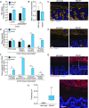
RELMβ limits entry of Gram-negative bacteria into the colon inner mucus layer. (A) Quantification of total colonic luminal and tissue-associated bacteria by Q-PCR determination of 16S rRNA gene copy number in cohoused wild-type and RELMβ-deficient (Retnlb−/−) mice. (B and C) MUC2 expression is not altered in RELMβ-deficient mice. (B) Q-PCR analysis of colonic Muc2 transcripts. (C) Immunofluorescence detection of the mucus layer in colons of wild-type and Retnlb−/− mice with Ulex europaeus agglutinin-I (UEA-I), which detects mucus glycans (34). (Scale bars: 50 µm.) (D) Q-PCR quantification of 16S gene copy number from specific bacterial groups. Bacteria were recovered from colonic tissue and analyzed using taxon-specific 16S rDNA primers. (E) Immunofluorescence detection of lipoteichoic acid (LTA) in colonic tissues indicates that spatial segregation of Gram-positive bacteria is not markedly impacted by RELMβ deficiency. (F) Q-PCR quantification of specific bacterial groups at the colonic mucosal surface. Values for each bacterial group are expressed relative to 16S rDNA levels in wild-type mice. (G) Immunofluorescence detection of Helicobacter species at the colon surface. (H) Helicobacter+ particles per square micrometer in the colon inner mucus layer. Quantification of particle density was performed using ImageJ from five fluorescent images from three mice of each genotype. For the 16S analyses, four mice per genotype were analyzed for each experiment, and Q-PCR assays were repeated in triplicate within each experiment. Means ± SD are plotted. Statistics were performed with Student’s t test; **P < 0.01; ***P < 0.001; ns, not significant. All tissues were counterstained with DAPI (blue), and antibody isotype controls are shown in Fig. S8. (Scale bars: 25 µm.)
Fig. S8.
Fig. S8.
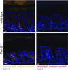
Isotype controls for immunofluorescence detection of bacteria. Colon tissues from wild-type and Retnlb−/− mice were detected with mouse IgG (isotype control for anti-LTA detection shown in Fig. 3E) and rabbit IgG (isotype control for anti-Helicobacter detection shown in Fig. 3G). (Scale bars: 25 µm.)
Fig. S6.
Fig. S6.
Determination of bacterial 16S copy number in small intestine, colon, MLN, and spleen from wild-type and Retnlb−/− mice. (A) Q-PCR determination of 16S rRNA gene copy number in the distal small intestinal (ileal) lumen, ileal tissue, MLN, and spleen of cohoused wild-type and Retnlb−/− mice. (B–F) Q-PCR analysis of 16S gene copy numbers from specific bacterial groups. Bacteria were recovered from the ileal lumen (B), ileal tissue (C), MLN (D), spleen (E), and colon lumen (F), and analyzed using taxon-specific 16S rDNA primers. (G) Q-PCR quantification of specific subgroups of Proteobacteria in the colon lumen. Values for each bacterial group are expressed relative to the 16S rDNA levels in wild-type mice. Four mice per genotype were analyzed for each experiment, and Q-PCR assays were repeated in triplicate within each experiment. Means ± SD are plotted. Statistics were performed with Student’s t test; *P < 0.05; ns, not significant; nd, not detected.
Fig. S7.
Fig. S7.
Phylogenetic analysis of 16S rRNA from tissue-associated and lumen microbial communities. Operational taxonomic units with an average of 100 reads and populations greater than or equal to 1% were included in the graphical analysis. (A and B) Phylum-level analysis of tissue-associated (A) and luminal (B) colonic bacteria. (C and D) Family-level analysis of tissue-associated (C) and luminal (D) colonic bacteria.
Fig. 4.
Fig. 4.
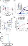
Human resistin (hRETN) is a bactericidal protein. (A) Human resistin (hRETN) bactericidal activity. Purified recombinant hRETN was added to midlogarithmic phase bacteria for 2 h, and numbers of surviving bacteria were quantified by dilution plating. Means ± SD are plotted. (B) hRETN permeabilizes bacterial membranes. C. rodentium was treated with increasing concentrations of hRETN, and PI uptake was measured over 2 h. The assay was performed twice and was repeated in triplicate within each experiment. (C) hRETN disrupts carboxyfluorescein (CF)-loaded PC:PS liposomes. Liposomes were treated with increasing concentrations of hRETN, and dye efflux was monitored over time. The 1.0% octyl glucoside (OG) was added at the end to disrupt remaining liposomes. Dye efflux is expressed as a percentage of maximal release by OG. (D) hRETN membrane-disrupting activity is superior to the membrane-disrupting activity of C terminus of mRELMβ. CF-loaded PC:PS liposomes (100 µM) were incubated with varying concentrations of full-length hRETN or the mRELMβ N or C terminus, and initial rates of liposome dye efflux as a function of hRETN concentrations are plotted. Assays were done in triplicate, and means ± SD are shown. (E) hRETN forms a multimeric complex in the presence of liposomes. The 10 μM full-length hRETN was incubated with 100 mM PC:PS liposomes and cross-linked with bis(sulfosuccinimdyl) suberate. Cross-linked complexes were solubilized in detergent and resolved by size exclusion chromatography. Statistics were performed with Student’s t test; *P < 0.05.

"V体育平台登录" References

    1. Sonnenburg JL, Bäckhed F. Diet-microbiota interactions as moderators of human metabolism. Nature. 2016;535:56–64. - PMC - PubMed
    1. Johansson MEV, et al. Composition and functional role of the mucus layers in the intestine. Cell Mol Life Sci. 2011;68:3635–3641. - PMC - PubMed
    1. Johansson ME, et al. The inner of the two Muc2 mucin-dependent mucus layers in colon is devoid of bacteria. Proc Natl Acad Sci USA. 2008;105:15064–15069. - PMC - PubMed
    1. Vaishnava S, et al. The antibacterial lectin RegIIIgamma promotes the spatial segregation of microbiota and host in the intestine. Science. 2011;334:255–258. - PMC - PubMed
    1. Okumura R, et al. Lypd8 promotes the segregation of flagellated microbiota and colonic epithelia. Nature. 2016;532:117–121. - PubMed

Publication types

LinkOut - more resources