"V体育平台登录" Skip to main page content
U.S. flag

An official website of the United States government

Dot gov

The VSports app下载. gov means it’s official. Federal government websites often end in . gov or . mil. Before sharing sensitive information, make sure you’re on a federal government site. .

Https

The site is secure. The https:// ensures that you are connecting to the official website and that any information you provide is encrypted and transmitted securely V体育官网. .

. 2017 Nov 6;214(11):3449-3466.
doi: 10.1084/jem.20170412. Epub 2017 Sep 21.

A multidimensional blood stimulation assay reveals immune alterations underlying systemic juvenile idiopathic arthritis

Affiliations

"V体育官网入口" A multidimensional blood stimulation assay reveals immune alterations underlying systemic juvenile idiopathic arthritis

Alma-Martina Cepika et al. J Exp Med. .

Abstract

The etiology of sporadic human chronic inflammatory diseases remains mostly unknown. To fill this gap, we developed a strategy that simultaneously integrates blood leukocyte responses to innate stimuli at the transcriptional, cellular, and secreted protein levels. When applied to systemic juvenile idiopathic arthritis (sJIA), an autoinflammatory disease of unknown etiology, this approach identified gene sets associated with specific cytokine environments and activated leukocyte subsets. During disease remission and off treatment, sJIA patients displayed dysregulated responses to TLR4, TLR8, and TLR7 stimulation. Isolated sJIA monocytes underexpressed the IL-1 inhibitor aryl hydrocarbon receptor (AHR) at baseline and accumulated higher levels of intracellular IL-1β after stimulation. Supporting the demonstration that AHR down-regulation skews monocytes toward macrophage differentiation, sJIA monocytes differentiated in vitro toward macrophages, away from the dendritic cell phenotype. This might contribute to the increased incidence of macrophage activation syndrome in these patients. Integrated analysis of high-dimensional data can thus unravel immune alterations predisposing to complex inflammatory diseases. VSports手机版.


Figures

Figure 1.
Figure 1.
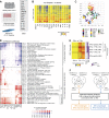
Transcriptional landscape of healthy adult blood stimulated with 15 stimuli. (A) Experimental workflow. (B) Hierarchical cluster of the 11,894 DETs in healthy adult blood cultured in vitro for 6 h in 16 conditions. Data are normalized to the medium control for each donor and averaged per stimulus. The median number of replicates per stimulus was eight, with an interquartile range of three. Samples from 13 donors were processed in five independent experiments. (C) Principal-component analysis based on the 11,894 DETs identified in B. Samples are colored by stimulus. (D) Hierarchical clustering of the reference module fingerprints. Module score is expressed as a percentage (transparency scale) of transcripts twofold over- (red) or underexpressed (blue). Data are normalized to the medium control for each donor. Fingerprints are averaged by stimulus. (E) Hierarchical clustering of the DETs in IFN-α versus IFN-γ stimulations. Induction was defined by significance analysis (ANOVA, P < 0.05) and normalized expression >1.25. When induced by both IFN-α and IFN-γ, transcripts were further separated based on the differential magnitude of expression using a twofold threshold (IFN-α = IFN-γ, IFN-α > IFN-γ, IFN-γ > IFN-α). (F) Identification of uncataloged type I and type II IFN-inducible transcripts. Venn diagrams represent the overlap between IFN-inducible transcripts in our dataset and in the INTERFEROME database, including both H. sapiens and Mus musculus organisms (normalized fold change >1.25). Transcripts uncataloged in the INTERFEROME were further subjected to IPA where transcripts connected directly or indirectly to IFNs were filtered out. IFN-α– and IFN-γ–induced transcripts were analyzed separately. PMA/I, PMA/ionomycin.
Figure 2.
Figure 2.
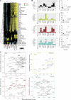
Multidimensional assessment of healthy adult blood responses to stimulation by microarray, FACS, and multiplex cytokine analysis. (A) Experimental workflow. (B) Hierarchical cluster of surface and intracellular proteins measured by FACS after 6-h stimulation. gMFI ratios were scaled per marker and cell subset by setting the medium reference control to 0 and the condition with maximum change to 1 or −1 (up or down). (C) Hierarchical clustering of 30 secreted proteins measured by Luminex. (D) Eigengene profiles of four WGCNA modules. The data represent log2-transformed ratios of stimulated samples normalized to medium control. (E) Heatmap representing the transcript overlap between WGCNA modules and the reference modules from Fig. 1 D. HKSA, heat-killed Staphylococcus aureus; HKSE, heat-killed Salmonella enterica.
Figure 3.
Figure 3.
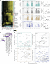
Integration of microarray, FACS and multiplex cytokine measurements by weighted gene coexpression network analysis. (A) Hierarchical cluster of the correlation matrix between WGCNA module eigengenes (x axis) and FACS (orange) or Luminex (green) measurements (y axis). (B) Area charts representing the module eigengene for four modules linked to leukocyte subset activation as quantified by CD69 expression. Charts were overlaid with boxplots representing the eigengene’s best FACS or Luminex correlate. Pearson correlations between eigengenes and protein measurements are represented as x–y charts on the right. (C) x–y plots representing the MM (x axis, R value) versus GS (y axis, R value) analysis for the four modules from B and their best protein correlate. MM and GS represent the correlation of each gene to the module eigengene and the protein expression, respectively. Genes highly representative of the module functional annotation were highlighted in bold font. Genes are duplicated when several probes were detected within the selected range of MM and GS. HKSA, heat-killed Staphylococcus aureus; HKSE, heat-killed Salmonella enterica.
Figure 4.
Figure 4.
Hyperresponsiveness to TLR4 and TLR8 ligands in inactive untreated sJIA patients. (A) Experimental workflow. (B) Hierarchical cluster of the 4,480 DETs between healthy controls and sJIA patient groups ex vivo (baseline; top). Hierarchical cluster of the 323 transcripts overexpressed ≥1.5-fold in healthy blood stimulated with IL-1β for 6 h (ref. Fig. 1; center). Data are normalized to medium controls for each donor. Hierarchical cluster of the same 323 transcripts in ex vivo signatures (bottom). Ex vivo blood samples were processed in two independent experiments (C) Dot plots representing the raw expression of IL1B and CASP1 transcripts, white blood cells (WBC), neutrophil, monocyte, and lymphocyte absolute counts, displayed by patient group. Horizontal lines indicate the median, whiskers the interquartile range. (D) Hierarchical cluster of the 14,575 DETs in stimulated blood from sJIA patients and pediatric healthy controls. Samples from 18 donors were processed in 16 independent experiments. (E) Box plot representing the molecular distance to medium (MDTM) derived from the 14,575 DETs identified in D, displayed per stimulus and patient group. The MDTM is calculated for each sample as the sum of absolute normalized fold changes ≥2 in the list of transcripts considered. Horizontal lines indicate the median. Boxes represent the interquartile range, and whiskers nonoutlier range (H, healthy). HKSA, heat-killed Staphylococcus aureus; HKSE, heat-killed Salmonella enterica.
Figure 5.
Figure 5.
Loss of balance between proinflammatory and IFN responses in sJIA patients. (A) Hierarchical cluster of the module–trait correlation matrix obtained by WGCNA in stimulated sJIA and control blood dataset. (B) Bar charts representing the eigengene profiles of four WGCNA modules that display distinct transcriptional differences between healthy controls and sJIA patient groups. Bar charts are overlaid with box plots of the eigengenes’ best FACS correlate (right; y axis). Pearson correlations between eigengenes and protein measurements are represented as x–y charts on the right (H, healthy). (C) Hierarchical cluster of the overlap between WGCNA modules and the reference modules. (D) x–y plots representing the MM (x axis) versus GS (y axis) analysis for the four modules from B and their best protein correlate. HKSA, heat-killed Staphylococcus aureus; HKSE, heat-killed Salmonella enterica.
Figure 6.
Figure 6.
Monocytes from sIU patients accumulate IL-1β after LPS stimulation, underexpress AHR gene at baseline, and differentiate into macrophages in vitro. (A) Experimental workflow. Monocytes from each donor were isolated and cultured independently. (B) gMFI ratios of intracellular IL-1β in monocytes for indicated conditions (left). Data are normalized to each donor’s baseline gMFI. **, P < 0.01, Mann–Whitney test. Histogram overlay of intracellular IL-1β in representative healthy and sJIA monocytes (right). (C) Concentration of secreted IL-1β in the supernatants of cultured monocytes. (D) x–y chart of DEGs between stimulated sJIA and healthy monocytes. Values represent FPKM ratios of healthy LPS to healthy medium (x axis) versus sJIA LPS to sJIA medium (y axis). Genes with P < 0.05 were filtered for an absolute log2(ratio) difference ≥1.5. (E) x–y chart of DEGs between sJIA and healthy monocytes ex vivo. Genes with P < 0.05 were filtered for an absolute log2 difference sJIA-healthy ≥1.5. Genes with absolute log2(ratio) (D) or log2 (E) FPKM values <1 were removed. (F) Expression of ACVR2A gene measured by RNA-seq and quantitative RT-PCR. The levels of activin A protein in supernatants of monocytes stimulated for 24 h with LPS are shown on the right. Activin A was not detectable in unstimulated conditions or at 6 h LPS. (G) Expression of AHR gene measured by RNA-seq and quantitative RT-PCR. (H) Expression of AHR gene measured by quantitative RT-PCR in an independent cohort of sJIA patients and controls (n = 5). (I) Monocytes from the second cohort were differentiated in vitro for 5 d; the phenotype of monocyte-derived cells was determined by flow cytometry as macrophage (moMac; CD16+CD1a) or DC (moDC; CD16CD1a+; left). Representative plots from one healthy control and one patient are shown in the middle. The ratio of the percentage of moMac cells to the percentage of moDC cells is shown on the right. Cultures were performed in three independent experiments. H, healthy. In dot plots, horizontal lines indicate the median and whiskers the interquartile range. In box plots, horizontal lines indicate the median, boxes the interquartile range, and whiskers the nonoutlier range. P-values for flow cytometry and quantitative RT-PCR data were calculated using Mann–Whitney U test.

Comment in

References

    1. Allantaz F., Chaussabel D., Stichweh D., Bennett L., Allman W., Mejias A., Ardura M., Chung W., Smith E., Wise C., et al. . 2007. Blood leukocyte microarrays to diagnose systemic onset juvenile idiopathic arthritis and follow the response to IL-1 blockade. J. Exp. Med. 204:2131–2144. 10.1084/jem.20070070 - DOI - PMC - PubMed
    1. Alsina L., Israelsson E., Altman M.C., Dang K.K., Ghandil P., Israel L., von Bernuth H., Baldwin N., Qin H., Jin Z., et al. . 2014. A narrow repertoire of transcriptional modules responsive to pyogenic bacteria is impaired in patients carrying loss-of-function mutations in MYD88 or IRAK4. Nat. Immunol. 15:1134–1142. 10.1038/ni.3028 - DOI - PMC - PubMed
    1. Anders S., Pyl P.T., and Huber W.. 2015. HTSeq--a Python framework to work with high-throughput sequencing data. Bioinformatics. 31:166–169. 10.1093/bioinformatics/btu638 - DOI - PMC - PubMed
    1. Bessede A., Gargaro M., Pallotta M.T., Matino D., Servillo G., Brunacci C., Bicciato S., Mazza E.M., Macchiarulo A., Vacca C., et al. . 2014. Aryl hydrocarbon receptor control of a disease tolerance defence pathway. Nature. 511:184–190. 10.1038/nature13323 - DOI - PMC - PubMed
    1. Blankley S., Graham C.M., Howes A., Bloom C.I., Berry M.P., Chaussabel D., Pascual V., Banchereau J., Lipman M., and O’Garra A.. 2014. Identification of the key differential transcriptional responses of human whole blood following TLR2 or TLR4 ligation in-vitro. PLoS One. 9:e97702 10.1371/journal.pone.0097702 - DOI - PMC - PubMed

MeSH terms (VSports在线直播)