Skip to main page content
U.S. flag

An official website of the United States government

Dot gov

The . gov means it’s official. Federal government websites often end in . gov or . mil VSports app下载. Before sharing sensitive information, make sure you’re on a federal government site. .

Https

The site is secure V体育官网. The https:// ensures that you are connecting to the official website and that any information you provide is encrypted and transmitted securely. .

Review
. 2018 Apr:76:163-178.
doi: 10.1016/j.semcdb.2017.08.055. Epub 2017 Sep 8.

Mitochondrial cytochrome c oxidase biogenesis: Recent developments

Affiliations
Review

Mitochondrial cytochrome c oxidase biogenesis: Recent developments (VSports手机版)

Alba Timón-Gómez et al. Semin Cell Dev Biol. 2018 Apr.

"VSports" Abstract

Mitochondrial cytochrome c oxidase (COX) is the primary site of cellular oxygen consumption and is essential for aerobic energy generation in the form of ATP. Human COX is a copper-heme A hetero-multimeric complex formed by 3 catalytic core subunits encoded in the mitochondrial DNA and 11 subunits encoded in the nuclear genome VSports手机版. Investigations over the last 50 years have progressively shed light into the sophistication surrounding COX biogenesis and the regulation of this process, disclosing multiple assembly factors, several redox-regulated processes leading to metal co-factor insertion, regulatory mechanisms to couple synthesis of COX subunits to COX assembly, and the incorporation of COX into respiratory supercomplexes. Here, we will critically summarize recent progress and controversies in several key aspects of COX biogenesis: linear versus modular assembly, the coupling of mitochondrial translation to COX assembly and COX assembly into respiratory supercomplexes. .

Keywords: COX assembly factor; COX1; COX2; Mitochondrial cytochrome c oxidase; Respiratory supercomplex V体育安卓版. .


Conflict of interest statement

Conflicts of interest

The authors declare that they do not have any conflict of interest.

V体育官网入口 - Figures

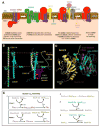
Figure 1
Figure 1. Mitochondrial cytochrome c oxidase: structure, redox metal cofactors and overall assembly pathway
(A and B) Ribbon diagrams of high-resolution structures of (A) the thirteen-subunit enzyme from bovine (Bos taurus) heart mitochondria [6, 127] (PDB 1OCC). The COX catalytic core subunits are colored in blue (COX1), red (COX2) and pink (COX3). The nucleus-encoded COX subunits are colored in light green. In (B) most of the bovine protein has been removed to show only the redox-active metal centers. A covalent bond between Tyr-371 and the CuB ligand His-378 is present but not shown in this structure. Detailed explanations of the CuA and CuB centers are in the text. (C) The schematic represents a simplified model for the process of monomeric COX assembly based on the modular model recently reported [9]. The catalytic core subunits form subassembly modules with other subunits and assembly factors prior to be incorporated into the main assembly pathway. In general, following their insertion into the inner membrane, COX1 and COX2 are stabilized by substrate-specific chaperones and matured by addition of metal cofactors. Following COX1 maturation, the nuclear DNA-encoded COX4 and COX5A, forming a module argued to be stabilized by the assembly factor HIGD1A are added to COX1 prior incorporation of the COX2-containing, and subsequent a set of three assembly factors (PET100, PET117 and MR-1S) that promote the incorporation of the COX3-containing module and the NDUFA4 subunit to form the COX holoenzyme or Complex IV (CIV). Only a few COX biogenetic factors are represented.
Figure 2
Figure 2. COX1 maturation and assembly line
(A) Model depicting the roles of COX14 (alias C12orf62), COA3 (alias MITRAC12, CCDC56, or COX25) and CMC1 in controlling post-translational events in COX1 biogenesis [14]. According to the model, newly synthesized COX1 would first bind COX14 and COA3, followed by CMC1. CMC1 would promote COX1 stability during or before COX1 maturation and would be released from the growing COX1 complex before the incorporation of COX4-1 and COX5A and additional assembly factors such as SURF1 and MITRAC7 (see explanation in the text). (B) Model for copper delivery to COX1 by COX11 showing a COX11-COX11 homodimer (model 5, cluster 2 of ref. [58]) hovering above fully folded COX1, taken from the structure of the bovine COX (PDB 5b1b). Tethering of the antiparallel dimer by the transmembrane helices of the COX11 monomers (upper ends are shown in gold) dictates that the four S-two Cu(I) cluster of the COX11 dimer (Cu(I) ions in yellow, Cys ligands in red) will face the membrane surface. Linkers of 15 amino acid between the transmembrane helices and the headgroup of the dimer will keep the COX11 Cu cluster near the membrane surface. CuB of fully assembled COX1 is shown in magenta while the position of the CuB ligands His-290 and His-291 are shown in pink. Cys35 in bacterial COX11 (Cys-121 of human COX11 and Cys-111 of yeast COX11) is not shown, but Cys-35 of each monomer will be present near the exit of each COX11 transmembrane helix from the upper surface of the membrane. It can be seen that His-290 and His-291 need move only a short distance toward the upper surface of COX1 in order for COX11 to transfer Cu(I) to these histidines, by the mechanism proposed in section 2.2.3. (C) Model for COX19 regulation of copper binding to and transfer from COX11 by maintaining Cys-35 in a reduced state. Models of yeast COX11 and COX19 with relevant residues shown as sticks. In COX11, the three relevant cysteines are labeled (the asterisk denotes that numbering of S. meliloti COX11 is based on PDB entry 1SO9). The red, green and blue segments correspond to those identified as interacting with COX19 by Bode et al [61] (in that order along the protein sequence). The red and green segments are the most hydrophobic and conserved, and thus most likely candidates for interaction. In COX19, the putative interaction surface would be a conserved hydrophobic patch built around two TyrLeu dipeptides in the yeast protein. 3D visualization available online at http://lucianoabriata.altervista.org/modelshome.html
Figure 3
Figure 3. COX2 maturation and assembly line
(A) Model depicting the roles of COX20 and COX18 in COX2 stabilization and membrane insertion [15, 16]. The release of COX18 from the complex coincides with the incorporation of the CuA maturation module formed by the copper chaperones SCO1 and SCO2 and the CX9C protein COA6. (B) Model depicting a possible action of COX20 on COX2 membrane insertion. Left: Acting at an earlier stage, COX20 could stabilize COX2’s TM helices when it is inserted in the membrane only through TM1 while TM2 and the globular domain are on the mitochondrial matrix. Right: At a later stage, COX20’s TM helices could stabilize COX2’s TM helices when they are both inserted in the membrane, before and/or during and/or after flipping of COX2 through the IMS, before its assembly onto the growing oxidase. In the globular domain of COX2 (purple) the purple spheres indicate the location of the CuA site, but note that at these stages the copper ions have actually not been loaded yet. 3D visualizations of these models are available online at http://lucianoabriata.altervista.org/modelshome.html. (C) Models for mammalian COX2 metallation by SCO1-SCO2. The schemes depict five published proposals for the assembly of CuA (explanations are given in section 3 and reviewed in [26]). The possible involvement of COA6 is not depicted. (D) A possible structural model for the electron and/or copper transfer reactions between apo- or copper-SCO and apo-COX2 proteins (cyan and yellow, respectively) built from the x-ray structure of the disulfide-mixed complex between Bradyrhizobium japonicum TlpA and COX2 (also available in 3D online). The depicted apo-COX2 is from B. japonicum (PDB ID 4TXV chain B) and the SCO protein is human SCO2 (PDB ID 2RLI). A structure of T. thermophilus copper-loaded COX2 (PDB ID 1EHK) is shown for the sake of comparing the compactness of the CuA-binding loop. All spheres are relevant cysteines, while other copper ligands are shown as sticks.
Figure 4
Figure 4. Contact sites of COX (complex IV or CIV) subunits with subunits from complexes CI and CIII in the context of the mammalian respirasome
(A) Ribbon diagrams of high-resolution structures of the tight form of the CI-CIII2-CIV respirasome from ovine (Ovis aries) heart mitochondria [102] (PDB 5J4Z). The three component complexes are colored in light blue (CI), light yellow (CIII) and light green (CIV). In deep blue are colored CI subunits in proximity and proposed to interact with CIV subunits, colored in deep green. (B) Magnified view of deep blue colored CI subunits (ND5 and NDUFB7) proposed to interact with deep green colored CIV subunits COX8 and COX7C [102]. (C) Different view of the structure presented in (A), rotated 156°, with deep yellow colored CIII subunits in proximity and potentially interacting with a CIV subunit colored in deep green. (D) Magnified view of deep yellow colored CIII subunits (UQCRC1, UQCR11 and UQCRB) of the active CIII monomer proposed to interact with the deep green colored CIV subunit COX7A [102].

References

    1. Havugimana PC, Hart GT, Nepusz T, Yang H, Turinsky AL, Li Z, Wang PI, Boutz DR, Fong V, Phanse S, Babu M, Craig SA, Hu P, Wan C, Vlasblom J, Dar VU, Bezginov A, Clark GW, Wu GC, Wodak SJ, Tillier ER, Paccanaro A, Marcotte EM, Emili A. A census of human soluble protein complexes. Cell. 2012;150(5):1068–81. - PMC - PubMed
    1. Fernandez-Vizarra E, Tiranti V, Zeviani M. Assembly of the oxidative phosphorylation system in humans: what we have learned by studying its defects. Biochim Biophys Acta. 2009;1793(1):200–11. - "VSports注册入口" PubMed
    1. Soto IC, Fontanesi F, Liu J, Barrientos A. Biogenesis and assembly of eukaryotic cytochrome c oxidase catalytic core. Biochim Biophys Acta. 2012;1817(16):883–97. - PMC - PubMed
    1. Pierron D, Wildman DE, Huttemann M, Markondapatnaikuni GC, Aras S, Grossman LI. Cytochrome c oxidase: evolution of control via nuclear subunit addition. Biochim Biophys Acta. 2012;1817(4):590–7. - PMC - PubMed
    1. Balsa E, Marco R, Perales-Clemente E, Szklarczyk R, Calvo E, Landazuri MO, Enriquez JA. NDUFA4 is a subunit of complex IV of the mammalian electron transport chain. Cell Metab. 2012;16(3):378–86. - PubMed

V体育官网 - Publication types

MeSH terms

Substances