Skip to main page content
U.S. flag

An official website of the United States government

Dot gov

The . gov means it’s official. Federal government websites often end in . gov or . mil. Before sharing sensitive information, make sure you’re on a federal government site. VSports app下载.

Https

The site is secure. The https:// ensures that you are connecting to the official website and that any information you provide is encrypted and transmitted securely V体育官网. .

. 2017 Nov;7(11):1238-1247.
doi: 10.1158/2159-8290.CD-17-0538. Epub 2017 Aug 22.

Constitutive Signaling from an Engineered IL7 Receptor Promotes Durable Tumor Elimination by Tumor-Redirected T Cells

Affiliations

Constitutive Signaling from an Engineered IL7 Receptor Promotes Durable Tumor Elimination by Tumor-Redirected T Cells

Thomas Shum (VSports app下载) et al. Cancer Discov. 2017 Nov.

VSports手机版 - Abstract

Successful adoptive T-cell immunotherapy of solid tumors will require improved expansion and cytotoxicity of tumor-directed T cells within tumors. Providing recombinant or transgenic cytokines may produce the desired benefits but is associated with significant toxicities, constraining clinical use. To circumvent this limitation, we constructed a constitutively signaling cytokine receptor, C7R, which potently triggers the IL7 signaling axis but is unresponsive to extracellular cytokine. This strategy augments modified T-cell function following antigen exposure, but avoids stimulating bystander lymphocytes. Coexpressing the C7R with a tumor-directed chimeric antigen receptor (CAR) increased T-cell proliferation, survival, and antitumor activity during repeated exposure to tumor cells, without T-cell dysfunction or autonomous T-cell growth. Furthermore, C7R-coexpressing CAR T cells were active against metastatic neuroblastoma and orthotopic glioblastoma xenograft models even at cell doses that had been ineffective without C7R support. C7R may thus be able to enhance antigen-specific T-cell therapies against cancer. Significance: The constitutively signaling C7R system developed here delivers potent IL7 stimulation to CAR T cells, increasing their persistence and antitumor activity against multiple preclinical tumor models, supporting its clinical development. Cancer Discov; 7(11); 1238-47. ©2017 AACR. This article is highlighted in the In This Issue feature, p VSports手机版. 1201. .


Conflict of interest statement

Disclosures of Potential Conflicts of Interest: Baylor College of Medicine (TS, CMR, SG, and BO) has filed a patent application on constitutively active cytokine receptors for cell therapy.

Figures

Figure 1
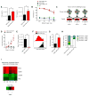
Figure 1. Constitutive signaling from C7R activates STAT5 in T-cells but does not support autonomous cell expansion
(A) Schematic comparisons of IL-7 bound to the natural IL-7 receptor composed of heterodimerized IL7Rα and γc, compared to the engineered C7R homodimerized receptor. (B,C) Transduction efficiency of Δ34 and C7R (representative of 3) in (B) CD4 and (c) CD8 T-cells relative to non-transduced (NT) cells. (D,E) Representative flow cytometric comparison of phosphorylated STAT5 (pSTAT5) in (D) CD4 and (E) CD8 T-cells that were transduced with Δ34 or C7R. Cells were cultured without IL-15 and IL-7 for 24–72 hours before analysis. (F,G) Average pSTAT5 MFI values when repeating the experiments in D and E with multiple donors. (H,I) Quantitated in-vitro persistence of Δ34 or C7R transduced (H) CD4 or (I) CD8 T-cells cultured in cytokine-free complete cell culture media starting 9–12 days after PBMC activation, without further antigen stimulus. Live cells were counted weekly using trypan-blue exclusion. X-axis denotes the number of days after IL-15 and IL-7 were withdrawn from culture media. Area under the curve (AUC) values were compared with the two-tailed t-test: 10.5 ± 0.6616 (CD8 Δ34), 56.37 ± 7.972 (CD8 C7R), p<0.05; 10.22 ± 1.694 (CD4 Δ34) and 31.36 ± 2.590 (CD4 C7R), p<0.05. *P<0.05, **P<0.01, ***P<0.001 (two-tailed paired t-test, FI). Graphs FI represent averages from different donors ± SEM (n=3).
Figure 2
Figure 2. C7R enhances GD2-CAR T-cell activity during serial tumor challenge
(A) Cytokines secreted by GD2-CAR T-cells or GD2-CAR.C7R T-cells 24 hours after co-culture with LAN-1 tumor cells was determined by ELISA. (B) 4-hour luciferase-based cytotoxicity assay of T-cells killing LAN-1 tumor cells. (C) Serial co-culture schematic. The first co-culture (CC1) was initiated with 1×10⁶ GD2-CAR or GD2-CAR.C7R T-cells together with 0.5×10⁶ LAN-1 GFP-FFluc tumor cells for 7 days, in the absence of IL-15 or IL-7. For the second and third co-cultures (CC2 and CC3), T-cells were harvested from the previous co-culture and then replated in new culture medium with fresh tumor cells at the same 2:1 E:T ratio. (D) Cumulative expansion of GD2-CAR or GD2-CAR.C7R T-cells during serial co-culture. Arrows indicate timepoints of T-cell re-stimulation with tumor cells. (E) LAN-1 tumor cells remaining after CC3 with GD2-CAR T-cells and GD2-CAR.C7R T-cells, respectively. (F,G) For proliferation analysis, GD2-CAR and GD2-CAR.C7R T-cells collected at the end of CC1 were labeled with Cell Trace Violet before being rechallenged during CC2. (F) Histogram overlay represents data from a representative donor. (G) The experiment in F was repeated with multiple donors and the division indices compiled from the GD2-CAR and GD2-CAR.C7R proliferation histograms. (H) For survival analysis, GD2-CAR and GD2-CAR.C7R T-cells were stained with Annexin V and 7-AAD after 2 serial tumor challenges with LAN-1 tumor cells. Bar graphs show the frequencies of T-cells staining positive for Annexin V, 7-AAD, both, or neither. The Annexin V(+)7-AAD(−) and Annexin V(−)7-AAD(+) mean comparisons were n.s. (I) After the end of CC2, tumors were labeled with GD2-specific antibody and magnetically separated from the CAR T-cells. Total RNA was isolated from T-cells and gene expression analysis was subsequently performed using the Human Immunology Panel Version 2 and nCounter Analysis System (Nanostring). The displayed heat map shows genes with log2 fold changes (GD2-CAR.C7R/GD2-CAR) that had P values less than 0.02. Data was generated from 5 donors (10 paired samples). *P<0.05, **P<0.01, (two-tailed paired t-test, A, B, D, E, G, H). Graphs A, B, C, E, G, H represent averages from different donors ± SEM (n=6, A, D, E; n=3, G, H).
Figure 3
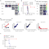
Figure 3. C7R enhances adoptive T-cell immunotherapy against metastatic and intracranial malignancies
(A) and (B) 1×10⁶ CHLA-255 FFluc cells were injected i.v. into female NSG mice, followed 7 days later by 1×10⁶ T-cells expressing an irrelevant CAR, GD2-CARΔ.C7R, GD2-CAR, or GD2-CAR.C7R. (A) Representative bioluminescent images of neuroblastoma growth over time. (B) Kaplan Meier survival analysis of CHLA-255 FFluc challenged mice. (C,D) To track T-cell migration and persistence, in a parallel experiment, 1×10⁶ CHLA-255 cells were injected intravenously into NSG mice, followed 7 days later by 1×10⁶ GFP-FFluc T-cells co-expressing GD2-CAR or GD2-CAR.C7R. (C) Sequential bioluminescent imaging of T-cells (D) Quantitated bioluminescent signal of T-cells over time (E) 1×10⁵ U373 GFP-FFluc cells were injected intracranially into male SCID mice. 7 days later, 1×10⁴ T-cells expressing EphA2-CARΔ.C7R, EphA2-CAR, or EphA2-CAR.C7R were intracranially injected into the tumor. Quantitated U373 GFP-FFluc bioluminescence from each treatment group is displayed over time. (F) Kaplan Meier survival analysis of U373 GFP-FFluc challenged mice after treatment with T-cells. *P<0.05 (Welch’s t-test, D). 5 mice were used for all groups. Graph D represent averages from experiment replicates ± SEM. In the experiment depicted in D, one mouse in the GD2-CAR.C7R group died during imaging on day 5 for reasons unrelated to tumor burden, therefore averaged bioluminescent signal in GD2-CAR.C7R for days 9–16 are computed from n=4.
Figure 4
Figure 4. C7R-CAR T-cells can be deleted using the iC9 suicide switch
(A) T-cells doubly transduced with GD2-CAR.C7R and iC9-CD19t vectors were selected for iC9 expression using CD19-specific Miltenyi beads. Cells were then incubated with AP20187 in complete culture media for 24 hours and then stained with Annexin V and 7-AAD. Bar graphs show relative frequencies of T-cells staining positive for Annexin V, 7-AAD, both, or neither. Annexin V(+)7-AAD(−) and Annexin V(−)7-AAD(+) comparisons were n.s. (B,C) LAN-1 tumors were established subcutaneously in NSG mice for 8 days before 1×10⁶ T-cells transduced with GFP-FFluc and with GD2-CARΔ.C7R, GD2-CAR.C7R, or GD2-CAR.C7R + iC9-CD19t were infused intravenously. GD2-CARΔ.C7R was used as the same control in B and C. Tumor volumes were measured over time. 2 mice in the GD2-CARΔ.C7R group were euthanized after Day 21 due to tumor burden, and on Day 24 the tumor sizes of the remaining 3 mice were compared with those in the GD2-CAR.C7R and GD2-CAR.C7R + iC9-CD19t groups. Mean tumor volume at 32 days after T-cell infusion: 236 ± 11 mm3 for GD2-CAR.C7R, 196 ± 18 mm3 for GD2-CAR.C7R + iC9-ΔCD19t, n.s. (p = 0.1857). (D) Bioluminescent signal of GD2-CAR.C7R T-cells (with and without iC9-CD19t) from the tumor site was quantitated over time. Red arrows indicate initiation of AP20187 dosing on Day 28 every 24 hours for a total of 3 doses. *P<0.05, **P<0.01 (two-tailed t-test, A,D; Welch’s t-test, B,C). n=5 mice per group. The graph in A represents averages from different donors ± SEM (n=3).

References

    1. Maude SL, Frey N, Shaw PA, Aplenc R, Barrett DM, Bunin NJ, et al. Chimeric Antigen Receptor T Cells for Sustained Remissions in Leukemia. N Engl J Med. 2014:1507–17. - PMC - PubMed
    1. Lim WA, June CH. Cell. Vol. 168. Elsevier Inc; 2017. The Principles of Engineering Immune Cells to Treat Cancer; pp. 724–40. - PMC - PubMed
    1. Kershaw MH, Westwood JA, Darcy PK. Gene-engineered T cells for cancer therapy. Nat Rev Cancer Nature Publishing Group. 2013;13:525–41. - PubMed
    1. Overwijk WW, Theoret MR, Finkelstein SE, Surman DR, de Jong LA, Vyth-Dreese FA, et al. Tumor regression and autoimmunity after reversal of a functionally tolerant state of self-reactive CD8+ T cells. J Exp Med. 2003;198:569–80. - "V体育官网入口" PMC - PubMed
    1. Sportès C, Babb RR, Krumlauf MC, Hakim FT, Steinberg SM, Chow CK, et al. Phase I study of recombinant human interleukin-7 administration in subjects with refractory malignancy. Clin Cancer Res. 2010;16:727–35. - PMC - PubMed

MeSH terms