"VSports最新版本" Skip to main page content
U.S. flag

An official website of the United States government

Dot gov

The . gov means it’s official. Federal government websites often end in . gov or VSports app下载. mil. Before sharing sensitive information, make sure you’re on a federal government site. .

Https

The site is secure. The https:// ensures that you are connecting to the official website and that any information you provide is encrypted and transmitted securely. V体育官网.

Review
. 2017 Aug 16;5(1):86.
doi: 10.1186/s40168-017-0285-3.

Schrödinger's microbes: Tools for distinguishing the living from the dead in microbial ecosystems

Affiliations
Review

V体育官网 - Schrödinger's microbes: Tools for distinguishing the living from the dead in microbial ecosystems

Joanne B Emerson et al. Microbiome. .

Abstract

While often obvious for macroscopic organisms, determining whether a microbe is dead or alive is fraught with complications. Fields such as microbial ecology, environmental health, and medical microbiology each determine how best to assess which members of the microbial community are alive, according to their respective scientific and/or regulatory needs. Many of these fields have gone from studying communities on a bulk level to the fine-scale resolution of microbial populations within consortia. For example, advances in nucleic acid sequencing technologies and downstream bioinformatic analyses have allowed for high-resolution insight into microbial community composition and metabolic potential, yet we know very little about whether such community DNA sequences represent viable microorganisms. In this review, we describe a number of techniques, from microscopy- to molecular-based, that have been used to test for viability (live/dead determination) and/or activity in various contexts, including newer techniques that are compatible with or complementary to downstream nucleic acid sequencing VSports手机版. We describe the compatibility of these viability assessments with high-throughput quantification techniques, including flow cytometry and quantitative PCR (qPCR). Although bacterial viability-linked community characterizations are now feasible in many environments and thus are the focus of this critical review, further methods development is needed for complex environmental samples and to more fully capture the diversity of microbes (e. g. , eukaryotic microbes and viruses) and metabolic states (e. g. , spores) of microbes in natural environments. .

Keywords: DNA sequencing; Flow cytometry; Infectivity; Live/dead; Low biomass; Metagenomics; Microbial ecology; PMA; RNA; Viability; qPCR. V体育安卓版.


Figures
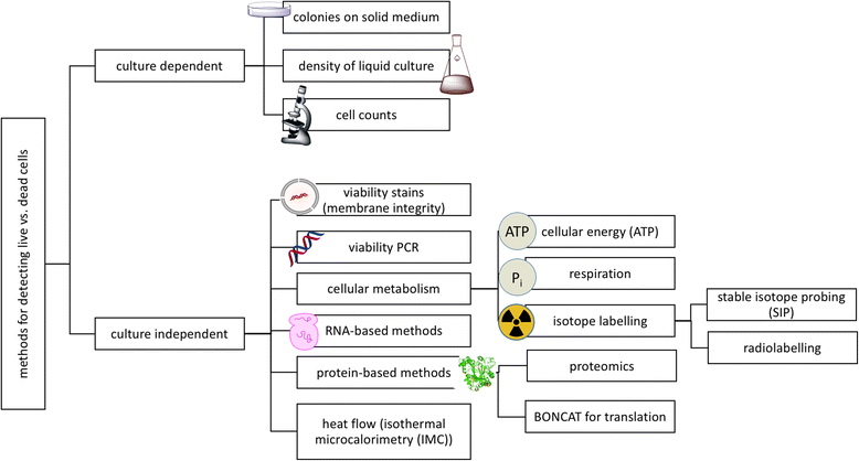
Fig. 1
Fig. 1
Overview of techniques to distinguish live from dead microbes. Both culture-dependent and culture-independent methods offer a variety of approaches, examples of which are categorized here, with culture-independent methods described further in the text
Fig. 2
Fig. 2
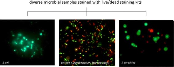
Example of Live/Dead staining kits applied to two bacterial samples and a eukaryotic sample. (A) A pure culture of E. coli was grown in LB medium overnight at 37 °C to an OD660 of 0.4. The cells were incubated with 100 mM H2O2 for 1 h at 37 °C. The sample was then stained with the LIVE/DEAD BacLight Bacterial Kit-L-7007 (Invitrogen, Grand Island, NY, USA) for microscopy according to the manufacturer’s instructions. A 10-μL aliquot was examined by fluorescence microscopy on a Carl Zeiss Axioskop using a filter with an excitation 488 nm and emission 528 nm. The live cells fluoresce green. (B) A developing biofilm on a glass slide created by incubating the slide in a solution containing three bacterial species: (1) Serratia marcescens ATCC 14756, (2) Corynebacterium xerosis ATCC 373, and (3) Staphylococcus epidermis ATCC 14990. It was stained using the LIVE/DEAD BacLight bacterial viability kit (PI/SYTO) [Molecular Probes]. Here, the live cells fluoresce green while the dead cells fluoresce red. (C) Yeast cells were stained with the LIVE/DEAD Yeast Viability Kit L-7009 (FUN 1 cell stain). The yeast were grown overnight in Sabouraud medium at 28 °C and then incubated with 100 μM H2O2 for 1 h. The samples were stained with FUN 1 cell stain according to the manufacturer’s instructions. The cells (10 μl aliquots) were viewed under a fluorescence microscope Axioskop (Carl Zeiss) with an excitation 489 nm and emission 539 nm. In contrast to the images of bacteria, here, the live cells form red fluorescent structures, while the dead cells are distinguished by a diffuse, green fluorescence. E. coli and S. cerevisiae micrographs were obtained by coauthor Balk, and the mixed bacterial micrograph was obtained by coauthors Adams and Lymperopoulou
Fig. 3
Fig. 3
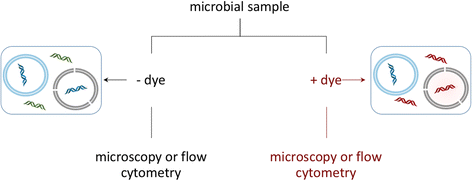
Live/dead staining workflow, propidium iodide (PI) example. In this technique, the sample is divided in two. One sample (left side) is stained with a total nucleic acid stain and used for cell enumeration, in which the live (blue membrane) and dead (black membrane) cells cannot be distinguished from each other, resulting in a stain of all nucleic acids. In the propidium iodide (PI) stained sample, the stain permeates compromised cell membranes, staining both cells presumed to be dead or in the process of dying (black membrane) and extracellular DNA or DNA, with PI-stained DNA colored red. Live cells with intact membranes (blue membrane) are not stained. In both types of samples, localization of stains within cells allows for enumeration, with stained free DNA relegated to background fluorescence. A comparison of counts from stained and unstained samples can be used to estimate the number of living cells. Alternatively, a single sample can be prepared with both a total nucleic acid stain and propidium iodide for counts of living and dead cells in the same preparation (not shown)
Fig. 4
Fig. 4
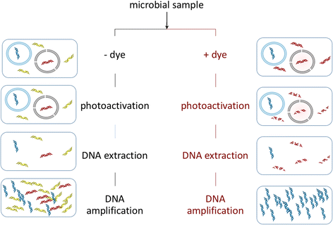
Viability PCR workflow (e.g., using EMA, PMA, or similar dyes). The initial sample is divided in two. One sample (left side) remains untreated, leaving total DNA—including extracellular DNA (yellow) and DNA in living (blue DNA, blue membrane) and dead (red DNA, black membrane) cells—relatively intact and available for downstream applications. The other sample (right side) is stained with a viability dye that binds to free DNA and to DNA in cells with compromised membranes. Upon photoactivation in the treated sample, bound DNA is degraded, such that it is no longer a suitable template for amplification. After amplification, a comparison of treated versus untreated samples can reveal relative proportions and/or types of living and dead microorganisms (e.g., via qPCR and/or DNA sequencing, respectively)
Fig. 5
Fig. 5
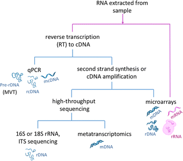
Summary of RNA-based techniques. Techniques that use RNA directly have pink pathway lines, and those using complementary DNA (cDNA, after retrotranscription) and double-stranded DNA (DNA, after second-strand synthesis or amplification) are colored blue. MVT is molecular viability testing
Fig. 6
Fig. 6
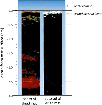
Autoradiography. The incorporation of radiolabelled isotopes by actively metabolizing organisms subsequently detected at the community level with scintillation counting, or at the individual level with microautoradiography, allows the precise identification of not only actively metabolizing members of an ecosystem, but metabolic type. Here, Rothschild and Mancinelli [201] sought to identify the location of the actively photosynthesizing members of a laminated microbial mat sample without destroying the fabric of the mat. Whirlpak® bags containing mat samples and water supplemented with radiolabelled 1 μCi/ml NaH14CO3 (New England Nuclear NEC 086H) were sealed and returned to the collection pond to incubate under in situ temperature and light levels, and then formalin was added to kill cells. In the lab, the samples were washed in acidified water, sliced to a thickness of ~2 mm with a gel slicer, and then frozen between two glass plates, which were removed prior to autoradiography. The frozen mats were exposed to X-ray film for 2–14 weeks at −80 °C. The developed film was placed in a photographic enlarger and used as a negative to print the image on the right and stands in contrast to the photograph of the frozen mat on the left. The white areas in the autoradiography panel correspond to acid-stable 14C incorporated into the mat sample, indicating the actively photosynthesizing community members
Fig. 7
Fig. 7
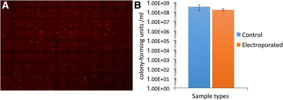
A transiently “dead” microbe. Competent E. coli (NEB5α cells competent cells, cat # c2987, New England Biolabs, Ipswitch, MA, USA) were thawed on ice. For a control sample, 2 μL of cells was added to 98 μL LB culture medium, 100 μL propidium iodide added, and the mixture allowed to stain for 5 min at room temperature. The experimental E. coli NEB5α (25 μL) was added to an electroporation cuvette previously cooled to 5 °C and electroporated at 2500 V twice. The cells were then diluted in LB and stained as with the control cells. a Fluorescence microscopy showing that almost all cells stained positive for propidium iodide treatment. b Colony-forming units showing no significant difference between controls and electroporated samples

References

    1. Koch R. Über di neuen Untersuchungsmethoden zum Nachweis der Mikrokosmen in Boden, Luft und Wasser. Vortrag auf dem XI Deutschen Arztetag in Berlin, Vereinsblatt für Deutschland, Komnussions-Verlag von FCW Vogel, Leipzig. 1883; 137:274–84.
    1. Bogosian G, Bourneuf EV. A matter of bacterial life and death. EMBO Rep. 2001;2(9):770–4. - PMC - PubMed
    1. Committee on the Forward Contamination of Mars. Preventing the forward contamination of Mars. vol ISBN: 0-309-65262-1. Division on Engineering and Physical Sciences. Washington, DC: National Research Council; 2006.
    1. Guarnieri V, Gaia E, Battocchio L, Pitzurra M, Savino A, Pasquarella C, et al. New methods for microbial contamination monitoring: an experiment on board the MIR orbital station. Acta Astronaut. 1997;40(2-8):195–201. - "V体育2025版" PubMed
    1. Koster W, Egli T, Ashbolt N, Botzenhart K, Burlion N, Endo T et al. Analytical methods for microbiological water quality testing. In: Dufour A, Snozzi M, Koster W, Bartram J, Ronchi E, Fewtrell L, editor. Microbial safety of drinking water: Improving approaches and methods. Geneva: World Health Organization. 2003. p. 237-92.

Publication types