V体育平台登录 - Skip to main page content
U.S. flag

An official website of the United States government

Dot gov

The . gov means it’s official. Federal government websites often end in . gov or . mil VSports app下载. Before sharing sensitive information, make sure you’re on a federal government site. .

Https

The site is secure V体育官网. The https:// ensures that you are connecting to the official website and that any information you provide is encrypted and transmitted securely. .

. 2017 Sep 1;121(6):e22-e36.
doi: 10.1161/CIRCRESAHA.117.310803. Epub 2017 Jul 25.

Paracrine Effects of the Pluripotent Stem Cell-Derived Cardiac Myocytes Salvage the Injured Myocardium (V体育2025版)

Affiliations

Paracrine Effects of the Pluripotent Stem Cell-Derived Cardiac Myocytes Salvage the Injured Myocardium

Atsushi Tachibana et al. Circ Res. .

Abstract

Rationale: Cardiac myocytes derived from pluripotent stem cells have demonstrated the potential to mitigate damage of the infarcted myocardium and improve left ventricular ejection fraction. However, the mechanism underlying the functional benefit is unclear VSports手机版. .

Objective: To evaluate whether the transplantation of cardiac-lineage differentiated derivatives enhance myocardial viability and restore left ventricular ejection fraction more effectively than undifferentiated pluripotent stem cells after a myocardial injury. Herein, we utilize novel multimodality evaluation of human embryonic stem cells (hESCs), hESC-derived cardiac myocytes (hCMs), human induced pluripotent stem cells (iPSCs), and iPSC-derived cardiac myocytes (iCMs) in a murine myocardial injury model. V体育安卓版.

Methods and results: Permanent ligation of the left anterior descending coronary artery was induced in immunosuppressed mice V体育ios版. Intramyocardial injection was performed with (1) hESCs (n=9), (2) iPSCs (n=8), (3) hCMs (n=9), (4) iCMs (n=14), and (5) PBS control (n=10). Left ventricular ejection fraction and myocardial viability, measured by cardiac magnetic resonance imaging and manganese-enhanced magnetic resonance imaging, respectively, was significantly improved in hCM- and iCM-treated mice compared with pluripotent stem cell- or control-treated mice. Bioluminescence imaging revealed limited cell engraftment in all treated groups, suggesting that the cell secretions may underlie the repair mechanism. To determine the paracrine effects of the transplanted cells, cytokines from supernatants from all groups were assessed in vitro. Gene expression and immunohistochemistry analyses of the murine myocardium demonstrated significant upregulation of the promigratory, proangiogenic, and antiapoptotic targets in groups treated with cardiac lineage cells compared with pluripotent stem cell and control groups. .

Conclusions: This study demonstrates that the cardiac phenotype of hCMs and iCMs salvages the injured myocardium effectively than undifferentiated stem cells through their differential paracrine effects VSports最新版本. .

Keywords: cell therapy; cytokines; magnetic resonance imaging; manganese; myocardial infarction; pluripotent stem cells. V体育平台登录.


Figures (V体育安卓版)

Figure 1
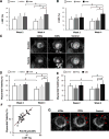
Figure 1. Effects of hESCs, iPSCs, hCMs and iCMs on LVEF and myocardial viability
(A) LVEF of hESCs, hCMs, and control at week 2 and 4 after cell transplantation. LVEF is significantly greater in hCMs at week 4 vs. control or hESCs at week 4 or hCMs at week 2. (B) LVEF of iPSCs, iCMs, and control at week 2 and 4 after cell transplantation. LVEF is significantly greater in iCMs at week 4 vs. control or iPSCs at week 4 or iCMs at week 2. (C) Short axis acquisitions during diastole and systole at mid-LV. The hCM and iCM treated mice exhibited higher contractility compared to hESC, iPSC, and control groups. (D) Myocardial viability of hESCs, hCMs, and control at week 2 and 4 after cell transplantation. Myocardial viability is significantly greater in hCMs at week 4 vs. control and hESCs at week 4 or hCMs at week 2. (E) Myocardial viability of iPSCs, iCMs, and control at week 2 and 4 after cell transplantation. Myocardial viability is significantly greater in hCMs at week 4 vs. control and hESCs at week 4 or hCMs at week 2. Conversely, the control group had significantly reduced myocardial viability between week 2 and 4. (F) There is a significant positive correlation between LVEF and myocardial viability (R=0.76, p<0.001). (G) Myocardial viability in the PIR as visualized by MEMRI, highlighted with red arrows. Data are presented as mean ± S.E.M., *p<0.05.
Figure 2
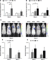
Figure 2. Stem cell engraftment measured by BLI
(A) BLI signals of hESCs, hCMs, and control at week 2 and 4. The hCM group demonstrated sustained engraftment throughout the 4-week period while the hESC group showed low levels of engraftment or cell death by week 2. (B) Similarly, the iCM group indicated greater cell engraftment at week 2 and 4 vs. control. The iPSC group had measureable engraftment signals at week 2 but showed no evidence of sustained engraftment at week 4. (C) Robust engraftment in hCMs and iCMs at week 2 decreased by week 4. (D) Absolute cell numbers were estimated in hESCs and hCMs, and (E) in iPSCs and iCMs. Data are presented as mean ± S.E.M., *p<0.05.
Figure 3
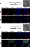
Figure 3. Immunofluorescence staining at the site of hCMs and iCMs engraftment
(A) Sections of hCM-treated myocardia were stained with human nuclear antibody and human cardiac troponin T at the site of injection. Hoechst 33342 was used to visualize nuclei (blue). Co-localization identified transplanted hCMs at week 4. (B) The iCM group also demonstrated cell engraftment at week 4. However, engraftment volume is significantly lower than the volume of increased myocardial viability in both hCM and iCM groups.
Figure 4
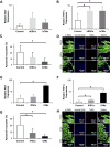
Figure 4. Anti-apoptotic effects of stem cell therapy in the PIR of the murine myocardium
(A) Relative gene expression of Akt1 and (B) TNF-α in the hESC, hCM and control groups. There was no significant difference in Akt1 expression; there was, however, a trend towards TNF-α upregulation in the hCM group compared to the control group. (C) Analysis of TUNEL-stained slides localized at the PIR indicated significantly less DNA fragmentation, and thus less apoptosis, in the hCM group compared to the control group. (D) Representative IHC images. (E) Akt1 and (F) TNF-α gene expression is significantly upregulated in iCM group compared to the control and the iPSC groups. (G) IHC confirmed a significant fewer TUNEL-positive cells in the PIR of iCM-treated mice compared to iPSC and control groups. (H) Representative IHC images. Data are presented as mean ± S.E.M., ‡p<0.3, *p<0.05.
Figure 5
Figure 5. Pro-migratory effects of stem cell therapy in the PIR of the murine myocardium
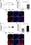
(A) Relative gene expression of connexin-43 in the hESC, hCM and control groups. There is an upward trend in hCMs vs. control. (B) IHC indicates signficant overexpression of CXCR4, a downstream effector of pro-migration cytokines and mRNA in the hCM group compared to the hESC group. (C) Representative IHC images. (D) Connexin-43 gene trends upwards in the iCM group. (E) CXCR4 is significantly overexpressed in the iCM group. (F) Representative IHC images. Data are presented as mean ± S.E.M., ‡p<0.3, *p<0.05.
Figure 6
Figure 6. Pro-angiogenic effects of stem cell therapy in the PIR of the murine myocardium
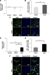
(A) RT-PCR of the PIR showed no significant difference in VEGF expression in the hCM group compared to the hESC or control groups. (B) However, IHC of CD31 showed significant overexpression of CD31 in the hCM-treated vs. hESC-treated PIR. (C) Representative IHC images. (D) There was upregulation of VEGF in the iCM group compared to the iPSC and control groups, suggesting increased angiogenesis, which was confirmed by (E) IHC of CD31. (F) Representative IHC images. Data are presented as mean ± S.E.M., ‡p<0.3, *p<0.05.
Figure 7
Figure 7. Salvage vs. regeneration of the stem cell therapy in the PIR
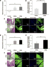
(A) MLC2v gene expression was greater in the hCM group compared to the control and had an upward trend compared to the hESC group. (B) The PIR was identified using H&E staining and analyzed for MLC2v expression. (C) Representative H&E and IHC images for MLC2v and EdU. (D) MLC2v was overexpressed in the iCM group compared to the control group. (E) There was significant MLC2v expression in the iCM group compared to the iPSC group. (F) Representative H&E and IHC images for MLC2v and EdU. Data are presented as mean ± S.E.M., ‡p<0.3, *p<0.05.
Figure 8
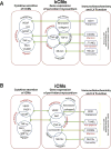
Figure 8. Schematic of the Paracrine Mechanism of hCMs and iCMs Salvage of the Injured Myocardium
Summary of the correlation among immunohistology, significantly up-regulated gene expression, and the associated cytokine productions in the myocardium treated with (A) hCMs and (B) iCMs.

"V体育官网" References

    1. Yancy CW, Jessup M, Bozkurt B, Butler J, Casey DE, Drazner MH, Fonarow GC, Geraci SA, Horwich T, Januzzi JL, Johnson MR, Kasper EK, Levy WC, Masoudi FA, McBride PE, McMurray JJ, Mitchell JE, Peterson PN, Riegel B, Sam F, Stevenson LW, Tang WH, Tsai EJ, Wilkoff BL, MEMBERS WC, Guidelines ACoCFAHATFoP 2013 accf/aha guideline for the management of heart failure: A report of the american college of cardiology foundation/american heart association task force on practice guidelines. Circulation. 2013;128:e240–327. - PubMed
    1. Mozaffarian D, Benjamin EJ, Go AS, Arnett DK, Blaha MJ, Cushman M, de Ferranti S, Després JP, Fullerton HJ, Howard VJ, Huffman MD, Judd SE, Kissela BM, Lackland DT, Lichtman JH, Lisabeth LD, Liu S, Mackey RH, Matchar DB, McGuire DK, Mohler ER, Moy CS, Muntner P, Mussolino ME, Nasir K, Neumar RW, Nichol G, Palaniappan L, Pandey DK, Reeves MJ, Rodriguez CJ, Sorlie PD, Stein J, Towfighi A, Turan TN, Virani SS, Willey JZ, Woo D, Yeh RW, Turner MB, Subcommittee AHASCaSS Heart disease and stroke statistics–2015 update: A report from the american heart association. Circulation. 2015;131:e29–322. - PubMed
    1. Bishop JE, Lindahl G. Regulation of cardiovascular collagen synthesis by mechanical load. Cardiovasc Res. 1999;42:27–44. - PubMed
    1. Kirk JA, Cingolani OH. Thrombospondins in the transition from myocardial infarction to heart failure. J Mol Cell Cardiol. 2015 - PMC - PubMed
    1. Dimmeler S, Zeiher AM, Schneider MD. Unchain my heart: The scientific foundations of cardiac repair. J Clin Invest. 2005;115:572–583. - PMC - PubMed

MeSH terms (VSports在线直播)