Skip to main page content
U.S. flag

An official website of the United States government

Dot gov

The . gov means it’s official. Federal government websites often end in . gov or . mil. Before sharing sensitive information, make sure you’re on a federal government site. VSports app下载.

Https

The site is secure. The https:// ensures that you are connecting to the official website and that any information you provide is encrypted and transmitted securely. V体育官网.

. 2017 Jul 3;114(27):E5434-E5443.
doi: 10.1073/pnas.1705759114. Epub 2017 Jun 19.

"VSports注册入口" Multiple layers of heterogeneity and subset diversity in human MAIT cell responses to distinct microorganisms and to innate cytokines

Affiliations

V体育官网 - Multiple layers of heterogeneity and subset diversity in human MAIT cell responses to distinct microorganisms and to innate cytokines

Joana Dias et al. Proc Natl Acad Sci U S A. .

Abstract

Mucosa-associated invariant T (MAIT) cells are a large innate-like T-cell subset in humans defined by invariant TCR Vα7. 2 use and expression of CD161. MAIT cells recognize microbial riboflavin metabolites of bacterial or fungal origin presented by the monomorphic MR1 molecule. The extraordinary level of evolutionary conservation of MR1 and the limited known diversity of riboflavin metabolite antigens have suggested that MAIT cells are relatively homogeneous and uniform in responses against diverse microbes carrying the riboflavin biosynthesis pathway. The ability of MAIT cells to exhibit microbe-specific functional specialization has not been thoroughly investigated. Here, we found that MAIT cell responses against Escherichia coli and Candida albicans displayed microbe-specific polyfunctional response profiles, antigen sensitivity, and response magnitudes. MAIT cell effector responses against E. coli and C. albicans displayed differential MR1 dependency and TCR β-chain bias, consistent with possible divergent antigen subspecificities between these bacterial and fungal organisms VSports手机版. Finally, although the MAIT cell immunoproteome was overall relatively homogenous and consistent with an effector memory-like profile, it still revealed diversity in a set of natural killer cell-associated receptors. Among these, CD56, CD84, and CD94 defined a subset with higher expression of the transcription factors promyelocytic leukemia zinc finger (PLZF), eomesodermin, and T-bet and enhanced capacity to respond to IL-12 and IL-18 stimulation. Thus, the conserved and innate-like MAIT cells harbor multiple layers of functional heterogeneity as they respond to bacterial or fungal organisms or innate cytokines and adapt their antimicrobial response patterns in a stimulus-specific manner. .

Keywords: MAIT cells; MR1; T cells; immunity; microbial immunity V体育安卓版. .


Conflict of interest statement

The authors declare no conflict of interest.

Figures
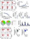
Fig. 1.
Fig. 1.
Distinct polyfunctional profile and MR1 dependency in the MAIT cell response against E. coli and C. albicans. (A) MAIT cells were stimulated with E. coli or C. albicans-fed monocytes for 24 h and in the presence of anti-CD28 mAb. Representative example of the CD69 and IFNγ, TNF, or IL-17 expression by MAIT cells after stimulation with the microbes (red) or at baseline conditions (gray). (B) Frequency of CD69+IFNγ+ MAIT cells after stimulation with different doses of E. coli (0.01–300 cfu per monocyte) and C. albicans (0.01–30 cfu per monocyte). (C) Down-regulation of the TCR Vα7.2 segment upon stimulation with E. coli or C. albicans, represented as the Vα7.2 geometric MFI of the stimulated CD161hi cells normalized to the one of the unstimulated control. (D) Frequency of MAIT cells expressing IFNγ, TNF, or IL-17. The microbial dose selected for each donor was the one inducing the highest frequency of CD69+IFNγ+ MAIT cells. (E) Polyfunctional profile of the MAIT cells responding to the microbes. The pie charts and the bar plot show the frequency of MAIT cells expressing CD69 and all different combinations of cytokines. (F) MAIT cells were stimulated for 18 h with E. coli-fed monocytes or 24 h with C. albicans-fed monocytes in the presence of anti-CD28 mAb and MR1-blocking mAb or isotype ctrl. Representative example of the expression of CD69 and IFNγ or TNF by MAIT cells after stimulation with the microbes and in the presence of MR1-blocking mAb (gray) or isotype ctrl (red). (G) Proportion of MR1-dependent IFNγ and TNF expression out of the total IFNγ and TNF expression by MAIT cells upon stimulation with E. coli or C. albicans. Graphs show the mean ± SEM (B–D) and mean + SEM (E, bar plot), and the box and whisker plots show the median, the 10th and 90th percentiles, and the interquartile range (G). Data are from 8 and 4–7 donors for E. coli and C. albicans, respectively (A–E) and from 8 and 9 donors for E. coli and 11 donors for C. albicans for IFNγ and TNF production, respectively (F and G). The unpaired t test or Mann–Whitney test was used (D, E, and G) to detect significant differences across multiple, unpaired samples. CA, C. albicans; cyt, cytokine; EC, E. coli. *P < 0.05; **P < 0.01; ***P < 0.001.
Fig. 2.
Fig. 2.
TCR Vβ use in MAIT cells influences the response to microbial stimulus. (A) Distribution of the 24 Vβ segments in the CD161hi Vα7.2+ MAIT, CD161Vα7.2+, CD161Vα7.2, and CD161+Vα7.2 populations. Each pie slice represents the median frequency of cells expressing each TCR Vβ segment. (B) Range of expression (maximum minus minimum frequency of MAIT cells expressing each Vβ segment) of the nine Vβ segments selected for further functional experiments. (C) Representative example of the strategy used to investigate the influence of the Vβ segment in MAIT cell responses. The MR1-dependent expression of IFNγ and TNF in each Vβ subpopulation was calculated and compared with that of the total MAIT cell population. (D) Frequency of MR1-dependent CD69+IFNγ+ or CD69+TNF+ CD8+ and CD8CD4 MAIT cells defined by each Vβ segment normalized to that of the total CD8+ and CD8CD4 MAIT cells, after stimulation with E. coli or C. albicans-fed monocytes for 18 h or 24 h, respectively, and in the presence of anti-CD28 mAb. Due to fluorochrome overlap in the anti-Vβ antibodies, the staining for each TCR Vβ segment was done separately (nine different samples per stimulation and per donor) and the Wilcoxon’s test or paired t test was therefore used to detect significant differences between the paired samples (total vs. Vβx+ MAIT cells from the same sample). Box and whisker plots show the median, the 10th and 90th percentiles, and the interquartile range. Data are from 16 to 20 donors (A and B) and from 6 to 9 and 8–11 donors (C and D, for E. coli and C. albicans, respectively). *P < 0.05; **P < 0.01; ***P < 0.001.
Fig. S1.
Fig. S1.
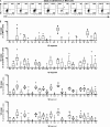
TCR Vβ use in MAIT cells and other T cells. (A) Representative example from 16 to 20 donors of the gating strategy to identify each Vβ segment on MAIT cells. (B) Frequency of MAIT, CD161Vα7.2+, CD161Vα7.2, and CD161+Vα7.2 cells expressing each Vβ segment. Box and whisker plots show the median, the 10th and 90th percentiles, and the interquartile range. Data are from 16 to 20 donors.
Fig. S2.
Fig. S2.
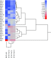
MAIT cells cluster separately from other T cells based on the TCR Vβ repertoire. Heat map and dendrogram based on the median frequency of cells expressing each Vβ segment. Data are from 16 to 20 donors.
Fig. S3.
Fig. S3.
The MAIT cell response against microbial stimuli is influenced by the TCR Vβ use. Frequency of MR1-dependent CD69+TNF+ (Upper) and MR1-dependent CD69+IFNγ+ (Lower) MAIT cells upon stimulation with (A) E. coli-fed monocytes or (B) C. albicans-fed monocytes. Lines represent individual donors. The Wilcoxon’s test or paired t test was used to detect significant differences between the paired samples. *P < 0.05; **P < 0.01; ***P < 0.001.
Fig. 3.
Fig. 3.
Rare TCR Vβ segments are associated with lower proliferative capacity of MAIT cells in vitro. MAIT cells in the PBMC sample were labeled with CTV and cultured for 5 d in the presence of E. coli (bacterial dose of 10), IL-2, and anti-CD28 mAb. (A) Representative example of the CTV dilution in Vβx+ (black histogram) and total (solid gray) CD8+ and CD8CD4 MAIT cells after 5 d in culture. (B) Correlation between the frequency of Vβx+CD8+ and CD8CD4 MAIT cells in the initial PBMC sample and the geometric MFI of the CTV staining in Vβx+CD8+ and CD8CD4 MAIT cells normalized to that of total CD8+ and CD8CD4 MAIT cells after 5 d of proliferation. Correlation was calculated using the Spearman’s test. Data are from five to seven donors.
Fig. 4.
Fig. 4.
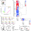
Surface immuno-proteome and transcription factor analysis of human MAIT cells. (A) Representative flow cytometry identification of the CD161hi Vα7.2+ MAIT, CD161Vα7.2+, CD161Vα7.2, and CD161+Vα7.2 cell populations. (B) Heat map and dendrogram based on the mean frequency of cells expressing a restricted group of proteins from the surface immuno-proteome dataset. The proteins for which the range of expression was equal or higher than 10% between any two T-cell populations were included. (C) PCA on the whole surface immuno-proteome dataset with the four T-cell populations plotted against principal components 1 and 2. (D) Representative example of the expression of Eomes, PLZF, FOXP3, Runx3, Helios, T-bet, and RORγt in CD161hi Vα7.2+ MAIT (red), CD161Vα7.2+ (blue), CD161Vα7.2 (green), and CD161+Vα7.2 cells (orange). The fluorescence minus one (FMO) control (black) is also shown. (E) Heat map and dendrogram based on the geometric MFI of the transcription factor stainings in MAIT, CD161Vα7.2+, CD161Vα7.2, and CD161+Vα7.2 cells. (F) Representative example of the CD3 and MR1 5-OP-RU tetramer staining in lymphocytes and CD161 and Vα7.2 expression in CD3+ MR1 5-OP-RU+ cells. (G) Frequency of CD161+Vα7.2+, CD161+Vα7.2, CD161Vα7.2+, and CD161Vα7.2 within the CD3+ MR1 5-OP-RU+ cell population in PBMCs from 10 healthy donors. (H) Representative example of the expression of CD195, CD244, CD218a, and CD319 on CD161hi MR1 5-OP-RU+ MAIT cells (red) and non-MAIT T cells (gray). Data are from three donors (A–C), eight donors for all transcription factors except for Runx3 (three donors) and FOXP3 (five donors) (D and E), and 10 donors (F–H).
Fig. S4.
Fig. S4.
Identification of cell populations. (A) Gating strategy representative of three donors to identify MAIT, CD161Vα7.2+, CD161Vα7.2, and CD161+Vα7.2 cells by flow cytometry in the immuno-proteome dataset. (B) Percent variability explained by each principal component and cumulatively. (C) Representative example from 10 donors of MR1 5-OP-RU and MR1 6-FP tetramer staining in CD161hi Vα7.2+ MAIT (red), CD161+ Vα7.2 (orange), and CD161Vα7.2+ (blue) cells.
Fig. 5.
Fig. 5.
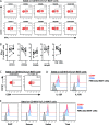
MAIT cells expressing innate receptors respond stronger to IL-12 and IL-18 stimulation. (A) Representative example of immuno-proteome receptors with high (>90%, Upper), low (<10%, Lower), and intermediate (between 10% and 90%, Middle) expression on CD161hi Vα7.2+ MAIT cells (red). (B) Representative example of the identification of CD161hi MR1 5-OP-RU+ MAIT cell population (red) and non-MAIT T cells (gray) by flow cytometry. (C) Representative example of immuno-proteome receptors with intermediate expression on CD161hi MR1 5-OP-RU+ MAIT cells (red) and non-MAIT T cells (gray). (D) MAIT cells in the PBMC sample were stimulated for 24 h with IL-12 and IL-18. Representative example of the production of IFNγ in the MAIT cell subsets defined by the expression of CD56, CD84, and CD94. (E) Frequency of MAIT cells expressing IFNγ after 24 h stimulation with IL-12 and IL-18. MAIT cells were divided into groups expressing or not expressing CD56, CD84, and CD94. (F) Frequency of CD94+ MAIT cells, at baseline conditions and after IL-12 and IL-18 stimulation. Data are from 3 donors (A), 10 donors (B and C), and 9 donors (D–F). Lines represent individual donors. The Wilcoxon’s test or paired t test was used to detect significant differences between the paired samples. **P < 0.01.
Fig. S5.
Fig. S5.
MAIT cell subset responses to innate cytokine stimulus and further characterization of CD56+ and CD56 MAIT cells. (A) MAIT cells in the PBMC sample were stimulated with IL-12 and IL-18 for 24 h. Representative example of IFNγ production in the MAIT cell subsets defined by the expression of CD101, CD328, CD226, NKp80, and CD8α. (B) Frequency of MAIT cells expressing IFNγ after 24-h stimulation with IL-12 and IL-18. MAIT cells were divided into groups expressing or not CD101, CD328, CD226, and NKp80, as well as in CD4CD8 double-negative (DN) and CD4CD8+ MAIT cells. Lines represent individual donors. Representative example of the expression of (C) CD94 on MAIT cells before and after 24-h stimulation with IL-12 and IL-18 and of (D) IL-12R and IL-18R and (E) Eomes, PLZF, Helios, and T-bet in resting CD56+ (red) and CD56 (blue) MAIT cells. The fluorescence minus one (FMO) control in total MAIT cells (black) is also shown. Data are from (A–C) 8–9 donors and (D and E) 9 donors, except for PLZF (8 donors). Lines represent individual donors. The Wilcoxon’s test or paired t test was used to detect significant differences between the paired samples.
Fig. 6.
Fig. 6.
Surface phenotype and transcription factor analyses of CD56+ and CD56 MAIT cells. (A) Heat map and dendrogram based on the mean frequency of cells expressing a restricted group of proteins from the surface immuno-proteome dataset. The proteins for which the difference in expression between CD56+ and CD56 MAIT cells was equal or higher than 10% were included. (B) Representative example of the expression of perforin and (C) geometric MFI of the perforin staining, in resting CD56 and CD56+ MAIT cells. Geometric MFI of the staining of (D) IL-12R and IL-18R and (E) PLZF, Eomes, Helios, and T-bet in MAIT cells at baseline conditions. Data are from 3 donors (A), 7 donors (B and C), and 8–9 donors (D and E). Lines represent individual donors. The Wilcoxon’s test or paired t test was used to detect significant differences between the paired samples. *P < 0.05; **P < 0.01; ***P < 0.001; ****P < 0.0001.
Fig. 7.
Fig. 7.
Factors contributing to the heterogeneity in MAIT cell responses to microbial antigens and innate cytokine stimuli.

References

    1. Dusseaux M, et al. Human MAIT cells are xenobiotic-resistant, tissue-targeted, CD161hi IL-17-secreting T cells. Blood. 2011;117:1250–1259. - PubMed
    1. Martin E, et al. Stepwise development of MAIT cells in mouse and human. PLoS Biol. 2009;7:e54. - PMC - PubMed
    1. Treiner E, et al. Selection of evolutionarily conserved mucosal-associated invariant T cells by MR1. Nature. 2003;422:164–169. - PubMed
    1. Fergusson JR, et al. CD161 defines a transcriptional and functional phenotype across distinct human T cell lineages. Cell Reports. 2014;9:1075–1088. - PMC - PubMed
    1. Lepore M, et al. Parallel T-cell cloning and deep sequencing of human MAIT cells reveal stable oligoclonal TCRβ repertoire. Nat Commun. 2014;5:3866. - PubMed (V体育ios版)

Publication types

MeSH terms