"VSports最新版本" Skip to main page content
U.S. flag

An official website of the United States government

Dot gov

The . gov means it’s official. Federal government websites often end in . gov or VSports app下载. mil. Before sharing sensitive information, make sure you’re on a federal government site. .

Https

The site is secure. The https:// ensures that you are connecting to the official website and that any information you provide is encrypted and transmitted securely. V体育官网.

. 2017 Oct 5;36(40):5567-5575.
doi: 10.1038/onc.2017.165. Epub 2017 Jun 5.

GATA3 targets semaphorin 3B in mammary epithelial cells to suppress breast cancer progression and metastasis

Affiliations

GATA3 targets semaphorin 3B in mammary epithelial cells to suppress breast cancer progression and metastasis

P Shahi et al. Oncogene. .

Abstract

Semaphorin 3B (SEMA3B) is a secreted axonal guidance molecule that is expressed during development and throughout adulthood. Recently, SEMA3B has emerged as a tumor suppressor in non-neuronal cells. Here, we show that SEMA3B is a direct target of GATA3 transcriptional activity. GATA3 is a key transcription factor that regulates genes involved in mammary luminal cell differentiation and tumor suppression. We show that GATA3 relies on SEMA3B for suppression of tumor growth. Loss of SEMA3B renders GATA3 inactive and promotes aggressive breast cancer development. Overexpression of SEMA3B in cells lacking GATA3 induces a GATA3-like phenotype and higher levels of SEMA3B are associated with better cancer patient prognosis VSports手机版. Moreover, SEMA3B interferes with activation of LIM kinases (LIMK1 and LIMK2) to abrogate breast cancer progression. Our data provide new insights into the role of SEMA3B in mammary gland and provides a new branch of GATA3 signaling that is pivotal for inhibition of breast cancer progression and metastasis. .


Conflict of interest statement

Conflict of Interest

Authors declare no conflict of interest.

Figures

Figure 1
Figure 1
In silico analysis of breast cancer databases. (a) Analysis of TCGA database. Heat map indicating gene expression analysis for GATA3, SEMA3B and MYC in multiple breast cancer samples. (N = 597) (b) Analysis of TCGA database via Regulome explorer. Data present a collection of proteins including GATA3 that may reside in the same protein network as SEMA3B. (c) Analysis of SEMA3B expression via the GSE9014 database. SEMA3B expression is significantly lowered in the invasive breast carcinoma samples. (Normal: N = 6, Invasive breast carcinoma: N = 53, P = 1.03E-21) (d) TCGA analysis indicating significantly lowered SEMA3B expression in triple negative breast cancer samples. (Normal: N = 244, Triple negative: N = 49, P = 4.06E-9) (e) Analysis of SEMA3B expression in PAM50 subtypes using the METABRIC database. Luminal breast cancers possess higher levels of SEMA3B expression than the basal subtype, P = <0.0001, Normal: N = 202, Luminal A: N = 721, Luminal B: N = 492. HER2: N = 240, Basal: N = 331) (f) Analysis of breast cancer patient survival (31). Tumor samples exhibiting lower SEMA3B expression show poorer patient prognosis. (Concordance index = 62.3, Log-rank equal curves P = 1.6E-4, R^ 2 = 0.005/0.941, Risk groups hazard ratio = 2.4, P = 2.61E-6)
Figure 2
Figure 2
GATA3 directly controls SEMA3B expression. (a) ChIP analysis indicating GATA3 transcription factor binding to SEMA3B promoter in EpH4.9, T47D and MDA-MB-231 cells. (b) qPCR analysis of GATA3 and SEMA3B expression in MDA-MB-231 cells, N = 6. Overexpression of GATA3 in MDA-MB-231 cells results in elevation of SEMA3B levels. (c) Western blot analysis of MDA-MB-231 cell lysate. Overexpression of GATA3 enhances SEMA3B expression. (d) Immunostaining of MDA-MB-231 (control) and GATA3 overexpressing MDA-MB-231 cells. Overexpression of GATA3 upregulates SEMA3B levels. Bar 50 μm. (e) qPCR analysis of SEMA3B in NMuMG cells, N = 2. Lowering GATA3 expression via siRNA results in downregulation of SEMA3B levels.
Figure 3
Figure 3
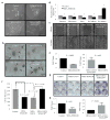
Inhibition of cellular migration and proliferation by GATA3 relies on presence of SEMA3B. (a) Bright-field images of MDA-MB-231 stable cell lines in 2D cell cultures. Bar, 50 μm. (b) Phase-contrast images of MDA-MB-231 stable cell lines in 3D cultures. Arrows indicate the presence of invadopodia moving outward from the colony. Bar, 200 μm. (c) Quantification of 3D Matrigel culture colonies. (N = 7) (d) qRT-PCR analysis of EMT-associated genes. RNA samples from MDA-MB-231 control and MDA-SEMA3B cells were collected. qRT-PCR analysis indicates that SEMA3B lowers EMT gene levels. SEMA3B enhanced E-cadherin levels compared to control. (e) Scratch assay analysis indicating difference in cellular migration in MDA-MB-231 stable cell lines. N = 6 (f) Quantification of cell infiltration of gaps between the two invading cell fronts. The gap was measured using ImageJ software. Y-axis indicates experimental group/control group. (N = 6). (g) Colony formation assay. MDA-MB-231 stable cell lines were cultured in 6-well plates to allow colony growth. Colonies were stained with crystal violet and counted. N = 12. (h) Graph representing the quantification of Giemsa stained colonies. (N = 12).
Figure 4
Figure 4
Loss of SEMA3B disrupts GATA3 tumor suppressive activity. (a) MDA-SEMA3B and (b) MDA-GATA3-SEMA3BKD stable cell lines where transplanted in 8-week-old female nude mice via orthotopic injection. Color photographs show a representative set of gross tumors from each transplanted group. Graphs indicates tumor measurement during the course of experiment, N = 10 for each group. (c) Immunostaining and quantification analysis for Ki67 expression in the tumor sections (N = 6). Bar, 100 μm. (d) Analysis of inguinal lymph node weight from transplanted mice, N = 10 for each group. (e) H&E analysis of lymph node sections indicating tumor metastasis. For each transplanted group, metastatic tumor cells to the lymph node was quantified (N = 6). Bar = 200 μm. (f) Western blot analysis indicating phosphorylation status of LIMK1 and LIMK2 in control and MDA-SEMA3B cell extracts. Total LIMK1 and LIMK2 indicate equal loading of samples. Protein bands 72 kDa.

References

    1. Luo Y, Raible D, Raper JA. Collapsin: a protein in brain that induces the collapse and paralysis of neuronal growth cones. Cell. 1993;75(2):217–27. - PubMed
    1. Neufeld G, Kessler O. The semaphorins: versatile regulators of tumour progression and tumour angiogenesis. Nat Rev Cancer. 2008;8(8):632–45. - "VSports在线直播" PubMed
    1. Staton CA, Shaw LA, Valluru M, Hoh L, Koay I, Cross SS, et al. Expression of class 3 semaphorins and their receptors in human breast neoplasia. Histopathology. 2011;59(2):274–82. - PubMed
    1. Kruger RP, Aurandt J, Guan KL. Semaphorins command cells to move. Nat Rev Mol Cell Biol. 2005;6(10):789–800. - PubMed
    1. Driessens MH, Hu H, Nobes CD, Self A, Jordens I, Goodman CS, et al. Plexin-B semaphorin receptors interact directly with active Rac and regulate the actin cytoskeleton by activating Rho. Curr Biol. 2001;11(5):339–44. - PubMed

MeSH terms

Substances