Skip to main page content
U.S. flag

An official website of the United States government

Dot gov

The . gov means it’s official. Federal government websites often end in . gov or . mil. Before sharing sensitive information, make sure you’re on a federal government site VSports app下载. .

Https

The site is secure V体育官网. The https:// ensures that you are connecting to the official website and that any information you provide is encrypted and transmitted securely. .

. 2017 Jul 21;292(29):12025-12040.
doi: 10.1074/jbc.M117.775981. Epub 2017 May 22.

Copper-zinc superoxide dismutase is activated through a sulfenic acid intermediate at a copper ion entry site

Affiliations

Copper-zinc superoxide dismutase is activated through a sulfenic acid intermediate at a copper ion entry site

Morgan M Fetherolf et al. J Biol Chem. .

Abstract

Metallochaperones are a diverse family of trafficking molecules that provide metal ions to protein targets for use as cofactors. The copper chaperone for superoxide dismutase (Ccs1) activates immature copper-zinc superoxide dismutase (Sod1) by delivering copper and facilitating the oxidation of the Sod1 intramolecular disulfide bond. Here, we present structural, spectroscopic, and cell-based data supporting a novel copper-induced mechanism for Sod1 activation. Ccs1 binding exposes an electropositive cavity and proposed "entry site" for copper ion delivery on immature Sod1. Copper-mediated sulfenylation leads to a sulfenic acid intermediate that eventually resolves to form the Sod1 disulfide bond with concomitant release of copper into the Sod1 active site. Sod1 is the predominant disulfide bond-requiring enzyme in the cytoplasm, and this copper-induced mechanism of disulfide bond formation obviates the need for a thiol/disulfide oxidoreductase in that compartment VSports手机版. .

Keywords: X-ray crystallography; chaperone; copper; enzyme activation; metalloenzyme; superoxide dismutase (SOD). V体育安卓版.


Conflict of interest statement

The authors declare that they have no conflicts of interest with the contents of this article

VSports app下载 - Figures
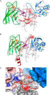
Figure 1.
Figure 1.
Structures of Sod1–Ccs1 complexes. In all panels, the Sod1 β-barrel is shown in green with the electrostatic loop orange and disulfide loop purple. Ccs1 D1 is blue, D2 is gray, and D3 is red. The Sod1 zinc ions are shown as orange spheres. A, the heterodimeric complex between human H46R/H48Q Sod1 and yeast E238A/E239A/R240A Ccs1 determined in the present work (Protein Data Bank code 5U9M). The reduced disulfide bond of H46R/H48Q Sod1 between Cys57 and Cys146 permits the displacement of the disulfide loop from the Sod1 β-barrel where it interacts extensively with residues from Ccs1 D2 and D3. The β-hairpin portion of D3 begins just downstream of Asn224 with Cys231 residing on the loop between the two short β-strands. B, the heterodimeric complex between yeast H48F Sod1 and yeast Ccs1 (Protein Data Bank code 1JK9 (19)). C, intercalation of the D3 β-hairpin between the Sod1 β-barrel and disulfide loop. Sod1 is shown as an electrostatic surface contoured at ±4kT. The reduced Cys57–Cys146 Sod1 disulfide bond exposes an electropositive “hole” near Cys146. Domains 1 (blue) and 2 (gray) of Ccs1 are shown as surfaces. The stabilizing positioning of the conserved Ccs1 tryptophan residues Trp222 and Trp237 from D3 are highlighted.
Figure 2.
Figure 2.
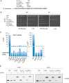
The spacing of the D3 CXC Cys residues is critical for Sod1 activation. A, mutations were made in the CXC motif of Ccs1 to alter the spacing to either a CC or a CXXC motif without disrupting downstream residues. B, viability tests were performed by plating cells on synthetic complete medium with or without lysine at 30 or 37 °C. C, Sod1 activity for various Ccs1 mutants was quantified; error bars represent S.D. D, the status of the disulfide bond was visualized by lysing cells in the presence of a PEG-maleimide alkylating agent that selectively reacts with free thiols.
Figure 3.
Figure 3.
Stalled Sod1–Ccs1 complexes. A, lysates from sod1Δ cells transformed with Sod1-Strep mutants were subject to affinity purification using Strep-Tactin resin where 2% (20 μg) of total input and 20% of total elution were loaded and analyzed by Western blotting. B, Strep affinity purification from ccs1Δ cells harboring Ccs1 CXC motif mutants. C, Strep affinity purification from ccs1Δ sod1Δ cells harboring Sod1 C146S-Strep and co-transformed with vectors encoding various Ccs1 CXC mutants. D and E, Strep affinity purification from lysates containing Ccs1 CXC spacing mutants coexpressing Sod1-Strep.
Figure 4.
Figure 4.
Sod1 can form an intermolecular disulfide with Ccs1. A, cell lysates from ccs1Δ sod1Δ cells co-transformed with WT or Ccs1 CXC mutants and WT Sod1-Strep or Sod1-Strep mutants were subjected to affinity purification where 2% (20 μg) of total input and 20% of total elution were loaded and analyzed by Western blotting. Reducing SDS-polyacrylamide (top) as well as non-reducing SDS-polyacrylamide (bottom) gels were run and blotted for Sod1 or Ccs1. B, WT Sod1-Strep or mutants were purified in the presence or absence of 500 μm BCS and analyzed for Ccs1 interaction. C, Sod1-Strep (WT) or various Sod1 mutations were affinity-purified, and metal ratios were determined by ICP-OES; error bars represent S.D.
Figure 5.
Figure 5.
Cysteine residues from Sod1 and/or Ccs1 form a novel Cu(I) entry site. Sod1 was purified from yeast lysates containing the indicated Sod1 and Ccs1 mutations. X-ray absorption spectroscopy was used to determine the copper coordination environment of Sod1. A, the CuK edge of WT samples along with WT Ccs1 + Sod1 H46R/H48Q and Ccs1 C17S/C20S/C27S/C64S/C159S + Sod1 H46R/H48Q. B, EXAFS region for WT samples. C, EXAFS region for sample containing WT Ccs1 + Sod1 H46R/H48Q. D, EXAFS region for sample containing C17S/C20S/C27S/C64S/C159S + Sod1 H46R/H48Q.
Figure 6.
Figure 6.
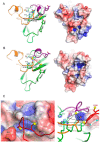
Electropositive cavity and copper ion entry site on immature Sod1. A, WT Sod1 (colored as in Figs. 1 and 2) shown in its disulfide-oxidized mature conformation (left). The electrostatic surface is contoured at ±4kT (right). B, the disulfide reduced, copper-free Sod1 molecule from the current Sod1–Ccs1 complex with residues 46 and 48 exchanged back to histidine for demonstrative purposes. The box in the right panel highlights an electropositive cavity on Sod1 made accessible through the intercalation of the Ccs1 D3 β-hairpin. C, the Cu(I) entry site on immature Sod1 (solid black lines) as shown here contains two cysteine sulfhydryl groups (Cys57 and Cys146 from Sod1) and one histidyl side chain from His48 or His120. The left panel shows a semitransparent electrostatic surface covering the entry site from the view of the Ccs1 D3 β-hairpin. The right panel is rotated back and counterclockwise (both ∼90°) from the left panel, and the electropositive cavity surface is shown to emphasize its depth and positioning with regard to the entry site ligands.
Figure 7.
Figure 7.
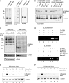
Copper-dependent sulfenylation at the Sod1 entry site and a role for reduced GSH. A, WT Sod1-Strep purified from yeast cells and treated with DMSO vehicle or dimedone at a final concentration of 20 mm. Coomassie staining of the purified Sod1 samples is in the left panel, and a Western blot using a cysteine sulfenic antibody or Sod1 antibody is in the right panels. B, Purified Sod1 with various mutations was treated with dimedone and visualized by Western blot analysis. C, WT cells or ctr1Δ cells were transformed with a Sod1-Strep-encoding plasmid. The cells were grown in the absence or presence of 150 μm BCS and subjected to Strep-Tactin affinity purification where 2% (20 μg) of total input and 40% of total elution were loaded and analyzed by Western blotting. D, in vitro activation of yeast Sod1. Zn–Sod1 (copper-free, disulfide-reduced) was mixed with either Cu–GSH, Cu–Ccs1, or apo-Ccs1 aerobically in 50 mm Tris, pH 7.6, 100 mm NaCl and loaded on the gel for an in-gel Sod1 enzymatic activity. Top panel, lane 1, Zn–Sod1; lane 2, Zn–Sod1 + Cu–GSH; lane 3, Zn–Sod1 + apo-Ccs1; lane 4, Zn–Sod1 + Cu–GSH + apo-Ccs1; lane 5, Zn–Sod1 + Cu–Ccs1. Bottom panel, the same Sod1 activation assay as the top panel but including the Ccs1 D1 MXCXXC (lanes 4 and 7) and D3 CXC (lanes 5 and 8) mutants. E, copper-mediated sulfenylation of yeast Sod1. Apo-Sod1 was preloaded with 1 molar eq of Zn(II) and mixed with yeast Ccs1 either preloaded with 1 molar eq of Cu(I), Co(II), or Zn(II). After mixing, the samples were incubated with 20 mm dimedone prior to loading on the gel. The top panel is the visualization of the dimedone-sulfenylated Sod1 adduct using the anti-sulfenic acid antibody, and the bottom panel shows the level of Sod1 protein as visualized with an anti-Sod1 antibody. F, copper-mediated sulfenylation of human Sod1. Apo-human Sod1 was preloaded with 1 molar eq of Zn(II) and incubated with yeast Ccs1 either preloaded with 1 molar eq of Cu(I), Co(II), or Zn(II). The conditions and two panels are as described in E.
Figure 8.
Figure 8.
Relative abundance of dimedone-modified Sod1 peptides calculated from the extracted ion chromatogram for each peptide across the five reactions. Reaction 1, 5 μm apo-Sod1, BCS buffer (50 mm Tris, pH 7.6, 100 mm NaCl, 0.5 mm tris(2-carboxyethyl)phosphine, 200 μm BCS). Reaction 2, 5 μm apo-Sod1, 5 μm H2O2, 5 mm dimedone, 20 μm Zn2SO4, 25 μm Cu(I)–yCcs1, BCS buffer. Reaction 3, 5 μm apo-Sod1, 25 μm H2O2, 5 mm dimedone, 20 μm Zn2SO4, 25 μm Cu(I)–yCcs1, BCS buffer. Reaction 4, 5 μm apo-Sod1, 5 mm dimedone, 20 μm Zn2SO4, 25 μm Cu(I)–yCcs1, BCS buffer. Reaction 5, 5 μm apo-Sod1, 20 μm Zn2SO4, 25 μm Cu(I)–yCcs1, BCS buffer. In A, 1–5 indicates yeast Sod1 reactions, and in B, 1–5 indicates human Sod1 reactions.

"VSports在线直播" References

    1. Culotta V. C., Klomp L. W., Strain J., Casareno R. L., Krems B., and Gitlin J. D. (1997) The copper chaperone for superoxide dismutase. J. Biol. Chem. 272, 23469–23472 - PubMed
    1. Fridovich I. (1989) Superoxide dismutases. An adaptation to a paramagnetic gas. J. Biol. Chem. 264, 7761–7764 - PubMed
    1. Arnesano F., Banci L., Bertini I., Martinelli M., Furukawa Y., and O'Halloran T. V. (2004) The unusually stable quaternary structure of human Cu,Zn-superoxide dismutase 1 is controlled by both metal occupancy and disulfide status. J. Biol. Chem. 279, 47998–48003 - PubMed (VSports app下载)
    1. Doucette P. A., Whitson L. J., Cao X., Schirf V., Demeler B., Valentine J. S., Hansen J. C., and Hart P. J. (2004) Dissociation of human copper-zinc superoxide dismutase dimers using chaotrope and reductant. Insights into the molecular basis for dimer stability. J. Biol. Chem. 279, 54558–54566 - PubMed
    1. Lindberg M. J., Normark J., Holmgren A., and Oliveberg M. (2004) Folding of human superoxide dismutase: disulfide reduction prevents dimerization and produces marginally stable monomers. Proc. Natl. Acad. Sci. U.S.A. 101, 15893–15898 - PMC - PubMed

Publication types

MeSH terms

"VSports手机版" Associated data