<u lang="EoplzeQ"></u><center lang="0HFu14Pd"></center> Skip to main page content
U.S. flag

An official website of the United States government

Dot gov

The . gov means it’s official. Federal government websites often end in . gov or VSports app下载. mil. Before sharing sensitive information, make sure you’re on a federal government site. .

Https

The site is secure. The https:// ensures that you are connecting to the official website and that any information you provide is encrypted and transmitted securely. V体育官网.

. 2017 Aug;95(8):873-886.
doi: 10.1007/s00109-017-1537-1. Epub 2017 May 18.

Elevated expression of CST1 promotes breast cancer progression and predicts a poor prognosis

Affiliations

Elevated expression of CST1 promotes breast cancer progression and predicts a poor prognosis

Da-Nian Dai et al. J Mol Med (Berl). 2017 Aug.

Erratum in

Abstract

Cystatin SN (CST1) belongs to the type 2 cystatin (CST) superfamily, which restricts the proteolytic activities of cysteine proteases. CST1 has been recently considered to be involved in the development of several human cancers. However, the prognostic significance and function of CST1 in breast cancer remains unknown. In the current study, we found that CST1 was generally upregulated in breast cancer at both mRNA and protein level. Furthermore, overall survival (OS) and disease-free survival (DFS) in the low CST1 expression subgroup were significantly superior to the high CST1 expression subgroup (OS, p < 0. 001; DFS, p < 0. 001), which indicated that CST1 expression level was closely correlated to the survival risk of these patients VSports手机版. Univariate and multivariate analyses demonstrated that CST1 expression was an independent prognostic factor, the same as ER status and nodal status. Next, CST1 overexpression promoted breast cancer cell proliferation, clonogenicity, migration, and invasion abilities. By contrast, knockdown of CST1 attenuated these malignant characteristics in breast cancer cells. Collectively, our study indicates that CST1 cannot only serve as a significant prognostic indicator but also as a potential therapeutic target for breast cancer. .

Key messages: High CST1 expression is negatively correlated with survival of breast cancer patients V体育安卓版. CST1 promotes cell proliferation, clone formation, and metastasis in breast cancer cells. CST1 is a novel potential prognostic biomarker and therapeutic target for breast cancer. .

Keywords: Breast cancer; CST1; Prognostic biomarker. V体育ios版.


Conflict of interest statement

The authors declare that they have no conflict of interest.

Figures (VSports最新版本)

Fig. 1
Fig. 1
Overexpression of CST1 in breast cancer. a CST1 was significantly upregulated in breast cancer tissues compared with normal breast tissues according to a microarray from TCGA database of Oncomine website. CST1 is marked by a red box. b, c CST1 mRNA and protein levels in 10 or 5 pairs of matched breast cancer and non-tumor tissues, respectively. d The CST1 protein level in a panel of breast cancer cell lines including MDA-MB-468, BT-474, MCF-7, MDA-MB-435, SKRr-3, MDA-MB-415, BT-549, MDA-MB-231 compared with the immortalized human breast epithelial cell 76N-tert. mRNA levels are presented as means ± SD and normalized to the housekeeping gene β-actin by qRT-PCR. N matched noncancerous tissue, T tumor tissue
Fig. 2
Fig. 2
CST1 expression analyzed by immunohistochemical staining. Negative CST1 staining in normal breast ductal epithelium (negative control) a ×100, b ×400, negative staining of CST1 in breast cancer tissue c ×100, d ×400, weak staining of CST1 in cytoplasm e ×100, f ×400, moderate staining of CST1 in cytoplasm g ×100, h ×400, and strong staining of CST1 in cytoplasm i ×100, j ×400
Fig. 3
Fig. 3
Kaplan–Meier estimates of the probability of survival for patients with breast cancer. a, b Five-year overall survival (OS) and disease-free survival (DFS) rates of 244 breast cancer patient were 65.3 and 62.5% respectively; c, d High CST1 expression level was significantly correlated to OS (p < 0.001) and DFS (p = 0.001) in all breast cancer patients
Fig. 4
Fig. 4
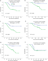
Kaplan–Meier estimates of the probability of survival for patients with breast cancer according to the receptor expression in classification. All patients were stratified according to ER status, PR status, and HER-2 status. a, b The overall survival (OS) rate of breast cancer patients in different ER status. c, d The overall survival (OS) rate of breast cancer patients in different PR status. e, f The overall survival (OS) rate of breast cancer patients in different HER-2 status
Fig. 5
Fig. 5
Kaplan–Meier estimates of the probability of survival for patients with breast cancer according to the general pathological indicators in classification. All patients were stratified according to tumor size, tumor differentiation, TNM classification, lymph node metastasis status, and histological grade. a, b The overall survival (OS) rate of breast cancer patients with different tumor sizes. c, d The overall survival (OS) rate of breast cancer patients in different tumor status. e, f The overall survival (OS) rate of breast cancer patients in different TNM classification. g, h The overall survival (OS) rate of breast cancer patients with different lymph node metastasis status. i The overall survival (OS) rate of breast cancer patients in histological grade
Fig. 6
Fig. 6
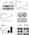
Overexpression of CST1 promotes proliferation in breast cancer cells. a Expression levels of CST1 protein after stable transfection with vector or CST1-expressing plasmids were detected by western blotting analysis. b Cell proliferation after CST1 overexpression in BT-549 cells was measured by MTT assay. c Cell proliferation after CST1 overexpression in MDA-MB-415 cells was measured by MTT assay. d Cell colony formation was determined in BT-549 and MDA-MB-415 cells expressing vector or CST1. e The number of colonies of BT-549 and MDA-MB-415 cells harboring vector or CST1. f The representative pictures of proliferation-related proteins after overexpression of CST1 in BT-549 and MDA-MB-415 cells, as determined by western blotting. Results are expressed as means ± SD (error bars). *p < 0.05, **p < 0.01, and ***p < 0.001 compared to vector
Fig. 7
Fig. 7
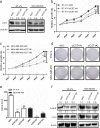
Knockdown of CST1 suppresses proliferation in breast cancer cells. a Knockdown of CST1 resulted in reduced CST1 protein expression in BT-474 and MDA-MB-468 cell lines. b Cell proliferation after CST1 knockdown in BT-474 cells was measured by MTT assay. c Cell proliferation after CST1 knockdown in MDA-MB-468 cells was measured by MTT assay. d Cell colony formation was determined in BT-474 and MDA-MB-468 cells transfected with NC- or CST1-targeting siRNAs. e The number of colonies after knockdown of CST1 in BT-474 and MDA-MB-468 cells. f The representative pictures of proliferation-related proteins after knockdown of CST1, as determined by western blotting. Results are expressed as means ± SD (error bars). *p < 0.05, **p < 0.01, and ***p < 0.001 compared to siNC
Fig. 8
Fig. 8
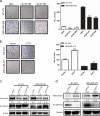
CST1 promotes migration in breast cancer cells. a The representative pictures and quantification of migration in BT-474 and MDA-MB-468 cells transfected with NC- or CST1-targeting (KD) siRNAs by transwell migration assay after the knockdown of CST1. b The representative pictures and quantification of cell migration in BT-549 and MDA-MB-415 cells harboring vector or pMSCV-CST1 plasmid by transwell migration assay. c The representative pictures of metastasis-related proteins after the knockdown of CST1, as determined by western blotting. d The representative pictures of metastasis-related proteins after the overexpression of CST1, as determined by western blotting. Migrating cells were identified using light microscope (×100). Results are expressed as means ± SD (error bars) from five viewing fields. *p < 0.05, **p < 0.01, and ***p < 0.001 compared to siNC or vector
Fig. 9
Fig. 9
CST1 promotes invasion of breast cancer cells. a The representative pictures and quantification of invasion of BT-474 and MDA-MB-468 cells transfected with NC- or CST1-targeting (KD) siRNAs by transwell invasion assay after the knockdown of CST1. b The representative pictures and quantification of cell invasion in BT-549 and MDA-MB-415 cells harboring vector or pMSCV-CST1 plasmid by transwell invasion assay. The invasive cells were identified using light microscope (×100). Results are expressed as means ± SD (error bars) from five viewing fields. *p < 0.05, **p < 0.01, and ***p < 0.001 compared to siNC or vector

References

    1. Global Burden of Disease Cancer C. Fitzmaurice C, Dicker D, Pain A, Hamavid H, Moradi-Lakeh M, MF MI, Allen C, Hansen G, Woodbrook R, et al. The global burden of cancer 2013. JAMA Oncol. 2015;1(4):505–527. doi: 10.1001/jamaoncol.2015.0735. - DOI - PMC - PubMed
    1. Gradishar WJ, Anderson BO, Balassanian R, Blair SL, Burstein HJ, Cyr A, Elias AD, Farrar WB, Forero A, Giordano SH, et al. Breast cancer version 2.2015. J Natl Compr Cancer Netw. 2015;13(4):448–475. doi: 10.6004/jnccn.2015.0060. - DOI - PubMed
    1. Yang Y, Im SA, Keam B, Lee KH, Kim TY, Suh KJ, Ryu HS, Moon HG, Han SW, Oh DY, et al. Prognostic impact of AJCC response criteria for neoadjuvant chemotherapy in stage II/III breast cancer patients: breast cancer subtype analyses. BMC Cancer. 2016;16(1):515. doi: 10.1186/s12885-016-2500-1. - DOI - PMC - PubMed
    1. Bergen ES, Tichy C, Berghoff AS, Rudas M, Dubsky P, Bago-Horvath Z, Mader RM, Exner R, Gnant M, Zielinski CC, et al. Prognostic impact of breast cancer subtypes in elderly patients. Breast Cancer Res Treat. 2016;157(1):91–99. doi: 10.1007/s10549-016-3787-y. - DOI - PMC - PubMed
    1. Loser R, Pietzsch J. Cysteine cathepsins: their role in tumor progression and recent trends in the development of imaging probes. Front Chem. 2015;3:37. doi: 10.3389/fchem.2015.00037. - DOI - PMC - PubMed

Publication types

LinkOut - more resources (VSports app下载)