Skip to main page content
U.S. flag

An official website of the United States government

Dot gov

The . gov means it’s official. Federal government websites often end in . gov or VSports app下载. mil. Before sharing sensitive information, make sure you’re on a federal government site. .

Https

The site is secure. The https:// ensures that you are connecting to the official website and that any information you provide is encrypted and transmitted securely V体育官网. .

. 2017 Jul 12;91(15):e00510-17.
doi: 10.1128/JVI.00510-17. Print 2017 Aug 1.

A Point Mutation in the Rhesus Rotavirus VP4 Protein Generated through a Rotavirus Reverse Genetics System Attenuates Biliary Atresia in the Murine Model

Affiliations

"V体育安卓版" A Point Mutation in the Rhesus Rotavirus VP4 Protein Generated through a Rotavirus Reverse Genetics System Attenuates Biliary Atresia in the Murine Model

Sujit K Mohanty (VSports最新版本) et al. J Virol. .

Abstract

Rotavirus infection is one of the most common causes of diarrheal illness in humans. In neonatal mice, rhesus rotavirus (RRV) can induce biliary atresia (BA), a disease resulting in inflammatory obstruction of the extrahepatic biliary tract and intrahepatic bile ducts. We previously showed that the amino acid arginine (R) within the sequence SRL (amino acids 445 to 447) in the RRV VP4 protein is required for viral binding and entry into biliary epithelial cells. To determine if this single amino acid (R) influences the pathogenicity of the virus, we generated a recombinant virus with a single amino acid mutation at this site through a reverse genetics system VSports手机版. We demonstrated that the RRV mutant (RRVVP4-R446G) produced less symptomatology and replicated to lower titers both in vivo and in vitro than those seen with wild-type RRV, with reduced binding in cholangiocytes. Our results demonstrate that a single amino acid change in the RRV VP4 gene influences cholangiocyte tropism and reduces pathogenicity in mice. IMPORTANCE Rotavirus is the leading cause of diarrhea in humans. Rhesus rotavirus (RRV) can also lead to biliary atresia (a neonatal human disease) in mice. We developed a reverse genetics system to create a mutant of RRV (RRVVP4-R446G) with a single amino acid change in the VP4 protein compared to that of wild-type RRV. In vitro, the mutant virus had reduced binding and infectivity in cholangiocytes. In vivo, it produced fewer symptoms and lower mortality in neonatal mice, resulting in an attenuated form of biliary atresia. .

Keywords: RRV; biliary atresia; cholangiocyte; reverse genetics. V体育安卓版.


"V体育官网入口" Figures

FIG 1
FIG 1
RRV VP4 structure. (A) Cryo-electron microscopy (cryoEM)-resolved rotavirus particle structure (PDB code 4V7Q) showing the location of the SRL motif on the surface of the outer capsid protein VP4. The SRL-containing motif (residues 444 to 450; VSRLYGL) is shown in red on the surface of the isolated chain BX of structure 4V7Q (shown using yellow surface and backbone rendering). (B) One-dimensional profile of the same chain, with red braids, green arrows, and blue segments representing helices, beta strands, and loops, respectively, and shaded boxes representing the solvent accessibility of individual amino acid residues (with black corresponding to fully buried and gray to partially exposed residues). The location of the SRL motif (residues 445 to 447; highlighted in yellow and circled) within the BX chain of structure 4V7Q is indicated, with mapping of all protein interaction interfaces shown in magenta. As can be seen, the SRL motif is located in a generally accessible region. (Adapted from reference with permission of the publisher.)
FIG 2
FIG 2
Blocking of viral binding by use of synthetic peptides. Cholangiocytes were pretreated with 1 mM synthetic peptides VSRLY, VSGLY, and VSKLY, followed by infection with WT RRV. The VSRLY peptide was able to inhibit binding of WT RRV, while VSGLY and VSKLY had no effect. *, P < 0.05.
FIG 3
FIG 3
Schematic representation of transcription plasmids carrying the full-length RRV VP4 gene. (A) Plasmid pBacT7-VP4(RRV) contains the authentic full-length VP4 gene cDNA of RRV, flanked by the T7 RNA polymerase promoter at the 5′ end and the HDV ribozyme at the 3′ end, followed by the T7 RNA polymerase terminator. Manipulation of the VP4 gene by means of silent mutations (positions are indicated by arrows below the sequences) was carried out in pBacT7-VP4(RRV): the mutant plasmids, pBacT7-VP4(RRVXbaI) and pBacT7-VP4(RRVR446G), contain a mutation creating a unique XbaI site and a mutation encoding an amino acid change from R to G, respectively. Numbers indicate the nucleotide positions in the RRV VP4 gene sequence. (B) Sequences at the 5′ and 3′ termini of the RRV VP4 gene in the transcription vectors. PT7, ribozyme, TT7, and UTR denote the T7 RNA polymerase promoter, HDV ribozyme, T7 RNA polymerase terminator, and untranslated region, respectively. ORF, open reading frame.
FIG 4
FIG 4
Rescue of viruses containing the cDNA-derived VP4 gene. (A) PAGE analysis of dsRNAs extracted from the rescued VP4 gene transfectants. Lane 1, dsRNAs from WT RRV; lane 2, dsRNAs from KU helper virus; lane 3, dsRNAs from KU-VP4(RRV). (B) PAGE analysis of dsRNAs extracted from single-gene reassortants. Lane 1, dsRNAs from WT RRV; lane 2, dsRNAs from strain TUCH; lane 3, dsRNAs from KU helper virus; lane 4, dsRNAs from KU-VP4(RRVR446G); lane 5, dsRNAs from RRVVP4-R446G.
FIG 5
FIG 5
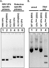
Site-specific mutations introduced into the genomes of the rescued VP4 gene transfectants. The VP4 gene of each virus was amplified by RT-PCR, using the primers shown in Table 2, to yield a 686-bp product. (A) The VP4 gene was amplified by use of RRV VP4-specific primers (lanes 1 to 3) and rotavirus VP4 degenerate primers (lanes 4 to 6). (B) The amplified fragments were digested with XbaI, followed by separation in a 2% agarose gel. Uncut WT VP4 (RRV) and VP4XbaI (RRV) were run in a 2% agarose gel (lanes 1 and 2), along with WT VP4 (RRV) and VP4XbaI (RRV) after digestion with XbaI (lanes 3 and 4).
FIG 6
FIG 6
Confirmation of site-specific mutations introduced into the genomes of the rescued VP4 gene transfectants. The full-length VP4 genes were directly sequenced, which confirmed the site-specific mutations introduced within the genomes of the infectious VP4 gene transfectants.
FIG 7
FIG 7
Viral binding and replication in murine cholangiocytes. (A) Percentages of RRV, RRVVP4-XbaI, and RRVVP4-R446G binding to murine cholangiocytes at 4°C. The quantity of bound virus was measured using FFA. (B) Quantification of infectious rotavirus strains in murine cholangiocytes at 24 and 48 h postinfection, measured using FFA. For both sets of experiments, values (n = 3) are expressed as mean FFU per milliliter, with standard errors; each assay was repeated three times. (C) Cholangiocytes were pretreated with 1 mM synthetic peptides TRTRVSRLY, DGEA, and GHRP, followed by infection with RRV or RRVVP4-R446G. The DGEA peptide was able to inhibit binding of both strains, while TRTRVSRLY was able to block only WT RRV binding. *, P < 0.05.
FIG 8
FIG 8
Viral binding and replication in MA104 and Caco-2 cells. Percentages of RRV, RRVVP4-XbaI, and RRVVP4-R446G binding to MA104 cells (A) and Caco-2 cells (B) at 4°C are shown. For both cell types, RRVVP4-R446G bound at a significantly lower percentage than that for RRV. Quantification of infectious rotavirus strains in MA104 cells (C) and Caco-2 cells (D) at 48 h postinfection revealed a similar pattern. For both sets of experiments (n = 3), values are expressed as mean FFU per milliliter, with standard errors; the assay was repeated three times.
FIG 9
FIG 9
In vivo effect of mutant virus in the mouse model of biliary atresia. (A) Symptoms were recorded for different groups of mice for 16 days. One hundred percent of the mice in the RRV (n = 21)- and RRVVP4-XbaI (n = 22)-treated groups showed symptoms, but only 80% of those in the RRVVP4-R446G-injected group (n = 23) showed symptoms, and this gradually decreased to 40% later. (B) Survival was noted to be significantly improved (85%) for mice injected with RRVVP4-R446G compared to that for mice injected with RRV (0%) or RRVVP4-XbaI (10%). *, P < 0.05.
FIG 10
FIG 10
Virus titers in organs following RRVVP4-R446G injection. (A) Viral titers from extrahepatic bile ducts harvested at 1 and 7 days postinfection were both significantly lower for mice injected with RRVVP4-R446G than for those injected with RRV or RRVVP4-XbaI (n = 6 per group). (B) Similarly, RRVVP4-R446G-injected mice expressed significantly lower viral titers in the intestine at 7 days postinfection than those for RRV-injected mice. *, P < 0.05.
FIG 11
FIG 11
Histologic evaluation of the liver and cholangiograms performed 12 days after infection with RRV and RRVVP4-R446G. (A) Hematoxylin and eosin staining of the liver after infection with RRV shows severe inflammation with an accumulation of inflammatory cells. (B) In contrast, mice injected with RRVVP4-R446G had mild inflammation. Magnification, ×10. (C and D) Cholangiograms demonstrating injection of methylene blue dye into the gallbladders of mice at 12 to 14 days postinoculation. The cholangiograms show the presence of biliary obstruction with RRV and the absence of biliary obstruction with RRVVP4-R446G. There were 7 mice in the RRV group and 8 in the RRVVP4-R446G group.
FIG 12
FIG 12
NK cell activation. (A and B) Flow cytometry of lymphocyte populations harvested from the livers of pups at 7 days postinfection illustrated a significant decrease in the number of activated NK cells present in the RRVVP4-R446G-injected mice compared to that for RRV-injected mice but a similar number compared to that for saline-injected mice. Red quadrants show the percentages of CD69+ cells in the CD49b+ population (2 livers per sample and 3 samples per strain). *, P < 0.05 (n = 3).

References

    1. Balistreri WF, Grand R, Hoofnagle JH, Suchy FJ, Ryckman FC, Perlmutter DH, Sokol RJ. 1996. Biliary atresia: current concepts and research directions. Summary of a symposium. Hepatology 23:1682–1692. - PubMed
    1. Bessho K, Bezerra JA. 2011. Biliary atresia: will blocking inflammation tame the disease? Annu Rev Med 62:171–185. doi:10.1146/annurev-med-042909-093734. - DOI - PMC - PubMed
    1. Riepenhoff-Talty M, Gouvea V, Evans MJ, Svensson L, Hoffenberg E, Sokol RJ, Uhnoo I, Greenberg SJ, Schakel K, Zhaori G, Fitzgerald J, Chong S, el-Yousef M, Nemeth A, Brown M, Piccoli D, Hyams J, Ruffin D, Rossi T. 1996. Detection of group C rotavirus in infants with extrahepatic biliary atresia. J Infect Dis 174:8–15. doi:10.1093/infdis/174.1.8. - DOI - PubMed
    1. Glaser JH, Balistreri WF, Morecki R. 1984. Role of reovirus type 3 in persistent infantile cholestasis. J Pediatr 105:912–915. doi:10.1016/S0022-3476(84)80076-1. - "VSports" DOI - PubMed
    1. Fjaer RB, Bruu AL, Nordbo SA. 2005. Extrahepatic bile duct atresia and viral involvement. Pediatr Transplant 9:68–73. doi:10.1111/j.1399-3046.2005.00257.x. - DOI - PubMed

Publication types

MeSH terms

LinkOut - more resources