Skip to main page content
U.S. flag

An official website of the United States government

Dot gov

The . gov means it’s official. Federal government websites often end in . gov or VSports app下载. mil. Before sharing sensitive information, make sure you’re on a federal government site. .

Https

The site is secure. The https:// ensures that you are connecting to the official website and that any information you provide is encrypted and transmitted securely V体育官网. .

. 2017 Aug 31;36(35):5075-5082.
doi: 10.1038/onc.2017.128. Epub 2017 May 1.

MYCN induces neuroblastoma in primary neural crest cells (VSports)

Affiliations

MYCN induces neuroblastoma in primary neural crest cells

R R Olsen (VSports app下载) et al. Oncogene. .

Abstract

Neuroblastoma (NBL) is an embryonal cancer of the sympathetic nervous system (SNS), which causes 15% of pediatric cancer deaths. High-risk NBL is characterized by N-Myc amplification and segmental chromosomal gains and losses. Owing to limited disease models, the etiology of NBL is largely unknown, including both the cell of origin and the majority of oncogenic drivers. We have established a novel system for studying NBL based on the transformation of neural crest cells (NCCs), the progenitor cells of the SNS, isolated from mouse embryonic day 9. 5 trunk neural tube explants. Based on pathology and gene expression analysis, we report the first successful transformation of wild-type NCCs into NBL by enforced expression of N-Myc, to generate phenotypically and molecularly accurate tumors that closely model human MYCN-amplified NBL. Using comparative genomic hybridization, we found that NCC-derived NBL tumors acquired copy number gains and losses that are syntenic to those observed in human MYCN-amplified NBL including 17q gain, 2p gain and loss of 1p36 VSports手机版. When p53-compromised NCCs were transformed with N-Myc, we generated primitive neuroectodermal tumors with divergent differentiation including osteosarcoma. These subcutaneous tumors were metastatic to regional lymph nodes, liver and lung. Our novel experimental approach accurately models human NBL and establishes a new system with potential to study early stages of NBL oncogenesis, to functionally assess NBL oncogenic drivers and to characterize NBL metastasis. .


"V体育2025版" Conflict of interest statement

The authors declare no conflict of interest.

Figures
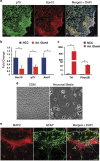
Figure 1
Figure 1
Cells isolated from trunk neural tube explants are NCCs by marker analysis and differentiation potential. (a) Day 9.5 embryos were isolated and dissected to remove the neural tube from a region caudal to the heart to the most caudal somite. Isolated neural trunks were treated with 1 mg/ml collagenase/dispase for 5 min, while being triturated in a Pasteur pipette. Neural tube explants were cultured in neural crest media (EMEM (Life Technologies, Waltham, MA, USA, 51200-038) with 3% chick embryo extract (US Biologicals, Salem, MA, USA, C3999) and 15% fetal bovine serum (Omega Scientific, Tarzana, CA, USA, FB-02)) on fibronectin-coated plates for 48 h, to allow NCC migration onto the plate before the neural tubes were removed. Immunofluorescence staining on cells isolated from neural tube explants using antibodies to the nerve growth factor receptor p75 (green, EMD Millipore, Billerica, MA, USA, 07-476) and the transcription factor Sox10 (red, ThermoFisher Scientific, Waltham, MA, USA, MAB2864), both markers of early neural crest. The merged image also includes DAPI nuclear staining. Scale bar=10 μm. (b) Quantitative real-time PCR was performed on a 7900HT Real Time PCR Machine (Applied Biosystems, Waltham, MA, USA) using Taqman Fast Advanced Master Mix (Life Technologies, 4444965). Expression of the early neural crest markers p75 and Sox10, and the migratory neural crest marker Ascl1 were measured relative to B2M in isolated NCCs compared with adult mouse adrenal gland, a differentiated SNS tissue (n=2–3). *P<0.001. (c) Quantitative real-time PCR comparing expression of Tyrosine hydroxylase (a marker of differentiated SNS) and Phox2B (an SNS lineage marker) in isolated NCCs versus adrenal gland tissue (n=2–3). *P<0.01. Results are expressed as the mean±s.d. We utilized a two-tail, unpaired Student’s t-test for all pair-wise comparisons (GraphPad Prism version 6) and analysis of variance where appropriate (GraphPad Prism). P-values <0.05 were considered statistically significant. TaqMan primers and probes (ThermoFisher Scientific) used include the following: p75 (Mm01309638_m1), Sox10 (Mm01300162_m1), Ascl1 (Mm04207567_g1), TH (Mm00447557_m1), Phox2B (Mm00435872_m1) and B2M (Mm0437762_m1). (d) Bright-field images of NCCs grown in chemically defined media (CDM) or neuronal differentiation media for 1 week. Scale bar=50 μm. (e) Immunofluorescence staining for markers of neurons (MAP2, Abcam, Cambridge, MA, USA, ab5392) and glia (GFAP, Cell Signaling Technology, Danvers, MA, USA, XP12389P) following differentiation of NCCs in neuronal differentiation media for 1 week. The merged image also includes DAPI nuclear staining. Scale bar=50 μm.
Figure 2
Figure 2
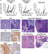
N-Myc transforms p53 compromised NCCs into metastatic primitive neuroectodermal tumors with divergent differentiation. (a) Approximately 10,000 p53mixed or p53+/− NCCs infected with N-Myc virus were mixed with 100 μl Matrigel (BD Biosciences, San Jose, CA, USA, 356237) and injected subcutaneously on the right flank of 6- to 8-week-old either Nu/Nu mice or C57Bl/6 mice (Charles River, Wilmington, MA, USA). Mice were injected on a rolling basis, to generate sufficient numbers of tumors for further analysis in a non-randomized, non-blinded manner. Eight Nu/Nu mice (left panel) and six C57Bl/6 mice (middle panel) were each flank injected with N-Myc transduced primary p53mixed NCCs pooled from p53+/− and p53−/− embryos (middle panel). Four C57Bl/6 mice were flank injected with N-Myc transduced primary p53+/− NCCs (right panel). Individual tumor volumes were measured over time as previously described. (b) Formalin-fixed tissue samples were prepared as previously described. Representative tumor histology (hematoxylin and eosin, H&E) is shown from a region positive for neuronal markers (upper left) and a region with osteosarcoma (upper right). Representative immunohistochemistry images show expression of neuronal markers. Antibodies used include TH (1:40, Vector, Burlingame, CA, USA, VP-T489), Synaptophysin (1:100, Spring Biosciences, Pleasanton, CA, USA, E2172), MAP2 (1:250, Millipore, Billerica, MA, USA, AB5622) and Phox2B (1:100, Abcam, ab183741). Scale bars=50 μM. (c) Representative histology (H&E) of lymph node, liver and lung metastasis with areas of metastasis highlighted in the insets. Scale bars=50 μM. This experiment was performed in strict accordance with the recommendations in the Guide for the Care and Use of Laboratory Animals of the National Institute of Health. Mice were sacrificed using CO2 asphyxiation followed by cervical dislocation. This research was approved by the St. Jude Children’s Research Hospital IACUC.
Figure 3
Figure 3
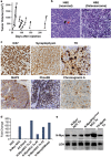
N-Myc transforms primary NCCs into NBL. (a) Approximately 10 000 wild-type NCCs infected with N-Myc were mixed with 100 ul Matrigel (BD Biosciences, 356237) and injected subcutaneously on the right flank of 6- to 8-week-old either Nu/Nu mice or C57Bl/6 mice (Charles River). Mice were injected on a rolling basis to generate sufficient numbers of tumors for further analysis in a non-randomized, non-blinded manner. Mice were killed and tumors isolated when they were at least 1000 mm3 or earlier if ulcerated. A total of 26 mice were flank injected with N-Myc transduced primary NCCs. (b) Representative hematoxylin and eosin (H&E) tumor histology from NCC tumors that were uniformly neuronal (left) or osteosarcoma (right). The black arrow identifies a ganglion cell and the red arrow identifies neuropil. Scale bar=50 μm. (c) Representative immunohistochemistry (IHC) images of neuronal tumors stained for Ki67 (1:200, ThermoFisher Scientific, RM-9106), Synaptophysin, TH, MAP2, Phox2B and Chromogranin A (1:1500, Immunostar, Hudson, WI, USA). Scale bar=50 μm. (d) Quantitative real-time PCR was performed on primary NCC, N-Myc virally transduced NCCs (N-Myc NCC), N-Myc NCC tumors (N-Myc/NCC Tu) and N-Myc; p53mixed NCC tumors (N-Myc; p53mixed NCC Tu) for NMYC (Mm00476449_m1) gene expression. Values indicate fold change over N-Myc expression in NCC, which is set at 1. (e) Protein was extracted from tumors derived from N-Myc NCCs and N-Myc; p53mixed NCCs, as well as isolated from the TH-MYCN mouse model of NBL by homogenizing with T-PER buffer (ThermoFisher Scientific) using a MicroTube homogenizer. Blots were probed with antibodies for N-Myc (Cell Signaling Technologies 9405) and as loading control lactate dehydrogenase (Abcam ab47010). Rabbit secondary (Cell Signaling Technologies 7074) and ECL Select (Amersham, Pittsburgh, PA, USA) were used to detect the signals. This experiment was performed in strict accordance with the recommendations in the Guide for the Care and Use of Laboratory Animals of the National Institute of Health. Mice were sacrificed using CO2 asphyxiation followed by cervical dislocation. This research was approved by the St. Jude Children's Research Hospital IACUC.
Figure 4
Figure 4
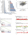
Mouse NCC-derived NBL recapitulates human NBL gene signatures and chromosomal changes. (a) ENRICHR analysis comparing top expressed genes from NCC-derived NBL to gene signatures of 917 cell lines in the Cancer Cell Line Encyclopedia (CCLE). Data is presented as average rank score (-Log10(pValue) for each cancer subtype. The number of cell lines represented in each cancer subtype is shown in parentheses and error is standard deviation. (b) The NCC-derived NBL gene signature in comparison to each of the individual NBL cell lines in the CCLE preferentially matches N-Myc amplified NBL (blue) than N-Myc wild type NBL (red). (c) Dot-blot comparing the log ratio of N-Myc amplified NBL (GEO data set: GSE32664) to human adrenal (GEO data set: GSE39716) versus the log ratio of N-Myc NCC-derived NBL (Affymetrix Mouse 2.0 ST arrays (ThermoFisher Scientific), GEO data set: GSE94914) versus mouse adrenal (GEO data set: GSE21829). (d) Genes differentially expressed between N-Myc NCC-derived NBL (Affymetrix Mouse 2.0 ST arrays, ThermoFisher Scientific: GSE94914) and mouse adrenal (GEO data set: GSE21829) share molecular signatures as determined by gene set enrichment analysis with gene sets downstream of CCND1 and CDK4 in primary NBLs (Molenaar) and cancer-specific genes shared between primary pediatric tumors and pediatric xenografts (Whiteford). (e) Average NCC-derived NBL tumor to normal liver genomic DNA ratio following array comparative genomic hybridization (aCGH). A log2ratio threshold of 0.3 was used to identify amplifications and −0.4 to identify deletions. NCC-derived tumors show amplification of the region syntenic to human Chromosome 17q23-qter on mouse chromosome 11. (f) NCC tumors have deletions in mouse chromosome 4 in the region syntenic to human 1p36. (g) NCC tumors have amplification of the region syntenic to human 2p13-15 on mouse chromosome 11. The positions of a subset of proposed NBL oncogenes and tumor suppressors from each region are indicated.

References

    1. Meany HJ, London WB, Ambros PF, Matthay KK, Monclair T, Simon T et al. Significance of clinical and biologic features in Stage 3 neuroblastoma: a report from the International Neuroblastoma Risk Group project. Pediatr Blood Cancer 2014; 61: 1932–1939. - "V体育官网入口" PubMed
    1. Wylie LA, Hardwick LJ, Papkovskaia TD, Thiele CJ, Philpott A. Ascl1 phospho-status regulates neuronal differentiation in a Xenopus developmental model of neuroblastoma. Disease Models Mech 2015; 8: 429–441. - PMC - PubMed
    1. Louis CU, Shohet JM. Neuroblastoma: molecular pathogenesis and therapy. Ann Rev Med 2015; 66: 49–63. - PMC - PubMed
    1. Simoes-Costa M, Bronner ME. Establishing neural crest identity: a gene regulatory recipe. Development 2015; 142: 242–257. - PMC - PubMed
    1. Karunasena E, McIver LJ, Bavarva JH, Wu X, Zhu H, Garner HR. 'Cut from the same cloth': Shared microsatellite variants among cancers link to ectodermal tissues-neural tube and crest cells. Oncotarget 2015; 6: 22038–22047. - PMC - PubMed

Publication types

MeSH terms