"V体育ios版" Skip to main page content
U.S. flag

An official website of the United States government

Dot gov

The . gov means it’s official. Federal government websites often end in . gov or VSports app下载. mil. Before sharing sensitive information, make sure you’re on a federal government site. .

Https

The site is secure V体育官网. The https:// ensures that you are connecting to the official website and that any information you provide is encrypted and transmitted securely. .

. 2017 Sep 1;19(9):1237-1247.
doi: 10.1093/neuonc/nox050.

Genomic profiles of low-grade murine gliomas evolve during progression to glioblastoma

Affiliations

VSports在线直播 - Genomic profiles of low-grade murine gliomas evolve during progression to glioblastoma

Mark Vitucci et al. Neuro Oncol. .

Abstract

Background: Gliomas are diverse neoplasms with multiple molecular subtypes. How tumor-initiating mutations relate to molecular subtypes as these tumors evolve during malignant progression remains unclear VSports手机版. .

Methods: We used genetically engineered mouse models, histopathology, genetic lineage tracing, expression profiling, and copy number analyses to examine how genomic tumor diversity evolves during the course of malignant progression from low- to high-grade disease V体育安卓版. .

Results: Knockout of all 3 retinoblastoma (Rb) family proteins was required to initiate low-grade tumors in adult mouse astrocytes. Mutations activating mitogen-activated protein kinase signaling, specifically KrasG12D, potentiated Rb-mediated tumorigenesis. Low-grade tumors showed mutant Kras-specific transcriptome profiles but lacked copy number mutations. These tumors stochastically progressed to high-grade, in part through acquisition of copy number mutations V体育ios版. High-grade tumor transcriptomes were heterogeneous and consisted of 3 subtypes that mimicked human mesenchymal, proneural, and neural glioblastomas. Subtypes were confirmed in validation sets of high-grade mouse tumors initiated by different driver mutations as well as human patient-derived xenograft models and glioblastoma tumors. .

Conclusion: These results suggest that oncogenic driver mutations influence the genomic profiles of low-grade tumors and that these, as well as progression-acquired mutations, contribute strongly to the genomic heterogeneity across high-grade tumors VSports最新版本. .

Keywords: genetically engineered mouse; glioblastoma; glioma; progression; transcriptome. V体育平台登录.


Figures

Fig. 1
Fig. 1
Initiating mutations influence low-grade tumor burden and transcriptomes. All T mice, with and without RP mutations, developed low-grade tumors (blue, grade II). Tumors in a subset (25%) of TRP−/− mice had progressed to high-grade (green, grade III) (A). Lineage tracing in TRP+/− mice showed tdTomato (red) tumor cells (B) that developed perineuronal satellitoses around NeuN+ (green) neurons (C). Hypercellular, hyperproliferative (Ki-67+ [green]) tumor foci were also evident (D) adjacent to areas of less proliferative tumor (E, DAPI [blue]). Morphometric analyses showed that initiating mutations affected tumor burden (one-way ANOVA P < 0.0001; *indicates t-test P < 0.05; NS, not significant) (F). PCA showed that tumor transcriptomes with (red) and without (blue) KrasG12D were distinct, while transcriptomes of histologically normal brain with (black with red outlines) and without (black) KrasG12D were indistinguishable (G). A KrasG12D-related gene signature derived from low-grade tumors was enriched in pre-glioblastoma (PG), but not neuroblastic (NB) or early progenitor-like (EPL) subtypes of human lower-grade astrocytomas (H). Scale bars 50 (B), 10 (C), 100 µm (D), and 50 µm (E).
Fig. 2
Fig. 2
Low-grade tumors stochastically progress to rapidly proliferative, lethal high-grade tumors. A contrast-enhancing tumor (A) developed focally in a TRP+/− mouse. This high-grade (B), T121-immunoreactive (C) tumor showed histological features of GBM (B), including endothelial proliferation (D). Widespread low-grade (BE), T121-immunoreactive (CF) tumor was evident elsewhere in the brain. Boxes in BC indicate corresponding images in DEF. Quantification of serial T2-weighted MRIs from 9 TRP+/− mice showed logarithmic increases in high-grade tumor volume with mean doubling of 3 ± 1 days (G). Median time to first appearance of high-grade tumors, median survival, and mean time to death after first high-grade tumor appearance were 119 ± 7 (range 84–154), 122 ± 2, and 14 ± 3 days, respectively (H). Scale bars 1 mm (BC) and 50 µm (DEF).
Fig. 3
Fig. 3
Initiating mutations influence CNA acquisition during malignant progression. Heatmaps of aCGH data showed minimal acquisition of CNA in lower-grade tumors, either at 2 months (Ai) or >1 year (Aii) after induction. All high-grade TR tumors showed gains of chromosome 6 (Aiii), but only 64%–72% of high-grade TRP+/− tumors show similar gains (Aiv, Fisher P = 0.15). Half of TR, but only 16%–20% of high-grade TRP+/− tumors showed chromosome 1 gains (Fisher’s P = 0.17). High-grade TRP−/− tumors acquired the least CNA and had the lowest frequency (13%–25%) of chromosome 6 gains (Av, Fisher P ≤ 0.04). Initiating mutations (B) significantly influenced the development of CNA in recurrently altered (>20% of samples, Supplementary Table S5) Rb, RTK/MAPK/PI3K, and p53 pathway genes (Fisher’s P ≤ 0.16). T astrocytes transfected with lentiviral vectors encoding Met showed increased receptor expression and phosphorylation (p-Met) by immunoblot (C). Met significantly increased T astrocyte proliferation by MTS assay in vitro (D).
Fig. 4
Fig. 4
Copy number landscapes of high-grade tumor models with and without p53 deletion are distinct. Heatmaps of aCGH data from 41 terminal high-grade TR±P tumors showed frequent CNA on chromosomes 1 and 6 (A). In contrast, 21 Rb1/Pten/p53 triple KO high-grade tumors (GSE22927, [5]) showed frequent CNA across the genome (B); aCGH heatmaps of 10 tumors from 7 TR±P mice that also harbored Trp53+/− deletion showed minimal CNA ~2 months after induction (C). However, GBM harvested from a terminally aged, neurologically symptomatic T;Trp53+/− mouse (D) showed a CNA pattern more similar to Rb1/Pten/p53 triple KO (B) than TR±P high-grade tumors (A).
Fig. 5
Fig. 5
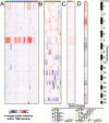
High-grade murine glioma transcriptomes are heterogeneous. Separation of low-grade tumors with (black boxes) and without mutant KrasG12D (open boxes) and S1 (red), S2 (blue), and S3 (green) high-grade tumor subtypes (circles) were preserved when transcriptome datasets from low- and high-grade TRP tumors were combined (A). In contrast to initiating mutations (Supplementary Figure S6), high-grade glioma subtype did not influence survival (B, log-rank P = 0.4). S1–S3 subtype correlated with human TCGA GBM subtype based on ClaNC (C, Fisher’s P = 2.2 × 10⁻¹²) as well as ssGSEA (D); ssGSEA also showed enrichment of distinct neural cell lineage signatures. Cultured astrocyte, oligodendrocyte precursor cell (OPC), and neuron signatures were enriched in S1, S2, and S3 tumors, respectively (E). Hierarchical clustering of an independent test set composed of 3 adult high-grade glioma GEM models with different initiating mutations showed that the 600-gene classifier accurately clustered samples according to their predicted S1–S3 subtypes (F). Consensus hierarchical clustering of high-grade TRP tumors with human patient-derived xenografts (PDX) and GBM (TCGA dataset) using 8916 overlapping genes identified 5 clusters (G). Sample annotation tracks include ClusterID, tumor type—GEM, PDX, GBM, and TCGA subtype for human GBM.
Fig. 6
Fig. 6
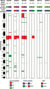
High-grade murine tumors pair show evidence of divergent genomic evolution. Two tumors harvested from distinct brain regions from 6 different mice showed different S1–S3 transcriptome subtypes, TCGA transcriptome subtypes, and CNA.

V体育官网 - References

    1. Miller CR, Perry A. Glioblastoma. Arch Pathol Lab Med. 2007;131(3):397–406. - PubMed
    1. Louis DN, Perry A, Reifenberger G, et al. The 2016 World Health Organization classification of tumors of the central nervous system: a summary. Acta Neuropathol. 2016;131(6):803–820. - "VSports" PubMed
    1. Brennan CW, Verhaak RG, McKenna A, et al. ; TCGA Research Network. The somatic genomic landscape of glioblastoma. Cell. 2013;155(2):462–477. - PMC - PubMed
    1. Cancer Genome Atlas Research Network. Comprehensive genomic characterization defines human glioblastoma genes and core pathways. Nature. 2008;455(7216):1061–1068. - "V体育官网入口" PMC - PubMed
    1. Verhaak RG, Hoadley KA, Purdom E, et al. ; Cancer Genome Atlas Research Network. Integrated genomic analysis identifies clinically relevant subtypes of glioblastoma characterized by abnormalities in PDGFRA, IDH1, EGFR, and NF1. Cancer Cell. 2010;17(1):98–110. - PMC - PubMed