Skip to main page content (V体育平台登录)
U.S. flag

An official website of the United States government

Dot gov

The . gov means it’s official VSports app下载. Federal government websites often end in . gov or . mil. Before sharing sensitive information, make sure you’re on a federal government site. .

Https

The site is secure. The https:// ensures that you are connecting to the official website and that any information you provide is encrypted and transmitted securely. V体育官网.

. 2017 Jun 13;8(24):39087-39100.
doi: 10.18632/oncotarget.16598.

Cytochrome P450 1B1 inhibition suppresses tumorigenicity of prostate cancer via caspase-1 activation

Affiliations

Cytochrome P450 1B1 inhibition suppresses tumorigenicity of prostate cancer via caspase-1 activation

Inik Chang et al. Oncotarget. .

Erratum in

Abstract

Cytochrome P450 1B1 (CYP1B1) is recognized as a universal tumor biomarker and a feasible therapeutic target due to its specific overexpression in cancer tissues. Despite its up-regulation in prostate cancer (PCa), biological significance and clinicopathological features of CYP1B1 are still elusive. Here, we show that overexpression or hyperactivation of CYP1B1 stimulated proliferative, migratory and invasive potential of non-tumorigenic PCa cells. Attenuation of CYP1B1 with its specific small hairpin (sh) RNAs greatly reduced proliferation through apoptotic cell death and impaired migration and invasion in PCa cells. Intratumoral injection of CYP1B1 shRNA attenuated growth of pre-existing tumors. The antitumor effect of CYP1B1 shRNA was also observed in prostate tumor xenograft mouse models. Among the genes altered by CYP1B1 knockdown, reduction of caspase-1 (CASP1) activity attenuated the antitumor effect of CYP1B1 inhibition. Indeed, CYP1B1 regulates CASP1 expression or activity. Finally, CYP1B1 expression was increased in higher grades of PCa and overall survival was significantly reduced in patients with high levels of CYP1B1 protein VSports手机版. CYP1B1 expression was reversely associated with CASP1 expression in clinical tissue samples. Together, our results demonstrate that CYP1B1 regulates PCa tumorigenesis by inhibiting CASP1 activation. Thus, the CYP1B1-CASP1 axis may be useful as a potential biomarker and a therapeutic target for PCa. .

Keywords: CYP1B1; caspase-1; prostate cancer; shRNA; tumorigenicity V体育安卓版. .


Conflict of interest statement

CONFLICTS OF INTEREST

The authors declare that they have no competing interests.

Figures

Figure 1
Figure 1. CYP1B1 promotes cellular transformation of RWPE-1 cells
(A and B) Endogenous expression of CYP1B1 protein (A) and mRNA (B) in PCa cells. Levels were determined by Western blot and qRT-PCR, respectively. (C–G) Effect of CYP1B1 overexpression on in vitro tumorigenicity. Ectopic expression of CYP1B1 in transfected RWPE-1 cells as examined by Western blot (C). Cell proliferation as determined by MTS assay (D). Colony formation as determined by crystal violet staining. Representative image of colonies (left panel) and quantification of stained colonies (right panel) are shown (E). Cell migration (F) and invasion (G) capability as determined by transwell migration and invasion assay, respectively. (H–L) Effect of DMBA treatment on in vitro tumorigenicity. Induction of CYP1B1 expression in RWPE-1 cells as determined by Western blot (H). Cell proliferation as determined by MTS assay (I). Colony formation as determined by crystal violet staining. Representative image of colonies (left panel) and quantification of stained colonies (right panel) are shown (J). Cell migration (K) and invasion (L) capability as determined by transwell migration and invasion assay, respectively. *p<0.05; **p<0.01.
Figure 2
Figure 2. CYP1B1 inhibition suppresses in vitro tumorigenicity
(A and B) Expression of CYP1B1 mRNA (A) and protein (B) in CYP1B1 shRNA or control shRNA expressing PC-3 cells. Levels were determined by qRT-PCR and Western blot, respectively. (C–H) Effect of CYP1B1 knockdown on in vitro tumorigenicity. Cell proliferation as determined by MTS assay at the indicated times. Representative images of cell morphology (left panel) and quantification of cell proliferation (right panel) are shown (C). Colony formation as determined by crystal violet staining. Representative image of colonies (left panel) and quantification of stained colonies (right panel) are shown (D). Apoptotic cell death as determined by flow cytometric analysis using double staining with Annexin V-FITC and 7-AAD. Representative biparametric histograms exhibiting cell (left panel) and quantification of apoptotic cells (right panel) are shown (E). Cell cycle progression as determined by DAPI staining (F). Cell migration (G) and invasion (H) capability as determined by transwell migration and invasion assay, respectively. Representative images (left panel) and quantification of assay (right panel) are shown. **p<0.01; ***p<0.001.
Figure 3
Figure 3. CYP1B1 inhibition suppresses in vivo tumor growth
(A to D) Effect of intratumoral injection of CYP1B1 shRNA on in vivo tumor growth. Volume of tumor established by PC-3 cells after administration of shRNAs for CYP1B1 or control. Representative image of tumors grown in mice and tumors extracted from individual mice (A) and quantification of tumor volume (B). Quantification of Ki67-positive cells in the control or CYP1B1 shRNA injected tumors. Representative image of section (left panel) and quantification of stained cells (right panel) are shown. Total 20 fields/sample were counted (C). CYP1B1 protein expression in tumors injected with control or CYP1B1 shRNA (D). (E–H) Effect of CYP1B1 shRNA knockdown on in vivo tumor growth using xenograft mouse model. Volume of tumor established by PC-3 cells stably expressing CYP1B1 shRNA #4-2 in xenografts. Representative image of tumors grown in mice and tumors extracted from individual mice (E) and quantification of tumor volume (F). Quantification of Ki67-positive cells in the control or CYP1B1 shRNA #4-2 expressing xenograft tumors. Representative image of section (left panel) and quantification of stained cells (right panel) are shown. Total 20 fields/sample were counted (G). CYP1B1 protein expression of control or CYP1B1 shRNA #4-2 expressing xenograft tumors (H). *p<0.05; **p<0.01; ***p<0.001.
Figure 4
Figure 4. CASP1 is a functional target of CYP1B1
(A–C) CASP1 mRNA (A), protein (B), and enzyme activity (C) in CYP1B1 shRNA stably expressing PCa cells. Levels were determined by qRT-PCR, Western blot and colorimetric assay, respectively. (D–G) Effect of CASP1 inhibition on the tumorigenicity of CYP1B1 shRNA stably expressing PCa cells. Cells were treated with Z-YVAD-fmk (100 μM) and proliferation (D), apoptotic cell death (E), migration (F), as well as invasion (G) were examined, respectively. (H) Effect of chemical inhibition of CYP1B1 on CASP1 activation. CASP1 activity was determined by colorimetric assay in PC-3 cells treated with the indicated concentration of TMS. (I) Effect of CASP1 inhibition on apoptotic cell death induced by chemical inhibition of CYP1B1. Apoptotic cell death was determined by flow cytometric analysis using double staining with Annexin V-FITC and 7-AAD in TMS-treated PC-3 cells with or without addition of Z-YVAD-fmk (100 μM). (J and K) Effect of CYP1B1 activation on CASP1 in RWPE-1 cells. CASP1 expression was determined by Western blot after CYP1B1 overexpression (J). CASP1 activity was examined by colorimetric assay in cells treated with the indicated concentration of DMBA (K). *p<0.05; **p<0.01; ***p<0.001.
Figure 5
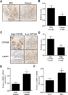
Figure 5. Association of CYP1B1 expression with clinicopathologic characteristics of PCa
(A–C) CYP1B1 expression in PCa tissues. Immunohistochemical staining of CYP1B1 protein in PCa specimens. Representative images showing immunoreactive CYP1B1 in BPH and cancer tissues (magnification: ×200) (A). Summary of CYP1B1 immunostaining score. Staining intensity was assessed as described in Materials and Methods (B). CYP1B1 mRNA expression in microdissected prostate tissues (C). (D) Correlation of CYP1B1 expression with clinicopathological characteristics of patients with PCa. †Data was not available for some samples. *p<0.05; ***p<0.001 (E) Kaplan-Meier survival curves for overall survival of patients with PCa. p=0.0111.
Figure 6
Figure 6. Inverse correlation between CYP1B1 and CASP1 expression
(A and B) Immunohistochemical staining of CASP1 protein in PCa specimens. Representative images showing immunoreactive caspase-1 in BPH and cancer tissues (magnification: × 200) (A). Summary of CASP1 immunostaining score (B). (C and D) Immunohistochemical staining of CYP1B1 and CASP1 protein in prostate cancer specimens. Representative images showing inverse correlation of CYP1B1 and CASP1 in BPH and cancer tissues (magnification: × 200) (C). Summary of CASP1 immunostaining score (D). (E and F) Expression of CASP1 mRNA in tumors injected with CYP1B1 shRNA (E) and PC-3/CYP1B1 shRNA #4-2 xenografts (F). *p<0.05.

References

    1. Murray GI. The role of cytochrome P450 in tumour development and progression and its potential in therapy. J Pathol. 2000;192:419–426. - VSports app下载 - PubMed
    1. Murray GI, Taylor MC, McFadyen MC, McKay JA, Greenlee WF, Burke MD, Melvin WT. Tumor-specific expression of cytochrome P450 CYP1B1. Cancer Res. 1997;57:3026–3031. - "VSports最新版本" PubMed
    1. Saini S, Hirata H, Majid S, Dahiya R. Functional significance of cytochrome P450 1B1 in endometrial carcinogenesis. Cancer Res. 2009;69:7038–7045. - PubMed
    1. Shatalova EG, Klein-Szanto AJ, Devarajan K, Cukierman E, Clapper ML. Estrogen and cytochrome P450 1B1 contribute to both early- and late-stage head and neck carcinogenesis. Cancer Prev Res (Phila) 2011;4:107–115. - PMC - PubMed
    1. Chang I, Mitsui Y, Fukuhara S, Gill A, Wong DK, Yamamura S, Shahryari V, Tabatabai ZL, Dahiya R, Shin DM, Tanaka Y. Loss of miR-200c up-regulates CYP1B1 and confers docetaxel resistance in renal cell carcinoma. Oncotarget. 2015;6:7774–7787. doi: 10.18632/oncotarget.3484. - DOI - PMC - PubMed

MeSH terms