Skip to main page content
U.S. flag

An official website of the United States government

Dot gov

The . gov means it’s official. Federal government websites often end in . gov or VSports app下载. mil. Before sharing sensitive information, make sure you’re on a federal government site. .

Https

The site is secure V体育官网. The https:// ensures that you are connecting to the official website and that any information you provide is encrypted and transmitted securely. .

. 2017 Mar 21;114(12):3163-3168.
doi: 10.1073/pnas.1621052114. Epub 2017 Mar 7.

Sensing of cell stress by human γδ TCR-dependent recognition of annexin A2 (VSports)

Affiliations

Sensing of cell stress by human γδ TCR-dependent recognition of annexin A2

Romain Marlin et al. Proc Natl Acad Sci U S A. .

"V体育ios版" Abstract

Human γδ T cells comprise a first line of defense through T-cell receptor (TCR) recognition of stressed cells. However, the molecular determinants and stress pathways involved in this recognition are largely unknown. Here we show that exposure of tumor cells to various stress situations led to tumor cell recognition by a Vγ8Vδ3 TCR. Using a strategy that we previously developed to identify antigenic ligands of γδ TCRs, annexin A2 was identified as the direct ligand of Vγ8Vδ3 TCR, and was found to be expressed on tumor cells upon the stress situations tested in a reactive oxygen species-dependent manner. Moreover, purified annexin A2 was able to stimulate the proliferation of a Vδ2neg γδ T-cell subset within peripheral blood mononuclear cells and other annexin A2-specific Vδ2neg γδ T-cell clones could be derived from peripheral blood mononuclear cells. We thus propose membrane exposure of annexin A2 as an oxidative stress signal for some Vδ2neg γδ T cells that could be involved in an adaptive stress surveillance VSports手机版. .

Keywords: cell stress surveillance; cytomegalovirus; gamma-delta T cells; innate-like lymphocytes; tumor immunology. V体育安卓版.


Conflict of interest statement

The authors declare no conflict of interest.

Figures

Fig. 1.
Fig. 1.
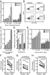
FMS-01 mAb inhibits 73R9 TCR-mediated recognition of U373MG. (A) CD69 expression by JRT3-73R9 cocultured 4 h with different target cells. Results shown are fold-increase of CD69 mean fluourescence intensity (MFI) in the presence of target cells versus in medium alone (horizontal dotted line). CEM, cell line donor’s initials. (B) CD69 expression by JRT3-73R9 (Vγ8Vδ3 TCR) and JRT3-26 (Vγ9Vδ3 TCR) cocultured 4 h with glioblastoma cells or with anti-CD3 mAb. (C) CD69 expression by JRT3-73R9 incubated with U373MG and with or without anti-Vγ8, anti-Vδ3, or anti-Vδ1 mAbs. In A–C bars represent the mean ± SEM of at least three independent experiments and Mann–Whitney test was used to compare conditions (*P < 0.05, ***P < 0.001). (D) CD69 expression by JRT3-73R9 incubated alone (Ctrl) or with U373MG cells in the presence or absence of a selection of hybridoma supernatants. (E) CD69 expression by JRT3 reporter cells expressing no TCR (JRT3 WT) or indicated Vδ2neg γδ TCRs, cultured in medium alone or with their own target cells (U373MG, HT29, SKW6.4). Supernatant of FMS-01 or control hybridoma (25% of culture volume) were added in indicated conditions. Data are representative of at least three independent experiments.
Fig. S1.
Fig. S1.
Comparative expression and contribution to clone 73R9 reactivity of FMS-01 antigen and anti–ILT-2. (A) Cell surface expression of FMS-01 antigen and ILT-2 on different tumor cell lines analyzed by flow cytometry (gray histograms). PE-conjugated goat anti-mouse IgM stained cells were used as negative controls (dotted-line histograms). MFI values are indicated. (B) CD107a expression on clone 73R9 after 4 h of culture with indicated tumor cell lines in the presence or not of FMS-01 hybridoma supernatant (25% of culture volume) or anti–ILT-2 mAb (10 µg/mL). Value of CD107a expression by the clone incubated in medium alone is represented as horizontal dotted line. (C) CD69 expression by JRT3-73R9 incubated with C91, 721.221, B-EBV, and Raji cell lines at the indicated E:T ratio. Value of CD69 expression by the JRT3-73R9 incubated in medium alone is represented as horizontal dotted line. Data are representative of at least three independent experiments.
Fig. 2.
Fig. 2.
CMV infection of glioblastoma cells enhances 73R9 TCR reactivity and FMS-01 expression. Activation of clone 73R9 (A) or JRT3-73R9 (B) by coculture with CMV-infected or uninfected glioblastoma cells. (C) Cell surface staining of CMV-infected (black line) and uninfected (gray histogram) U373MG with FMS-01, or with GAM IgM (dotted line). Results are mean ± SEM (A and B) or representative (C) of at least three experiments. Statistical significance was tested using the Willcoxon test, *P < 0.05 and **P < 0.005.
Fig. S2.
Fig. S2.
CMV infection, high confluence, and heat shock enhance FMS-01 expression and 73R9 TCR reactivity. (A) JRT3-73R9 activation by CMV-infected or uninfected U373MG with or without FMS-01 and/or control IgM. (B and C) U343MG monolayers were harvested at 60% (low) or 100% (high) of confluence before counting and incubation with JRT3-73R9. (D and E) U343MG monolayers were harvested and exposed to 42 °C heat-shock stress from 5 to 120 min before incubation with JRT3-73R9. (B and D) CD107a expression by clone 73R9 was evaluated after coculture at 1:1 ratio for 4 h with pretreated U343MG cells, in the absence or presence of FMS-01 mAb. (C and E) Staining of stressed cells with FMS-01 mAb. PE-conjugated goat anti-mouse IgM-stained cells were used as controls (dotted line). Results are representative of at least three independent experiments.
Fig. 3.
Fig. 3.
Stress stimulation of glioblastoma cells increases FMS-01 expression and 73R9 TCR reactivity. (A–C) U343MG monolayers were cultured from 24 h to 168 h in 0.1% O2 atmosphere, then detached, counted, and stained or incubated with T cells in normoxia. (D) U343MG monolayers were harvested at 60% (low confluence) or 100% (high confluence) of confluence before counting and incubation with JRT3-73R9. (E) U343MG cells were exposed to 42 °C from 5 to 120 min before incubation with JRT3-73R9. CD69 expression by JRT3-73R9 (A, D, E) or CD107a expression by clone 73R9 (B) were evaluated after coculture at 1:1 ratio for 4 h with pretreated U343MG cells, in the absence or presence of FMS-01 mAb. In A and E results are shown as fold-increase of CD69 MFI compared with negative control MFI (JRT3 in medium alone, horizontal dotted line). In B numbers indicate the percentage of cells in the gate. (C) Staining of stressed cells with FMS-01. Goat anti-mouse IgM-stained cells were used as controls (dotted line). Results represent the mean ± SEM (A and E) or are representative (B) of at least three independent experiments (*P < 0.05). (F–H) U373MG, U343MG, and U251MG cells were incubated for 5 d in hypoxia (F), at high confluence (G) or for 120 min at 42 °C heat (H). Cells were preincubated or not with 10 mM of NAC prior stress induction or cell detachment and FMS-01 surface expression was evaluated. Results of different experiments are shown as FMS-01 MFI fold-increase upon stress.
Fig. 4.
Fig. 4.
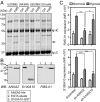
FMS-01 recognizes annexin A2. (A) Colloidal blue-stained SDS/PAGE of proteins immunoprecipitated with control IgM (Ctrl) or FMS-01 mAb (FMS) from glioblastoma or FMS-01 cells (Ctrl cells). Black arrow indicates the specific band. Heavy (Ig HC) and light chains (Ig LC) of antibodies used for immunoprecipitation are indicated. (B) Immunoblot analysis of EGTA eluates from U373MG, recombinant annexin 2 (His-tagged), and recombinant S100A10 (GST-tagged), detected with anti-annexin 2 mAb (Left), anti-S100A10 mAb (Center), or FMS-01 mAb (Right). Data are representative of at least two independent experiments. (C) Expression of FMS-01 ligand (Upper) and S100A10 (Lower) by U373MG transduced with scramble, annexin A2 (ANXA2), or S100A10 sh-RNAs and treated in normoxia or hypoxia. Data are represented as mean ± SEM of three independent experiments and two-way ANOVA test was used to compare conditions (*P < 0.05, **P < 0.005, ***P < 0.001).
Fig. S3.
Fig. S3.
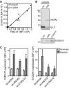
FMS-01 mAb recognizes annexin A2 on U343MG cells. (A) Linear regression analysis of S100A10 surface staining according to FMS-01 surface staining on different cell lines individually represented as dots. (B) Immunoblot analysis of U343MG cell proteins immunoprecipitated with control IgM (Ctrl), FMS-01 (FMS), or anti-S100A10 mAb, and detected with a commercial mAb to annexin A2 (Upper) or to S100A10 (Lower). Data are representative of at least two independent experiments. (C) Expression of FMS-01 ligand (Left) and S100A10 (Right) by U343MG cells transduced with scramble, annexin A2 (ANXA2), or S100A10 sh-RNAs and treated in normoxia or hypoxia. Data are represented as mean ± SEM of three independent experiments.
Fig. 5.
Fig. 5.
73R9 TCR recognizes annexin A2 CD69 expression by JRT3-73R9 incubated: (A) with or without EGTA eluates from highly confluent U373MG cells with or without FMS-01 mAb; (B) with U373 (Left) or U343 (Right) transduced with scramble, annexin A2 (ANXA2), or S100A10 sh-RNAs and pretreated in normoxia or hypoxia; (C) with or without increasing doses of recombinant soluble annexin A2 and/or S100A10 (Left), with anti-CD3 mAb, or with soluble annexin A6 (Right). (D) Clone 73R9 was activated with anti-CD3 mAb, soluble annexin A2 or A6, and CD107a membrane expression after 4 h (Left) or indicated cytokine secretion after 24 h (Right) were evaluated by flow cytometry. (E) Binding of annexin A2 (1.75 µM) to biotinylated 73R9 TCR or two control TCRs immobilized on streptavidin-coated flow cells at 2,153 RU, 2,175 RU, and 2,748 RU, respectively, or streptavidin alone, assessed by SPR and presented as resonance units (RU). (F) Detection of phosphorylated SLP-76 and ERK-1/2 in clone 73R9 incubated in the indicated conditions. All of the results are from at least three independent experiments and are shown as mean ± SEM (*P < 0.05, **P < 0.005).
Fig. S4.
Fig. S4.
Annexin A2, but not S100A10, is recognized by TCR 73R9. (A, Left) JRT3-73R9 was incubated with or without EGTA eluates from U373MG cells grown at low or high confluence. (Right) JRT3-73R9 (Vγ8Vδ3 TCR), JRT3 26 (Vγ9Vδ3 TCR), and JRT3 MAU (Vγ9Vδ1 TCR) were incubated with or without EGTA eluates from highly confluent U373MG cells. (B) JRT3-73R9 and JRT3 26 were incubated or not with increasing doses of recombinant annexin A2 or with anti-CD3. (C) JRT3-73R9 was incubated with or without increasing doses of annexin A2 in the presence or not of FMS-01 mAb. (D) Equilibrium affinity analysis of annexin A2 binding to the 73R9 TCR (Kd = 3.2 µM). (Inset) Scatchard plot of the same data (Kd = 3.0 µM). (E and F) Detection of phosphorylated SLP-76 and ERK-1/2 in clone LES (E), JRT3 73R9 (F, Left) and JRT3 26 (Right) incubated for 1 min in the indicated conditions. All of the results are shown as mean ± SEM of at least two independent experiments.
Fig. 6.
Fig. 6.
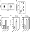
Annexin A2 stimulates Vδ2neg γδ T-cell proliferation. (A–C) CFSE [5-(and 6)-carboxyfluorescein diacetate succinimidyl ester] labeled Vδ2neg γδ T cells were cocultured with autologous PBMC in the presence of recombinant IL-2 with or without annexin A2 for 5 d. Results are presented as percentages of CFSE-low cells among Vδ2neg γδ T cells. (A) Representative dot-plot of flow cytometry staining, (B) percentages of proliferating Vδ2neg γδ T cells from independent donors (n = 7), and (C) comparison of annexin A2 and annexin A6 effects. (D) TNF-α production by two T-cell clones isolated from healthy donors and incubated for 24 h with soluble annexins A2 or A6. Results from C–E are mean ± SEM of at least two independent experiments.

References (V体育平台登录)

    1. Zinkernagel RM, Doherty PC. The discovery of MHC restriction. Immunol Today. 1997;18(1):14–17. - PubMed (V体育官网)
    1. Rock EP, Sibbald PR, Davis MM, Chien YH. CDR3 length in antigen-specific immune receptors. J Exp Med. 1994;179(1):323–328. - "V体育ios版" PMC - PubMed
    1. Chien YH, Meyer C, Bonneville M. γδ T cells: First line of defense and beyond. Annu Rev Immunol. 2014;32:121–155. - PubMed
    1. Born WK, Kemal Aydintug M, O’Brien RL. Diversity of γδ T-cell antigens. Cell Mol Immunol. 2013;10(1):13–20. - "VSports注册入口" PMC - PubMed
    1. Crowley MP, et al. A population of murine gammadelta T cells that recognize an inducible MHC class Ib molecule. Science. 2000;287(5451):314–316. - PubMed

Publication types

"V体育平台登录" MeSH terms

LinkOut - more resources