Skip to main page content
U.S. flag

An official website of the United States government

Dot gov

The . gov means it’s official VSports app下载. Federal government websites often end in . gov or . mil. Before sharing sensitive information, make sure you’re on a federal government site. .

Https

The site is secure. The https:// ensures that you are connecting to the official website and that any information you provide is encrypted and transmitted securely V体育官网. .

. 2017 Mar 6;13(3):e1006641.
doi: 10.1371/journal.pgen.1006641. eCollection 2017 Mar.

Analysis of the human monocyte-derived macrophage transcriptome and response to lipopolysaccharide provides new insights into genetic aetiology of inflammatory bowel disease (V体育2025版)

Affiliations

Analysis of the human monocyte-derived macrophage transcriptome and response to lipopolysaccharide provides new insights into genetic aetiology of inflammatory bowel disease

J Kenneth Baillie et al. PLoS Genet. .

Abstract

The FANTOM5 consortium utilised cap analysis of gene expression (CAGE) to provide an unprecedented insight into transcriptional regulation in human cells and tissues. In the current study, we have used CAGE-based transcriptional profiling on an extended dense time course of the response of human monocyte-derived macrophages grown in macrophage colony-stimulating factor (CSF1) to bacterial lipopolysaccharide (LPS). We propose that this system provides a model for the differentiation and adaptation of monocytes entering the intestinal lamina propria. The response to LPS is shown to be a cascade of successive waves of transient gene expression extending over at least 48 hours, with hundreds of positive and negative regulatory loops. Promoter analysis using motif activity response analysis (MARA) identified some of the transcription factors likely to be responsible for the temporal profile of transcriptional activation. Each LPS-inducible locus was associated with multiple inducible enhancers, and in each case, transient eRNA transcription at multiple sites detected by CAGE preceded the appearance of promoter-associated transcripts. LPS-inducible long non-coding RNAs were commonly associated with clusters of inducible enhancers. We used these data to re-examine the hundreds of loci associated with susceptibility to inflammatory bowel disease (IBD) in genome-wide association studies VSports手机版. Loci associated with IBD were strongly and specifically (relative to rheumatoid arthritis and unrelated traits) enriched for promoters that were regulated in monocyte differentiation or activation. Amongst previously-identified IBD susceptibility loci, the vast majority contained at least one promoter that was regulated in CSF1-dependent monocyte-macrophage transitions and/or in response to LPS. On this basis, we concluded that IBD loci are strongly-enriched for monocyte-specific genes, and identified at least 134 additional candidate genes associated with IBD susceptibility from reanalysis of published GWA studies. We propose that dysregulation of monocyte adaptation to the environment of the gastrointestinal mucosa is the key process leading to inflammatory bowel disease. .


Conflict of interest statement

The authors have declared that no competing interests exist.

Figures
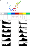
Fig 1
Fig 1. Transcription factor gene expression during stimulation of human monocyte derived macrophages by LPS.
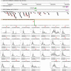
A. Sample-based network of transcription factor gene expression. Gene-based expression levels for all of the annotated transcription factors in the human genome [34] were extracted from the FANTOM5 data and averaged. Only transcription factor genes where at least one value (averaged across three replicates) was ≥ 20 TPM were included in the analysis. The results were entered into BioLayout Express3D to create a sample-to-sample network layout where the nodes (spheres) represent a single time point after stimulation with LPS (averaged over three replicates). Edges (lines between nodes) represent sample-to-sample correlations in overall expression pattern of transcription factor genes of R ≥ 0.90. The time points are group and color-coded as indicated, showing the clear progressive change in the transcriptional profile with time. B. Representative expression profiles for genes expressed at the different time points. Clusters of co-regulated transcription factor genes were identified based upon gene-to-gene network analysis with BioLayout Express3D (R ≥ 0.75; MCL inflation value 2.2) as described in Methods. The X axis shows the time points (in the order 0 min, 15 min, 30 min, 45 min, 1 hour, 1 hour 20 min, 1 hour 40 min, 2 hours, 2 hours 30min, 3 hours, 3 hours 30 min, 4 hours, 5 hours, 6 hours, 7 hours, 8 hours, 10 hours, 12 hours, 14 hours, 16 hours, 18 hours, 20 hours, 22 hours, 24 hours, 36 hours, 48 hours). Y axis shows the mean TPM value for the set of genes with that profile (averaged across three replicates). Note that each cluster has a characteristic temporal profile. The complete set of clusters and the names of the transcription factor genes within them is provided in S2 Table.
Fig 2
Fig 2. Transcript expression in human macrophages exposed to LPS.
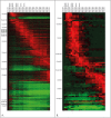
Heatmaps of transcript expression in human macrophages exposed to LPS depicting change from baseline, normalized to the maximum expression of each transcript. Red = increase from baseline, green = decrease from baseline. Only nodes included in a coexpression cluster are shown here. (A) All clustered transcripts in LPS-treated human monocyte-derived macrophages. Clusters containing more than 50 transcripts are labeled. (B) Transcripts contained within the clusters shown in (A) that initiate near a putative Crohn’s disease-associated SNP (p<10e-6, window size to transcription start site = 2000 bases).
Fig 3
Fig 3. The time course of activation of enhancers and promoter at the IL6 locus.
The core panel shows a genome browser view of the IL6 locus with the locations of FANTOM5 enhancers. The upper right panel shows the time course of induction of IL6 mRNA, detected by CAGE, which peaks around 3–4 hours and declines by 12 hours. The lower panels show the transient activity of the enhancers indicated, the majority of which peak around 1–2 hours and decline rapidly. Data are expressed at TPM, and are the average of the three replicates.
Fig 4
Fig 4. The time course of activation of enhancers and promoters at the CCL3/CLL4/CCL18 locus.
The core panel shows a genome browser view of the locus with the locations of FANTOM5 enhancers. The upper panel show the time course of induction of each of the mRNAs, detected by CAGE. Whereas CCL3 and CCL4 are coordinately-regulated, CCL18 follows a much slower time course and is still rising at 48 hours. The lower panels show the activity of the enhancers indicated. The lowest track shows the histograms of CAGE tags mapped to the region, with colours indicating direction of transcription; green to the right and purple to the left. Note that the entire regions shows evidence of bidirectional transcription initiation. Data are expressed as TPM, and are the average of the three replicates.
Fig 5
Fig 5. The time course of activation of enhancers and promoter at the TNFAIP3 locus.
The core panel shows a genome browser view of the TNFAIP3 locus with the locations of FANTOM5 enhancers. The lower right panel (p1@TNFAIP3) shows the time course of induction of TNFAIP3 mRNA, detected by CAGE, which peaks around 2 hours and declines to a new, elevated steady state by 8 hours. Other panels show the transient activity of the enhancers indicated, the majority of which peak around 1–2 hours and decline rapidly. Panel at bottom right shows the activity of the enhancer containing the SNP originally associated with CD susceptibility, 185kb upstream of the TNFAIP3 locus [2]. Data are expressed as TPM, and are the average of the three replicates.
Fig 6
Fig 6. Motif activity response analysis (MARA) of the response to LPS
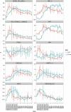
MARA analysis of promoters and enhancers was carried out as described in Materials and Methods. MARA averages the expression of all promoters (blue) and enhancers (red) that contain the consensus transcription factor binding motif. Activities are plotted as Z-scores (averages of activity divided by standard deviation) in each time point, with error bars representing the standard deviation of Z-scores.
Fig 7
Fig 7. Enrichment of macrophage-expressed or regulated genes in genomic regions associated with IBD susceptibility
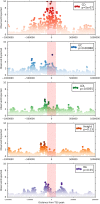
Each panel shows the enrichment of GWAS-associated variants in vicinity of genes meeting the expression criteria (specifically inducible in monocytes AND down regulated during differentiation to macrophages) for each of the traits/diseases shown. For 1000 bins spanning a region of 1Mb above and below all TSS meeting expression criteria, observed:expected ratios were calculated as the ratio of the absolute count of variants with p<1e06 for association with each trait, to the absolute count of all SNPs genotyped in the same study. p-values for enrichment were calculated for the whole set using PASCAL (see Methods).

V体育2025版 - References

    1. Kostic AD, Xavier RJ, Gevers D (2014) The microbiome in inflammatory bowel disease: current status and the future ahead. Gastroenterology 146: 1489–1499. 10.1053/j.gastro.2014.02.009 - DOI - PMC - PubMed
    1. Ellinghaus D, Jostins L, Spain SL, Cortes A, Bethune J, et al. (2016) Analysis of five chronic inflammatory diseases identifies 27 new associations and highlights disease-specific patterns at shared loci. Nat Genet 48: 510–518. 10.1038/ng.3528 - DOI - PMC - PubMed
    1. Richard-Miceli C, Criswell LA (2012) Emerging patterns of genetic overlap across autoimmune disorders. Genome Med 4: 6 10.1186/gm305 - DOI - PMC - PubMed
    1. Jostins L, Ripke S, Weersma RK, Duerr RH, McGovern DP, et al. (2012) Host-microbe interactions have shaped the genetic architecture of inflammatory bowel disease. Nature 491: 119–124. 10.1038/nature11582 - DOI (VSports手机版) - PMC - PubMed
    1. Liu JZ, van Sommeren S, Huang H, Ng SC, Alberts R, et al. (2015) Association analyses identify 38 susceptibility loci for inflammatory bowel disease and highlight shared genetic risk across populations. Nat Genet 47: 979–986. 10.1038/ng.3359 - DOI - PMC - PubMed

Publication types

MeSH terms

Substances