<small lang="w36eR7e"></small> <b dropzone="3k5il"></b> V体育官网 - Skip to main page content
U.S. flag

An official website of the United States government

Dot gov

The . gov means it’s official VSports app下载. Federal government websites often end in . gov or . mil. Before sharing sensitive information, make sure you’re on a federal government site. .

Https

The site is secure. The https:// ensures that you are connecting to the official website and that any information you provide is encrypted and transmitted securely. V体育官网.

. 2017 Apr;11(4):422-437.
doi: 10.1002/1878-0261.12045. Epub 2017 Mar 17.

ZKSCAN1 gene and its related circular RNA (circZKSCAN1) both inhibit hepatocellular carcinoma cell growth, migration, and invasion but through different signaling pathways

Affiliations

V体育ios版 - ZKSCAN1 gene and its related circular RNA (circZKSCAN1) both inhibit hepatocellular carcinoma cell growth, migration, and invasion but through different signaling pathways

Zhicheng Yao et al. Mol Oncol. 2017 Apr.

Erratum in (VSports注册入口)

Abstract (VSports)

There is increasing evidence that circular RNA (circRNA) are involved in cancer development, but the regulation and function of human circRNA remain largely unknown. In this study, we demonstrated that ZKSCAN1, a zinc finger family gene, is expressed in both linear and circular (circZKSCAN1) forms of RNA in human hepatocellular carcinoma (HCC) tissues and cell lines. Here, we analyzed a cohort of 102 patients and found that expression of both ZKSCAN1mRNA and circZKSCAN1 was significantly lower (P < 0. 05) in the HCC samples compared with that in matched adjacent nontumorous tissues by reverse transcription PCR (RT-PCR). The low expression level of ZKSCAN1 was only associated with tumor size (P = 0. 032), while the cirZKSCAN1 levels varied in patients with different tumor numbers (P < 0 VSports手机版. 01), cirrhosis (P = 0. 031), vascular invasion (P = 0. 002), or microscopic vascular invasion (P = 0. 002), as well as with the tumor grade (P < 0. 001). Silencing both ZKSCAN1mRNA and circZKSCAN1 promoted cell proliferation, migration, and invasion. In contrast, overexpression of both forms of RNA repressed HCC progression in vivo and in vitro. Silencing or overexpression of both forms of RNA did not interfere with each other. RNA-seq revealed a very different molecular basis for the observed effects; ZKSCAN1mRNA mainly regulated cellular metabolism, while circZKSCAN1 mediated several cancer-related signaling pathways, suggesting a nonredundant role for ZKSCAN1mRNA and circRNA. In conclusion, our results revealed two post-translational products (ZKSCAN1mRNA and circZKSCAN1) that cooperated closely with one another to inhibit growth, migration, and invasion of HCC. cirZKSCAN1 might be a useful marker for the diagnosis of HCC. .

Keywords: HCC; RNA-seq; ZKSCAN1; circular RNA; qRT-PCR. V体育安卓版.


"V体育安卓版" Figures

Figure 1
Figure 1
The mRNA and protein expression levels of ZKSCAN1, and circZKSCAN1 validation in human HCC tissues. (A) Western blot and RTPCR analysis of the expression level of ZKSCAN1 in three randomly selected hepatocellular carcinoma specimens and the paired adjacent nontumorous tissues. GAPDH was used as the reference in both analyses. N: adjacent nontumorous tissue; T: hepatocellular carcinoma tissue. (B) Divergent primers were used to amplify circular RNA in cDNA but not genomic DNA (gDNA). Convergent primers can amplify both circular RNA and linear RNA, GAPDH, and the linear control. (C) Sanger sequencing of a PCR product resulting from divergent primers demonstrating the head‐to‐tail splicing of this exon. (D) The predicted circular RNA is resistant to RNase R treatment. **P <0.01, in contrast to the linear control gene GAPDH, the circular RNA was resistant to RNase R treatment.
Figure 2
Figure 2
qRTPCR analysis of the expression of ZKSCAN1 and circZKSCAN1 and the ROC curves of ZKSCAN1 and circZKSCAN1. (A) The expression levels of circZKSCAN1 in each patient were significantly lower than those in the corresponding adjacent nontumorous tissues. < 0.001. N: adjacent nontumorous tissue; T: hepatocellular carcinoma tissue. (B) The expression levels of ZKSCAN1 in each patient were significantly lower than those in the corresponding nontumorous tissues. *< 0.05. N: adjacent nontumorous tissue; T: hepatocellular carcinoma tissue. (C) Comparisons of the ROC curves of circZKSCAN1,ZKSCAN1.
Figure 3
Figure 3
qRTPCR analysis of the expression of ZKSCAN1 and circZKSCAN1 with and/or without interferences in human HCC cell lines. (A) The expression levels of ZKSCAN1 in five HCC cell lines (Huh7 cell line, SMMC‐7721 cell line, BEL‐7402 cell line, HepG2 cell line, and Hep3B cell line) were significantly lower than those in the human normal hepatic L02 cell line. **< 0.001. (B) The expression levels of circZKSCAN1 in five HCC cell lines (Huh7 cell line, SMMC‐7721 cell line, BEL‐7402 cell line, Hep3B cell line, and HepG2 cell line) were significantly lower than those in the L02 cell line. **< 0.001. (C) The expression levels of ZKSCAN1 and circZKSCAN1 in the control, overexpression, and knockdown of circZKSCAN1 or ZKSCAN1 SMMC‐7721 cell line. (D) The expression levels of ZKSCAN1 and circZKSCAN1 in the control, overexpression, and knockdown of circZKSCAN1 or ZKSCAN1 in the HepG2 cell line.
Figure 4
Figure 4
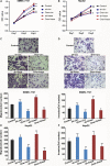
Cell proliferation, invasion, and migration of the SMMC‐7721 cell line and HepG2 cell line following the overexpression and knockdown of circZKSCAN1 or ZKSCAN1. (A) MTS method examining SMMC‐7721 and HepG2 (B) cell proliferation of five different treatments on days 1, 2, and 3. (C) Migration assay and invasion assay (D) of SMMC‐7721 cells. Migration assay (E) and invasion assay (F) of HepG2 cells transfected with different treatments. Images are representative of the cells invading one field. **< 0.001. Control: negative control; sh‐circ: knockdown treatment of circZKSCAN1; over‐circ: overexpression treatment of circZKSCAN1; sh‐linear: knockdown treatment of ZKSCAN1; over‐linear: overexpression treatment of ZKSCAN1.
Figure 5
Figure 5
Liver xenografts in each group at 5 weeks after subcutaneous implantation of five different SMMC‐7721 cells. The mean tumor volumes and weight in five nude mice in each group are shown at different time points (right). **< 0.001 compared with the controls. Control: negative control; sh‐circ: knockdown treatment of circZKSCAN1; over‐circ: overexpression treatment of circZKSCAN1; sh‐linear: knockdown treatment of ZKSCAN1; over‐linear: overexpression treatment of ZKSCAN1.
Figure 6
Figure 6
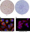
Cellular localization of ZKSCAN1 and circZKSCAN1. (A) Immunohistochemistry analysis of ZKSCAN1 in the normal (N) and HCC tissues (T). (B) FISH analysis of circZKSCAN1 cellular localization in the Human Protein Atlas Database (http://www.proteinatlas.org/). (C) FISH analysis of the cellular localization of circZKSCAN1 in this study.
Figure 7
Figure 7
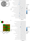
Differentially expressed genes in SMMC‐7721 cells after knockdown of circZKSCAN1 and ZKSCAN1. (A) Venn diagram of the number of differentially expressed genes identified in this study. (B) Heat map analysis of the number of differentially expressed genes identified in this study. Red indicates up‐regulated expression; green indicates down‐regulated expression. Scatter diagram of the enriched KEGG pathways in (C) knockdown of circZKSCAN1 and (D) ZKSCAN1 cells showing differentially expressed genes. The degree of enrichment was measured according to the Rich factor, Q‐value, and the number of genes that were enriched in one pathway. The Rich factor is the ratio between the number of differentially expressed genes enriched in one pathway and the number of GO annotations. The greater the value of the Rich factor, the higher the degree of enrichment. The Q‐value is a variant of the P‐value, for which lower numbers equate to significant enrichment. The y‐axis shows the name of the pathway, and the x‐axis shows the Rich factor. The point size indicates the number of differentially expressed genes in one pathway, and the color of the point denotes the range of the Q‐value.
Figure 8
Figure 8
qRTPCR analysis of the expression of 21 genes selected from the RNA‐seq results. Control: normal SMMC‐7721 cell line; sh‐circ: knockdown treatment of circZKSCAN1 in the SMMC‐7721 cell line; over‐circ: overexpression treatment of circZKSCAN1 in the SMMC‐7721 cell line; sh‐linear: knockdown treatment of ZKSCAN1 in the SMMC‐7721 cell line; over‐linear: overexpression treatment of ZKSCAN1 in the SMMC‐7721 cell line. The y‐axis shows the gene expression levels after normalization to the reference gene GAPDH. *P <0.05, compared with NC.

References

    1. Audic S and Claverie JM (1997) The significance of digital gene expression profiles. Genome Res 7, 986–995. - PubMed
    1. Bartel DP (2009) MicroRNAs: target recognition and regulatory functions. Cell 136, 215–233. - PMC - PubMed
    1. Burd CE, Jeck WR, Liu Y, Sanoff HK, Wang Z, Sharpless NE (2010) Expression of linear and novel circular forms of an INK4/ARF‐associated non‐coding RNA correlates with atherosclerosis risk. PLoS Genet 6, e1001233. - "V体育安卓版" PMC - PubMed
    1. Chen YT and Old LJ (1990) Cerebellar degeneration‐related antigen: a highly conserved neuroectodermal marker mapped to chromosomes X in human and mouse. Proc Natl Acad Sci U S A 87, 3077–3081. - PMC - PubMed
    1. van Dekken H, Tilanus HW, Hop WC, Dinjens WN, Wink JC, Vissers KJ and van Marion R (2009) Array comparative genomic hybridization, expression array, and protein analysis of critical regions on chromosome arms 1q, 7q, and 8p in adenocarcinomas of the gastroesophageal junction. Cancer Genet Cytogenet 189, 37–42. - PubMed

MeSH terms