Skip to main page content
U.S. flag

An official website of the United States government

Dot gov

The . gov means it’s official. Federal government websites often end in . gov or . mil. Before sharing sensitive information, make sure you’re on a federal government site VSports app下载. .

Https

The site is secure V体育官网. The https:// ensures that you are connecting to the official website and that any information you provide is encrypted and transmitted securely. .

. 2017 Feb 10;18(2):378.
doi: 10.3390/ijms18020378.

Photoluminescent Gold Nanoclusters in Cancer Cells: Cellular Uptake, Toxicity, and Generation of Reactive Oxygen Species

Affiliations

VSports注册入口 - Photoluminescent Gold Nanoclusters in Cancer Cells: Cellular Uptake, Toxicity, and Generation of Reactive Oxygen Species

Marija Matulionyte (V体育平台登录) et al. Int J Mol Sci. .

"V体育官网入口" Abstract

In recent years, photoluminescent gold nanoclusters have attracted considerable interest in both fundamental biomedical research and practical applications. Due to their ultrasmall size, unique molecule-like optical properties, and facile synthesis gold nanoclusters have been considered very promising photoluminescent agents for biosensing, bioimaging, and targeted therapy. Yet, interaction of such ultra-small nanoclusters with cells and other biological objects remains poorly understood. Therefore, the assessment of the biocompatibility and potential toxicity of gold nanoclusters is of major importance before their clinical application. In this study, the cellular uptake, cytotoxicity, and intracellular generation of reactive oxygen species (ROS) of bovine serum albumin-encapsulated (BSA-Au NCs) and 2-(N-morpholino) ethanesulfonic acid (MES)capped photoluminescent gold nanoclusters (Au-MES NCs) were investigated VSports手机版. The results showed that BSA-Au NCs accumulate in cells in a similar manner as BSA alone, indicating an endocytotic uptake mechanism while ultrasmall Au-MES NCs were distributed homogeneously throughout the whole cell volume including cell nucleus. The cytotoxicity of BSA-Au NCs was negligible, demonstrating good biocompatibility of such BSA-protected Au NCs. In contrast, possibly due to ultrasmall size and thin coating layer, Au-MES NCs exhibited exposure time-dependent high cytotoxicity and higher reactivity which led to highly increased generation of reactive oxygen species. The results demonstrate the importance of the coating layer to biocompatibility and toxicity of ultrasmall photoluminescent gold nanoclusters. .

Keywords: accumulation; breast cancer cells; gold nanoclusters; photoluminescence; reactive oxygen species; toxicity V体育安卓版. .


Conflict of interest statement

The authors declare no conflict of interest.

Figures

Figure 1
Figure 1
Normalized absorption, photoluminescence (PL) and photoluminescence excitation spectra of 2-(N-morpholino) ethanesulfonic acid (MES)-capped photoluminescent gold nanoclusters (Au-MES NCs) (top) and bovine serum albumin-encapsulated gold nanoclusters (BSA-Au NCs) (bottom) in deionized water.
Figure 2
Figure 2
Accumulation of photoluminescent BSA-Au NCs (λex = 488 nm) (A1,A2,C1,C2) and BSA-Alexa conjugate (λex = 488 nm) (B1,B2,D1,D2) in MCF-7 and MDA-MB-231 cells after 24 h of incubation, nuclei stained with Hoechst 33258 (λex = 405 nm). Scale bar is 30 μm.
Figure 3
Figure 3
Accumulation of photoluminescent BSA-Au NCs and BSA-Alexa conjugate in MCF-7 and MDA-MB-231 cells after 3, 6, and 24 h of incubation. Percentage of the cells that have accumulated BSA-Au NCs and BSA-Alexa conjugate (A); mean PL intensity (MPI) of BSA-Au NCs and BSA-Alexa conjugate per cell (B). Control represents autofluorescence of non-treated cells. Error bars show the standard deviations. * indicates significant differences between accumulation of BSA-Alexa 488 and BSA-Au NCs (p ≤ 0.05); # indicates significant differences between the MCF-7 and MDA-MB-231 cell lines (p ≤ 0.05).
Figure 4
Figure 4
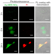
Accumulation of photoluminescent Au-MES NCs (λex = 405 nm) in MCF-7 breast cancer cells after 3, 6, and 24 h of incubation (green photoluminescence). Red fluorescence represents propidium iodide (PI) stained non-viable cells (λex = 488 nm). Yellow color in the merged pictures presents overlap of photoluminescence of Au-MES NCs and fluorescence of propidium iodide. Scale bar is 15 μm.
Figure 5
Figure 5
Accumulation of photoluminescent BSA-Au NCs (λex = 488 nm) (A1,A2), BSA-Alexa 488 conjugate (λex = 488 nm); (B1,B2), and photoluminescent Au-MES NCs; (C1,C2) in MDA-MB-231 cells. Cells were incubated with BSA-Au NCs and BSA-Alexa 488 conjugate for 24 h, with Au-MES NCs—for 6 h. In (A1,A2,B1,B2), nuclei were stained with Hoechst 33258 (λex = 405 nm). Scale bar is 25 μm.
Figure 6
Figure 6
Intracellular distribution of photoluminescent BSA-Au NCs in early endosomes (EE), late endosomes (LE) and lysosomes (Lys). Yellow colour represents co-localisation of green fluorescent protein (GFP) labeled endosomal compartments and accumulated BSA-Au NCs. Scale bar is 10 μm.
Figure 7
Figure 7
Cell viability of MCF-7 and MDA-MB-231 cells incubated with BSA-Au NCs, BSA, Au-MES NCs, and MES solutions. Error bars show the standard deviations. * indicates significant differences compared to the non-treated cells (Control) (p ≤ 0.05); # indicates significant differences between the MCF-7 and MDA-MB-231 cell lines (p ≤ 0.05).
Figure 8
Figure 8
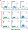
Apoptosis analysis of MDA-MB-231 cells after incubation with Au-MES NCs for 3 and 6 h (A); and BSA and BSA-Au NCs for 24 and 48 h (B) by flow cytometry, using Annexin V/ PI apoptosis assay. Only PI positive population (Q1) represents necrotic cells whereas only Annexin V positive population (Q3) are early apoptotic cells. Cells in late apoptosis take up both dyes (Q2). Q4 represent live cells. In the case of cells treated with BSA-Au NCs, population Q1 displays BSA-Au NCs positive cells.
Figure 9
Figure 9
Reactive oxygen species (ROS) generation in MCF-7 and MDA-MB-231 cancer cells after treatment with BSA-Au NCs, Au-MES NCs, BSA, and MES. The data normalized according to the non-treated cells (Control), tert-Butyl hydroperoxide (TBHP) treated cells were taken as a positive control. Error bars show the standard deviations. * indicates significant differences compared to the non-treated cells (p ≤ 0.05); # indicates significant differences between the MCF-7 and MDA-MB-231 cell lines (p ≤ 0.05).

References

    1. Liu J.L., Lu L.L., Xu S.Y., Wang L.Y. One-pot synthesis of gold nanoclusters with bright red fluorescence and good biorecognition abilities for visualization fluorescence enhancement detection of E. coli. Talanta. 2015;134:54–59. doi: 10.1016/j.talanta.2014.10.058. - DOI - PubMed
    1. Chen T.H., Tseng W.L. (Lysozyme type VI)-stabilized Au8 clusters: Synthesis mechanism and application for sensing of glutathione in a single drop of blood. Small. 2012;8:1912–1919. doi: 10.1002/smll.201102741. - DOI - PubMed
    1. Chen L.Y., Wang C.W., Yuan Z.Q., Chang H.T. Fluorescent gold nanoclusters: Recent advances in sensing and imaging. Anal. Chem. 2015;87:216–229. doi: 10.1021/ac503636j. - DOI - PubMed
    1. Shang L., Stockmar F., Azadfar N., Nienhaus G.U. Intracellular thermometry by using fluorescent gold nanoclusters. Angew. Chem. Int. Edit. 2013;52:11154–11157. doi: 10.1002/anie.201306366. - DOI - PubMed
    1. Chen D.Y., Luo Z.T., Li N.J., Lee J.Y., Xie J.P., Lu J.M. Amphiphilic polymeric nanocarriers with luminescent gold nanoclusters for concurrent bioimaging and controlled drug release. Adv. Funct. Mater. 2013;23:4324–4331. doi: 10.1002/adfm.201300411. - DOI

MeSH terms

"V体育官网入口" LinkOut - more resources