"V体育安卓版" Skip to main page content
U.S. flag

An official website of the United States government

Dot gov

The . gov means it’s official. Federal government websites often end in . gov or VSports app下载. mil. Before sharing sensitive information, make sure you’re on a federal government site. .

Https

The site is secure. The https:// ensures that you are connecting to the official website and that any information you provide is encrypted and transmitted securely. V体育官网.

. 2017 May 1;77(9):2512-2521.
doi: 10.1158/0008-5472.CAN-16-3242. Epub 2017 Feb 15.

"V体育官网入口" Mcl-1 Degradation Is Required for Targeted Therapeutics to Eradicate Colon Cancer Cells

Affiliations

Mcl-1 Degradation Is Required for Targeted Therapeutics to Eradicate Colon Cancer Cells

Jingshan Tong et al. Cancer Res. .

Abstract (VSports)

The Bcl-2 family protein Mcl-1 is often degraded in cancer cells subjected to effective therapeutic treatment, and defective Mcl-1 degradation has been associated with intrinsic and acquired drug resistance. However, a causal relationship between Mcl-1 degradation and anticancer drug responses has not been directly established, especially in solid tumor cells where Mcl-1 inhibition alone is insufficient to trigger cell death VSports手机版. In this study, we present evidence that Mcl-1 participates directly in determining effective therapeutic responses in colon cancer cells. In this setting, Mcl-1 degradation was induced by a variety of multikinase inhibitor drugs, where it relied upon GSK3β phosphorylation and FBW7-dependent ubiquitination. Specific blockade by genetic knock-in (KI) abolished apoptotic responses and conferred resistance to kinase inhibitors. Mcl-1-KI also suppressed the antiangiogenic and anti-hypoxic effects of kinase inhibitors in the tumor microenvironment. Interestingly, these same inhibitors also induced the BH3-only Bcl-2 family protein PUMA, which is required for apoptosis. Degradation-resistant Mcl-1 bound and sequestered PUMA from other prosurvival proteins to maintain cell survival, which was abolished by small-molecule Mcl-1 inhibitors. Our findings establish a pivotal role for Mcl-1 degradation in the response of colon cancer cells to targeted therapeutics, and they provide a useful rational platform to develop Mcl-1-targeting agents that can overcome drug resistance. Cancer Res; 77(9); 2512-21. ©2017 AACR. .


Conflict of interest statement

Conflict of interest: The authors have no conflict of interest.

Disclosure of Potential Conflicts of Interest

No potential conflicts of interest were disclosed.

Figures

Figure 1
Figure 1. Induction of Mcl-1 proteasomal degradation by inhibition of oncogenic kinase signaling
(A) Western blotting of Mcl-1 in HCT116 colon cancer cells treated for 24 hr with 10 nM TRAIL, 20 μM etoposide, 120 μM sulindac sulfide (sulindac), 20 μM sorafenib, 40 μM regorafenib, 1 μM UCN-01, 15 μM sunitinib, or 10 μM roscovitine. (B) Western blotting of Mcl-1 in HCT116 cells treated with 40 μM regorafenib or 20 μM sorafenib at indicated time points. (C) Western blotting of Mcl-1 in indicated colon cancer cell lines treated with 40 μM regorafenib for 24 hr. (D) Western blotting of Mcl-1 in HCT116 cells treated with control vehicle or 40 μM regorafenib for 1 hr, and then with 10 μg/ml of the translation inhibitor cyclohemide (CHX) at indicated time points. (E) Western blotting of Mcl-1 in HCT116 cells treated with 40 μM regorafenib or 20 μM sorafenib for 4 hr, with or without pretreatment with 5 μM MG132 for 30 min.
Figure 2
Figure 2. GSK3β-dependent Mcl-1 phosphorylation mediates its interaction with FBW7 and proteasomal degradation
(A) Western blotting of total and phosphorylated Mcl-1 (p-Mcl-1; Ser159/Thr163) in HCT116 cells treated with 40 μM regorafenib at indicated time points. (B) HCT116 cells pretreated with 5 μM of the proteasome inhibitor MG132 for 30 min were treated with 40 μM regorafenib for 24 hr. Immunoprecipitation (IP) was performed to pull down Mcl-1, followed by western blotting of indicated proteins. (C) WT and FBW7-KO HCT116 cells transfected with HA-ubiquitin and pretreated with 5 μM MG132 for 30 min were treated with 40 μM regorafenib for 4 hr. IP was performed to pull down Mcl-1, followed by western blotting of indicated proteins. (D) Western blotting of total and phosphorylated GSK3 (p-GSK3β; Ser9) and ERK (p-ERK; Thr202/Tyr204) in HCT116 cells treated with 40 μM regorafenib at indicated time points. (E) HCT116 cells pretreated with 5 μM MG132 for 30 min were treated with 40 μM regorafenib with or without the GSK3 inhibitor SB216736 for 24 hr. IP was performed to pull down Mcl-1, followed by western blotting of indicated proteins. (F) HCT116 cells transfected with HA-ubiquitin and pretreated with 5 μM of MG132 for 30 min were treated with 40 μM regorafenib with or without SB216736 for 4 hr. IP was performed to pull down Mcl-1, followed by western blotting of indicated proteins. (G) Western blotting of indicated proteins in HCT116 cells treated with 40 μM regorafenib with or without SB216736 for 48 hr. (H) Western blotting of indicated proteins in HCT116 cells treated with 25 μM of the ERK inhibitor PD98059 for 24 hr. (I) Western blotting of Mcl-1 in HCT116 cells with siRNA knockdown of CRAF, which was verified by RT-PCR.
Figure 3
Figure 3. Selectively blocking Mcl-1 phosphorylation by knock-in of a phosphorylation site mutant suppresses Mcl-1 degradation
(A) Schematic representation of the Mcl-1 genomic locus and the knock-in vector highlighting the putative phosphorylation sites of Mcl-1 that are involved in its degradation. (B) DNA sequencing of the targeted genomic regions in WT and Mcl-1 knock-in (Mcl-1-KI) HCT116 cells highlighting WT and corresponding mutant sequences. (C) Western blotting of Mcl-1 in untreated WT and two independent Mcl-1-KI cell lines. (D) Mcl-1 localization was analyzed by western blotting of mitochondrial and cytosolic fractions isolated from indicated cells. β-Actin and cytochrome oxidase subunit IV (COX IV) were used as a control for loading and fractionation. (E) Western blotting of Mcl-1 in WT and Mcl-1-KI cells treated with control vehicle or 40 μM regorafenib for 1 hr, and then with 10 μg/ml of the translation inhibitor cyclohemide (CHX) at indicated time points. (F) Western blotting of Mcl-1 in WT and Mcl-1-KI HCT116 cells treated with 40 μM regorafenib at indicated time points. (G) WT and Mcl-1-KI HCT116 cells pretreated with 5 μM MG132 for 30 min were treated with 40 μM regorafenib for 24 hr. IP was used to pull down Mcl-1, followed by western blotting of indicated proteins. (H) WT and Mcl-1-KI HCT116 cells transfected with HA-ubiquitin and pretreated with 5 μM MG132 for 30 min were treated with 5 μM regorafenib for 4 hr. IP was used to pull down Mcl-1, followed by western blotting of indicated proteins.
Figure 4
Figure 4. Blocking Mcl-1 degradation renders resistance to regorafenib and sorafenib by blocking the mitochondrial apoptotic pathway
(A) MTS analysis of viability of WT, Mcl-1-KI, and Mcl-1-KI with knockdown of Mcl-1 HCT116 cells treated with regorafenib or sorafenib at different concentrations for 72 hr. (B) Colony formation assay was done by seeding an equal number of WT and Mcl-1-KI HCT116 cells treated with 40 μM regorafenib or 20 μM sorafenib for 48 hr in 12-well plates, and staining of the attached cells with crystal violet after 14 days. Left, representative pictures of colonies; right, enumeration of colony numbers. ***, P < 0.001. (C) Western blotting of Mcl-1 and cleaved (C) caspases 3, 8 and 9 in WT and Mcl-1-KI HCT116 cells treated with 40 μM regorafenib or 20 μM sorafenib for 48 hr. (D) Cytochrome c release in cells treated with 40 μM regorafenib or 20 μM sorafenib was analyzed by western blotting of mitochondrial or cytosolic fractions isolated from treated cells. β-Actin and cytochrome oxidase subunit IV (COX IV) were used as a control for loading and fractionation, respectively. (E) Bax conformational change was detected by immunoprecipitation (IP) with anti-Bax 6A7 (activated) antibody followed by western blotting. (F) Bax multimerization in isolated mitochondria was analyzed by western blotting under non-denaturing conditions following DSP cross-link.
Figure 5
Figure 5. Mcl-1 degradation mediates sensitivity of CRC cells to kinase inhibitors through PUMA-dependent apoptosis
(A) Western blotting of Mcl-1 in WT and Mcl-1-KI HCT116 cells treated with indicated agents for 24 hr. (B) Apoptosis in WT and Mcl-1-KI HCT116 cells treated with indicated agents for 48 hr. (C) Western blotting of Mcl-1 and PUMA in HCT116 cells treated with indicated agents for 24 hr. (D) WT and Mcl-1-KI HCT116 cells were treated with 40 μM regorafenib for 24 hr. IP was performed to pull down Mcl-1, followed by western blotting of indicated proteins. (E) WT and Mcl-1-KI HCT116 cells transfected with V5-Bcl-XL were treated with 40 μM regorafenib for 24 hr. IP was performed to pull down V5 (Bcl-XL), followed by western blotting of indicated proteins. (F) Apoptosis in WT and Mcl-1-KI HCT116 cells treated with indicated agents with or without a combination with 5 μM UMI-77 or TW-37. (G) Western blotting of cleaved (C) caspase 3 in WT and Mcl-1-KI HCT116 cells treated as in (F). (H) Apoptosis in WT and Mcl-1-KI HCT116 cells infected with a low dose of adenovirus expressing full-length (Ad-PUMA) or BH3 domain-deleted PUMA (ΔBH3), and then treated with indicated agents. (I) Western blotting of cleaved (C) caspase 3 in WT and Mcl-1-KI HCT116 cells treated as in (H). In (A)-(H): UCN-01, 1 μM; sunitinib 15 μM; roscovitine, 10 μM; TRAIL, 10 nM; etoposide, 20 μM; sulindac sulfide, 120 μM. In (B), (F) and (H), apoptosis was analyzed by counting cells with condensed and fragmented nuclei after nuclear staining with Hoechst 33258.
Figure 6
Figure 6. Mcl-1 degradation contributes to the in vivo antitumor activity of regorafenib
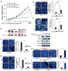
(A) Nude mice were injected s.c. with 4 × 10⁶ WT or Mcl-1-KI HCT116 cells. After 1 week, mice were treated with 30 mg/kg regorafenib daily by oral gavage (indicated by arrows), or the vehicle control cremephor EL/ethanol for 10 consecutive days. Tumor volume at indicated time points after treatment was calculated and plotted with statistical significance for indicated comparisons (n=7 in each group). (B) Representative tumors at the end of the experiment. (C–F) Nude mice with WT or Mcl-1-KI HCT116 tumors were treated with regorafenib as in (A) for 4 consecutive days. Tissue sections were analyzed by staining for TUNEL (C), active caspase 3 (D), CD31 (E), and Carbonic Anhydrase 9 (CA9) (F). Left, representative staining pictures; right, quantification of positive cells. (G) Western blotting of indicated proteins in WT and Mcl-1-KI HCT116 tumors from (C). (H) Tissue sections from WT HCT116 xenograft tumors with or without regorafenib treatment as in (A) for 1 day were analyzed by proximity ligation assay (PLA) to detect the interaction of Mcl-1 and FBW7. Left, representative PLA pictures, with a section without primary antibody staining as a negative control; right, quantification of PLA signals per field. (I) Tissue sections from WT and Mcl-1-KI HCT116 xenograft tumors treated with regorafenib as in (A) for 1 day were analyzed by PLA for the interaction of Mcl-1 and PUMA. Left, representative PLA pictures, with a section without primary antibody staining as a negative control; right, quantification of PLA signals per field. In (C), (D), (E), (F), (H), and (I), results were expressed as means ± s.d. of three independent experiments. Arrows indicate example cells with positive staining. Scale bars, 25 μm. NS, P >0.05, *, P <0.05, **, P <0.01, ***, P <0.001.

References

    1. Moldoveanu T, Follis AV, Kriwacki RW, Green DR. Many players in BCL-2 family affairs. Trends in biochemical sciences. 2014;39:101–11. - PMC - PubMed
    1. Bhola PD, Letai A. Mitochondria-Judges and Executioners of Cell Death Sentences. Mol Cell. 2016;61:695–704. - PMC (VSports app下载) - PubMed
    1. Thomas LW, Lam C, Edwards SW. Mcl-1; the molecular regulation of protein function. FEBS Lett. 2010;584:2981–9. - PubMed
    1. Perciavalle RM, Opferman JT. Delving deeper: MCL-1’s contributions to normal and cancer biology. Trends in cell biology. 2013;23:22–9. - PMC - PubMed
    1. Mojsa B, Lassot I, Desagher S. Mcl-1 ubiquitination: unique regulation of an essential survival protein. Cells. 2014;3:418–37. - PMC - PubMed

V体育平台登录 - Publication types

MeSH terms

VSports注册入口 - Substances

LinkOut - more resources