Skip to main page content
U.S. flag

An official website of the United States government

Dot gov

The . gov means it’s official. Federal government websites often end in VSports app下载. gov or . mil. Before sharing sensitive information, make sure you’re on a federal government site. .

Https

The site is secure. The https:// ensures that you are connecting to the official website and that any information you provide is encrypted and transmitted securely. V体育官网.

. 2017 Feb 28;114(9):E1698-E1706.
doi: 10.1073/pnas.1615771114. Epub 2017 Feb 13.

Galectin-3 directs antimicrobial guanylate binding proteins to vacuoles furnished with bacterial secretion systems

Affiliations

"V体育2025版" Galectin-3 directs antimicrobial guanylate binding proteins to vacuoles furnished with bacterial secretion systems

Eric M Feeley et al. Proc Natl Acad Sci U S A. .

Abstract

Many invasive bacteria establish pathogen-containing vacuoles (PVs) as intracellular niches for microbial growth. Immunity to these infections is dependent on the ability of host cells to recognize PVs as targets for host defense. The delivery of several host defense proteins to PVs is controlled by IFN-inducible guanylate binding proteins (GBPs), which themselves dock to PVs through poorly characterized mechanisms. Here, we demonstrate that GBPs detect the presence of bacterial protein secretion systems as "patterns of pathogenesis" associated with PVs. We report that the delivery of GBP2 to Legionella-containing vacuoles is dependent on the bacterial Dot/Icm secretion system, whereas the delivery of GBP2 to Yersinia-containing vacuoles (YCVs) requires hypersecretion of Yersinia translocon proteins. We show that the presence of bacterial secretion systems directs cytosolic carbohydrate-binding protein Galectin-3 to PVs and that the delivery of GBP1 and GBP2 to Legionella-containing vacuoles or YCVs is substantially diminished in Galectin-3-deficient cells. Our results illustrate that insertion of bacterial secretion systems into PV membranes stimulates Galectin-3-dependent recruitment of antimicrobial GBPs to PVs as part of a coordinated host defense program. VSports手机版.

Keywords: galectin; guanylate binding proteins; immunity-related GTPase; interferon; ubiquitin V体育安卓版. .


Conflict of interest statement

The authors declare no conflict of interest.

VSports最新版本 - Figures

Fig. 1.
Fig. 1.
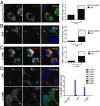
GBPs associate with LCVs independent of IRGs and ATG5. Confocal images of dsRED+ L. pneumophila (L.p.)-infected RAW 264.7 macrophages expressing GFP-mGBP1, GFP-mGBP2, or GFP-mGBP7 (A) and confocal images of IFN-γ–primed, L. pneumophila-infected BMDMs stained with anti-mGBP2 antibody at 2 hpi (B) are shown. Percentages of mGBP2+ LCVs in IFN-γ–primed BMDMs were quantified in 20-min intervals for the first 2 hpi (C). Frequencies of Irgb10+ and Irga6+ LCVs in IFN-γ–primed WT vs. Irgm1−/−Irgm3−/− BMDMs at 2 hpi are shown (D). Frequencies of mGBP2+ LCVs in IFN-γ–primed WT vs. Irgm1−/−Irgm3−/− (E) or ATG5−/− BMDMs (F) are shown. Error bars represent SDs. At least three independent experiments were performed. Statistical analysis was performed by unpaired two-tailed Student’s t test (***P < 0.005; n.s., not significant). (Magnification, 63×.)
Fig. S1.
Fig. S1.
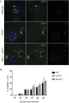
Recruitment of mGBP2 to LCVs is independent of autophagy and Irgm1/Irgm3 proteins. WT, ATG5−/−, ATG7−/−, Beclin−/− and Irgm1−/−Irgm3−/− BMMs were infected with dsRED+ L. pneumophila (L.p.) and stained with anti-mGBP2 (green). Confocal images for infected WT, ATG5−/−, and Irgm1−/−Irgm3−/− BMMs are shown in A. Percentages of mGBP2+ LCVs over the course of 2 h of infection in WT, ATG7−/−, and Beclin−/− BMMs are shown in B. At least 100 infected cells were counted per condition and experiment. Two independent experiments were performed. Error bars represent SDs. (Magnification, 63×.)
Fig. 2.
Fig. 2.
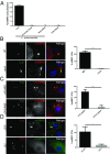
mGBP2 targets vacuoles containing live bacteria expressing bacterial secretion systems. Live or dead dsRED+ L. pneumophila (L.p.) or latex beads were fed to IFN-γ–primed BMDMs for 2 h. The percentages of mGBP2-positive phagocytosed bacteria or beads are depicted (A). IFN-γ–primed BMDMs were infected with GFP+ L. pneumophila (B), GFP+ Y. pseudotuberculosis (Y.p.) (C), or mCherry+ Y. pseudotuberculosis (D) of the indicated genotypes. Representative images and percentages of mGBP2+ PVs at 2 hpi are shown. A minimum of 200 infected cells was analyzed per condition and experiment. At least three independent experiments were performed. Error bars represent SDs. Statistical analysis was performed by one-way ANOVA with Tukey’s multiple comparison test (A) and unpaired two-tailed Student’s t test (BD; *P < 0.05 and ***P < 0.005). (Magnification, 63×.)
Fig. S2.
Fig. S2.
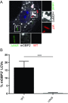
Host recognizes the presence of the Dot/Icm T4SS in a phagosome-intrinsic manner. WT BMDMs were asynchronously coinfected with dsRED+ WT and GFP+ ΔdotA L. pneumophila at a 1:1 ratio. Colocalization of mGBP2 with LCVs was assessed by immunofluorescence at 2 hpi, and representative confocal images are shown in A. Per experiment, a minimum of 100 dually infected (WT + ΔdotA) BMDMs were assessed for mGBP2 colocalization with LCVs (B). Data are from two independent experiments. Error bars represent SDs. ***P < 0.005. (Magnification, 63×.)
Fig. 3.
Fig. 3.
Galectin-3 colocalizes with dotA-expressing L. pneumophila (L.p.) and YopB-/YopD-hypersecreting Y. pseudotuberculosis. (Y.p.) YFP-Gal3–expressing iBMDMs were infected with dsRED+ L. pneumophila (A) or mCherry+ Y. pseudotuberculosis (B) of the indicated genotypes. The percentage of Gal3+ PVs at 2 hpi is depicted. A minimum of 200 infected cells were quantified per condition and experiment. At least three independent experiments were performed. Error bars represent SDs. Statistical analysis was performed by two-tailed Student’s t test (**P < 0.01 and ***P < 0.005). (Magnification, 63×.)
Fig. 4.
Fig. 4.
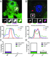
Galectin-3 and mGBP2 colocalize at PVs. YFP-Gal3–expressing iBMDMs were infected with dsRED+ WT L. pneumophila (L.p.) (A) or mCherry+ WT Y. pseudotuberculosis (Y.p.) (B) and stained for endogenous mGBP2 at 2 hpi. Representative colocalization analysis and line trace are shown. The percentage of LCVs (C) or YCVs (D) staining positive for Galectin-3 only, mGBP2 only, or dual-positive for Galectin-3 and mGBP2 are shown. A minimum of 100 infected cells were quantified per condition and experiment. At least three independent experiments were performed. Statistical analysis by two-tailed student’s t test for double-positive bacteria is shown (***P < 0.005). (Magnification, 63×.)
Fig. 5.
Fig. 5.
Galectin-3 and GBPs colocalize at sterilely damaged vesicles. YFP-Gal3–expressing iBMDMs were exposed to hypotonic shock, and the formation of Gal3+ puncta and the frequency of their colocalization with mGBP2 are shown (A). Endosomal damage in MEFs expressing YFP-Gal3 was induced by CPP treatment, and the quantification of Gal3+ puncta and colocalization with mGBP2 are shown by treatment (B). YFP-Gal3–expressing iBMDMs were damaged with LLOMe, and the formation of Gal3+ puncta and colocalization with mGBP2 were quantified (C). HEK 293T cells expressing YFP-Gal3 and hGBP-mCherry fusion proteins were treated with CPP or LLOMe. The percentages of YFP-Gal3 puncta colocalizing with individual hGBP orthologs are shown (D). A minimum of100 damaged cells were quantified per condition and experiment. HS, hypotonic shock. (Magnification, 63×.)
Fig. S3.
Fig. S3.
Hypotonic shock induces mGBP2 puncta. Cells were treated for hypotonic shock (H.S.) or left untreated. Each dot represents the mean number of Gal3 (A) or mGBP2 (B) punctae per cell and per experiment. Representative images of IFN-γ–primed iBMDMs are shown. Average of combined data from four experiments ± SEM is depicted. At least 100 cells were quantified per condition and experiment. *P < 0.05. (Magnification, 40×.)
Fig. 6.
Fig. 6.
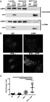
Galectin-3 and mGBP2 form protein complexes. iBMDMs transduced with YFP-Gal3 retrovirus or untransduced controls were damaged with LLOMe or left untreated, and the formation of GBP2/YFP-Gal3–containing protein complexes was assessed with by GFP-Trap IPs. Whole-cell lysates and unbound and IP fractions were immunoblotted against GBP2 and YFP (A). IFN-γ–primed and unprimed MEFs expressing YFP-Gal3 were treated with CPP or left untreated, and interaction was detected by PLA and quantified. A minimum of 15 fields were quantified per treatment. Represenative images (B) and quantification (C) are shown. At least three independent experiments were performed. Statistical analysis was performed by two-way ANOVA with Tukey’s multiple comparison test (***P < 0.005). NT, nontreated. (Magnification, 63×.)
Fig. 7.
Fig. 7.
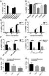
Galectin-3 promotes targeting of mGBP2 and p62 to LCVs and YCVs. WT iBMDM expressing individual Galectins as YFP-fusion proteins were infected with dsRED+ L. pneumophila. The percentages of Galectin+ LCVs were quantified at 2 hpi (A). WT iBMDMs were treated with shRNA against Galectin-3, -8, and -9, and the percentages of mGBP2+ LCVs were quantified at 2 hpi (B). WT and Gal3−/− BMDMs were infected with dsRED+ L. pneumophila (C) or mCherry+ ΔEJK Y. pseudotuberculosis (D), and the percentages of mGBP2+ PVs were quantified over time. Colocalization of GFP-mGBP1 and GFP-mGBP2 with ΔEJK YCVs was monitored in WT and Gal3−/− iBMDMs at 2 hpi (E). RAW 264.7 cells were treated with 2 mM BGN or DMSO control for 3 d before infection with Y. pseudotuberculosis ΔEJK. Percentages of mGBP2+ YCVs were quantified at the indicated time points (F). BMDMs of the indicated genotypes were infected with ΔEJK, and YCV colocalization with p62 (G) or ubiquitin (H) was assessed at 2 hpi. A minimum of 100 infected cells were counted per experimental condition and experiment. At least three independent experiments were performed except for the experiment shown in A, which was repeated once. Error bars represent SDs. Statistical analysis was performed by two-way ANOVA (B, G, and H), two-tailed Student’s t test (C, D, and E), and one-way ANOVA with Tukey’s multiple comparison test (F; *P < 0.05, **P< 0.01, and ***P < 0.005).
Fig. S4.
Fig. S4.
Expression of mGBP2 protein is comparable between WT and Gal3−/− BMDMs. Anti-mGBP2 and corresponding anti-actin immunoblots of protein lysates taken from IFN-γ–primed and unprimed WT, Gal3−/−, and GBPchr3−/− BMDMs are shown.
Fig. S5.
Fig. S5.
Inhibition of host O-glycosylation reduces targeting of mGBP2 to YCVs. RAW 264.7 cells were treated with 2 mM BGN or DMSO control for 3 d before infection with Y. pseudotuberculosis ΔHOJMEK. Percentages of mGBP2+ YCVs were quantified at indicated time points. **P < 0.01.
Fig. S6.
Fig. S6.
Irgm1 and Irgm3 promote ubiquitination of YCVs in BMDMs. WT and Irgm1−/−Irgm3−/− BMMs were infected with mCherry+ Y. pseudotuberculosis ΔEJK and stained with anti-mGBP2. Percentages of mGBP2+ YCVs at 2 hpi are shown.
Fig. S7.
Fig. S7.
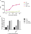
Galectin-3 is nonessential for cell-autonomous immunity toward L. pneumophila and Y. pseudotuberculosis in IFN-γ–primed BMDMs. WT and Gal3−/− BMDMs were primed with 100 U/mL IFN-γ overnight or left unprimed and were then infected with bioluminescent ΔflaA L. pneumophila. Luminescence as relative light units (RLU) is depicted over the course of 30 h of infection (A). Similarly, IFN-γ–primed WT and Gal3−/− BMDMs were infected with the indicated strains of Y. pseudotuberculosis at an MOI of 5, and bacterial burden was assessed by immunofluorescence microscopy at 8 hpi (B). Data are representative of at least two independent experiments.

References

    1. Alix E, Mukherjee S, Roy CR. Subversion of membrane transport pathways by vacuolar pathogens. J Cell Biol. 2011;195(6):943–952. - PMC - PubMed
    1. Juhas M, et al. Genomic islands: Tools of bacterial horizontal gene transfer and evolution. FEMS Microbiol Rev. 2009;33(2):376–393. - PMC - PubMed
    1. Zhao Y, Shao F. The NAIP-NLRC4 inflammasome in innate immune detection of bacterial flagellin and type III secretion apparatus. Immunol Rev. 2015;265(1):85–102. - PubMed
    1. Vance RE, Isberg RR, Portnoy DA. Patterns of pathogenesis: Discrimination of pathogenic and nonpathogenic microbes by the innate immune system. Cell Host Microbe. 2009;6(1):10–21. - PMC - PubMed
    1. Creasey EA, Isberg RR. Maintenance of vacuole integrity by bacterial pathogens. Curr Opin Microbiol. 2014;17:46–52. - PMC - PubMed

Publication types

"VSports" MeSH terms