Skip to main page content
U.S. flag

An official website of the United States government

Dot gov

The . gov means it’s official VSports app下载. Federal government websites often end in . gov or . mil. Before sharing sensitive information, make sure you’re on a federal government site. .

Https

The site is secure V体育官网. The https:// ensures that you are connecting to the official website and that any information you provide is encrypted and transmitted securely. .

. 2017 Jan 27:8:16.
doi: 10.3389/fimmu.2017.00016. eCollection 2017.

"VSports" Capability of Neutrophils to Form NETs Is Not Directly Influenced by a CMA-Targeting Peptide

Affiliations

Capability of Neutrophils to Form NETs Is Not Directly Influenced by a CMA-Targeting Peptide

VSports注册入口 - Christian Maueröder et al. Front Immunol. .

Abstract

During inflammatory reaction, neutrophils exhibit numerous cellular and immunological functions, notably the formation of neutrophil extracellular traps (NETs) and autophagy. NETs are composed of decondensed chromatin fibers coated with various antimicrobial molecules derived from neutrophil granules. NETs participate in antimicrobial defense and can also display detrimental roles and notably trigger some of the immune features of systemic lupus erythematosus (SLE) and other autoimmune diseases. Autophagy is a complex and finely regulated mechanism involved in the cell survival/death balance that may be connected to NET formation. To shed some light on the connection between autophagy and NET formation, we designed a number of experiments in human neutrophils and both in normal and lupus-prone MRL/lpr mice to determine whether the synthetic peptide P140, which is capable of selectively modulating chaperone-mediated autophagy (CMA) in lymphocytes, could alter NET formation. P140/Lupuzor™ is currently being evaluated in phase III clinical trials involving SLE patients. Overall our in vitro and in vivo studies established that P140 does not influence NET formation, cytokine/chemokine production, or CMA in neutrophils VSports手机版. Thus, the beneficial effect of P140/Lupuzor™ in SLE is apparently not directly related to modulation of neutrophil function. .

Keywords: NET formation; P140/Lupuzor; autophagy; murine models of lupus; neutrophils; systemic lupus erythematosus V体育安卓版. .


Figures
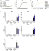
Figure 1
Figure 1
No influence of P140 or the control peptides scrambled P140 (ScP140) and 131-51 on neutrophil extracellular trap (NET) formation or production of reactive oxygen species (ROS) or cytokines/chemokines in human PMN. (A) Kinetics of relative SYTOX Green fluorescence in isolated human PMN after incubation with or without 100 ng/mL PMA. Graphs show data from one representative out of three normal healthy blood donors. (B) Intracellular ROS production or (C) cytokine/chemokine release into the supernatants upon incubation of isolated human PMN with varying concentrations of P140 or the control peptides ScP140 or 131-151. Baseline values of vehicle-treated neutrophils are subtracted. Bars in (C) show the means and SD of one representative out of two experiments.
Figure 2
Figure 2
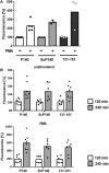
Plate reader-based quantification of neutrophil extracellular trap formation in mice treated with P140 and scrambled P140 (ScP140). (A) Relative SYTOX Green fluorescence in neutrophils isolated from the blood of P140- or ScP140-pretreated mice incubated for 2 h with or without 100 ng/mL PMA or 2 µg/mL ionomycin. Graphs show individual values and means, one dot represents one mouse. ns, not significant. (B) Area and fluorescence intensity of SYTOX Green+ events in neutrophils isolated from the blood of P140- or ScP140-pretreated mice incubated for 2 h with or without 100 ng/mL PMA or 2 µg/mL ionomycin. Plots show medians and individual values from one representative out of 6–8 mice. ***p < 0.001, as determined by Student’s t-test with Bonferroni post hoc test. ns, not significant.
Figure 3
Figure 3
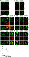
Morphological analysis and quantification of neutrophil extracellular traps (NETs) in P140 and scrambled P140 (ScP140)-treated mice by fluorescence microscopy. Representative immunofluorescence images of SYTOX Green-, neutrophil elastase (NE)-, and citrullinated histone H3 (citH3)-stained neutrophils isolated from ScP140- (A–C) and P140- (D–F) treated mice and incubated without external stimulus (A,D), with PMA (B,E) or with ionomycin (C,F). Left panels show staining controls incubated without (wo) primary antibody to NE or citH3. CY5 stands for control Cyanin 5 and OL stands for overlay. (G) Quantitative analysis of NETs. Scatter plots show individual values and mean of %NETs (defined as % PI+/NE+ cells with >3-fold mean nuclear size) from two mice.
Figure 4
Figure 4
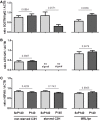
Analysis of neutrophil extracellular trap formation in starved C3H and in MRL/lpr mice treated with P140, scrambled P140 (ScP140), or 131-151 peptides. Neutrophils were isolated from the blood of C3H mice starved for 36 h (A) or MRL/lpr mice (B) pretreated with with injections of P140, ScP140, or the non-phosphorylated peptide 131-151 and incubated with SYTOX Green and with or without 100 ng/mL PMA. Graphs show individual values and means of relative SYTOX fluorescence after 2 h incubation (A) or 2 and 4 h incubation (B), respectively, normalized to starting values. One symbol represents one mouse. n = 3–8.
Figure 5
Figure 5
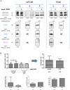
Effect of P140 peptide on autophagic flux evaluated by measuring MAP1LC3B-II levels in total splenocytes from non-starved mice and starved C3H mice and MRL/lpr mice. Cell lysates were resolved by SDS-PAGE, transferred onto polyvinylidene difluoride membranes before staining with anti-microtubule-associated-protein light chain 3b (MAP1LC3B) antibodies. When indicated, cells were treated (+) or not (−) during the last 4 h of the culture with 5 µg/mL pepstatin A and 5 µg/mL E64d to block lysosomal degradation. (A) MAP1LC3B-II levels were evaluated by densitometry and normalized to ACTB/β-actin. Histogram bars represent the means of individual experiments with SEM. *p < 0.05, using a Mann–Whitney U test to compare the data obtained in the presence or absence of protease inhibitors. (B) Representative immunoblot. ACTB was used as loading controls. (C) Autophagic flux as measured by comparing values of MAP1LC3B-II in the presence of lysosomal protease inhibitors divided by values of MAP1LC3B-II in the absence of lysosomal protease inhibitors. Histogram bars represent the means of individual experiments with SEM. *p < 0.05 using a Mann–Whitney test between the untreated and treated mice, in each condition. The groups were constituted of n = 6 for non-starved C3H mice, n = 4 for starved C3H mice, and n = 5 for MRL/lpr mice.
Figure 6
Figure 6
Effect of P140 peptide on autophagy evaluated by measuring SQSTM1, ATG12/5, and HSPA8 levels in total splenocytes from non-starved mice, starved C3H mice, and MRL/lpr mice. Three markers were followed, namely, (A) SQSTM1, (B) ATG12/5, and (C) HSPA8. The groups were constituted of n = 6 for non-starved C3H mice, n = 4 for starved C3H mice, and n = 5 for MRL/lpr mice. Histogram bars represent the means of individual experiments with SEM. *p < 0.05 using a Mann–Whitney test between the untreated and treated mice, in each condition.
Figure 7
Figure 7
Effect of P140 on autophagy in isolated splenic neutrophils. Similar experiments were performed as in Figures 5 and 6, but with isolated splenic neutrophils instead of total splenoctes. (A) Immunoblots of microtubule-associated-protein light chain 3b (MAP1LC3B), SQSTM1, HSPA8, and ATG12/5 antibodies with three independent mice for control peptide scrambled P140 (ScP140) and two for P140 peptide. The expression levels were quantified by densitometry and normalized to ACTB/β-actin. For MAP1LC3B-II, autophagic flux was measured by dividing the values of MAP1LC3B-II with lysosomal protein inhibitor by those of MAP1LC3B-II without lysosomal protein inhibitor. (B) Histogram representation of immunoblots from Figure 7A. nt, not tested.

References

    1. Baumann I, Kolowos W, Voll RE, Manger B, Gaipl U, Neuhuber WL, et al. Impaired uptake of apoptotic cells into tingible body macrophages in germinal centers of patients with systemic lupus erythematosus. Arthritis Rheum (2002) 46(1):191–201.10.1002/1529-0131(200201)46:1<191:AID-ART10027>3.0.CO;2-K - DOI - PubMed
    1. Hakkim A, Furnrohr BG, Amann K, Laube B, Abed UA, Brinkmann V, et al. Impairment of neutrophil extracellular trap degradation is associated with lupus nephritis. Proc Natl Acad Sci U S A (2010) 107(21):9813–8.10.1073/pnas.0909927107 - DOI (VSports在线直播) - PMC - PubMed
    1. Radic M, Marion TN. Neutrophil extracellular chromatin traps connect innate immune response to autoimmunity. Semin Immunopathol (2013) 35(4):465–80.10.1007/s00281-013-0376-6 - DOI - PubMed
    1. Smith CK, Kaplan MJ. The role of neutrophils in the pathogenesis of systemic lupus erythematosus. Curr Opin Rheumatol (2015) 27(5):448–53.10.1097/BOR.0000000000000197 - DOI - PubMed
    1. Li P, Li M, Lindberg MR, Kennett MJ, Xiong N, Wang Y. PAD4 is essential for antibacterial inate immunity mediated by neutrophil extracellular traps. J Exp Med (2010) 207(9):1853–62.10.1084/jem.20100239 - DOI - PMC - PubMed

LinkOut - more resources