"VSports在线直播" Skip to main page content
U.S. flag

An official website of the United States government

Dot gov

The . gov means it’s official. Federal government websites often end in . gov or . mil. Before sharing sensitive information, make sure you’re on a federal government site. VSports app下载.

Https

The site is secure. The https:// ensures that you are connecting to the official website and that any information you provide is encrypted and transmitted securely V体育官网. .

. 2017 Jan 25:17:14.
doi: 10.1186/s12935-017-0383-0. eCollection 2017.

MicroRNA-32 promotes cell proliferation, migration and suppresses apoptosis in breast cancer cells by targeting FBXW7

Affiliations

MicroRNA-32 promotes cell proliferation, migration and suppresses apoptosis in breast cancer cells by targeting FBXW7

Wei Xia et al. Cancer Cell Int. .

"V体育ios版" Abstract

Background: MicroRNAs are a class of small non-coding RNAs that are involved in many important physiological and pathological processes by regulating gene expression negatively. The purpose of this study was to investigate the effect of miR-32 on cell proliferation, migration and apoptosis and to determine the functional connection between miR-32 and FBXW7 in breast cancer. VSports手机版.

Methods: In this study, quantitative RT-PCR was used to evaluate the expression levels of miR-32 in 27 breast cancer tissues, adjacent normal breast tissues and human breast cancer cell lines. The biological functions of miR-32 in MCF-7 breast cancer cells were determined by cell proliferation, apoptosis assays and wound-healing assays. In addition, the regulation of FBXW7 by miR-32 was assessed by qRT-PCR, Western blot and luciferase reporter assays. V体育安卓版.

Results: MiR-32 was frequently overexpressed in breast cancer tissue samples and cell lines as was demonstrated by qRT-PCR V体育ios版. Moreover, the up-regulation of miR-32 suppressed apoptosis and promoted proliferation and migration, whereas down-regulation of miR-32 showed an opposite effect. Dual-luciferase reporter assays showed that miR-32 binds to the 3'-untranslated region of FBXW7, suggesting that FBXW7 is a direct target of miR-32. Western blot analysis showed that over-expression of miR-32 reduced FBXW7 protein level. Furthermore, an inverse correlation was found between the expressions of miR-32 and FBXW7 mRNA levels in breast cancer tissues. Knockdown of FBXW7 promoted proliferation and motility and suppressed apoptosis in MCF-7 cells. .

Conclusions: Taken together, the present study suggests that miR-32 promotes proliferation and motility and suppresses apoptosis of breast cancer cells through targeting FBXW7 VSports最新版本. .

Keywords: Apoptosis; Breast cancer; FBXW7; MiR-32; Proliferation. V体育平台登录.


Figures
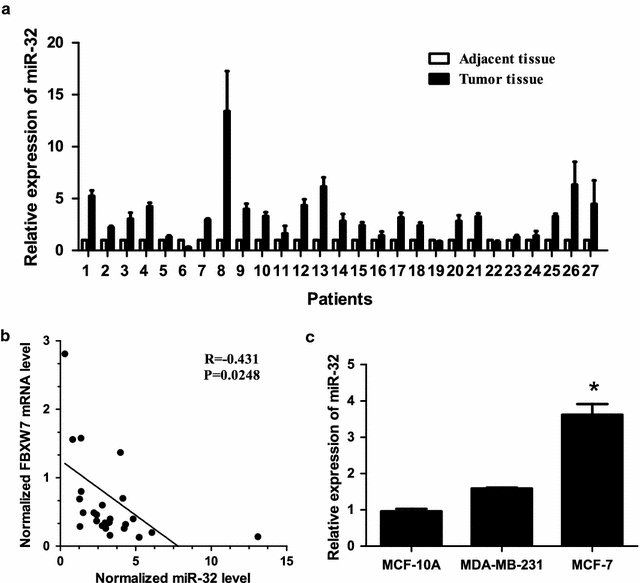
Fig. 1
Fig. 1
MiR-32 is up-regulated in breast cancer samples and cell lines. a Expression levels of miR-32 were up-regulated in 20 breast cancer tissues when compared with adjacent normal tissues. b In 19 breast cancer tissues and corresponding adjacent non-tumor tissues, miR-32 has a negative correlation with FBXW7 mRNA expression. c The expression level of miR-32 was up-regulated in MCF-7 and MDA-MB-231 cell lines when compared with MCF-10A. Bars represent the mean of three independent experiments performed three times ±SD. *P < 0.01
Fig. 2
Fig. 2
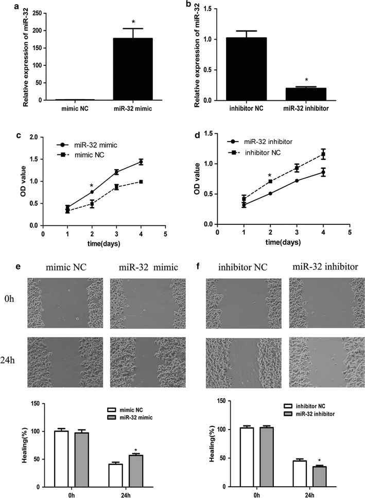
Effect of miR-32 expression on growth and migration in breast cancer cells. a, b QRT-PCR of miR-32 expression in mimic/inhibitor or NC transfected MCF-7 cells. U6 was used as an internal control. c, d MTT analysis of MCF-7 growth following transfection with miR-32 mimic/inhibitor or NC. MiR-32 mimic promotes cell proliferation and inhibitor suppresses cell growth. e, f Wound-healing assay showing that gain of miR-32 promotes cell migration and loss of miR-32 suppresses cell migration. Each assay was repeated three times. *P < 0.05
Fig. 3
Fig. 3
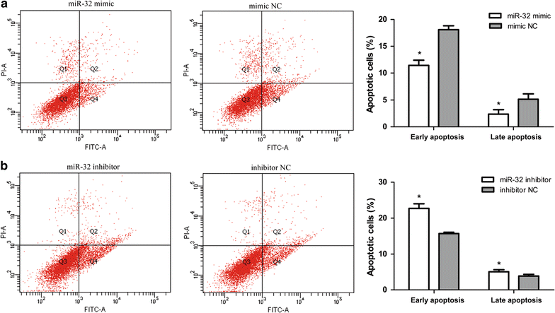
Effects of miR-32 expression on apoptosis in breast cancer cells. a, b MiR-32 mimic suppresses MCF-7 cell apoptosis and miR-32 inhibitor induces cell apoptosis. Cell apoptosis was analyzed by Annexin V-FITC analysis. Data represents the mean of three independent assays ±SD. *P < 0.05
Fig. 4
Fig. 4
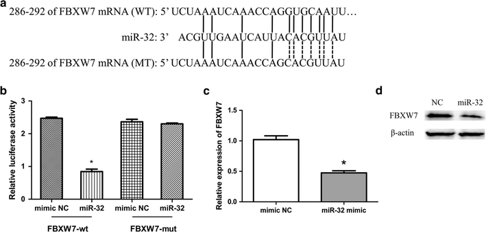
MiR-32 targeted FBXW7. a The miR-32 binding site in FBXW7 3′-UTR, located 286-292 bp upstream of the FBXW7 3′-UTR. b The relative luciferase activity in miR-32 mimic group and NC group. c The relative expression of FBXW7 in miR-32 mimic transfected group and NC group. Bars represent the mean of three independent experiments performed three times ±SD. d Western blot analysis of FBXW7 protein levels in miR-32 overexpressing and NC cells. *P < 0.05
Fig. 5
Fig. 5
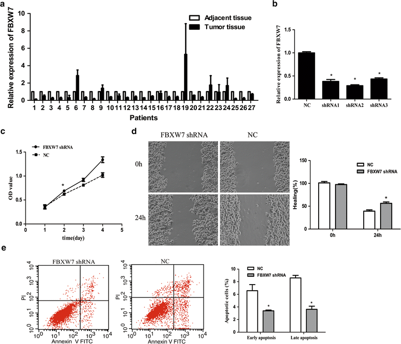
The effect of FBXW7 depletion in MCF-7 cells. a Expression level of FBXW7 was down-regulated in 19 breast cancer tissues compared with adjacent normal tissues. b The expression level of FBXW7 in transfected shRNA MCF-7 cells. c MTT analysis showed that FBXW7 depletion promotes cells proliferation. d Wound-healing assay showed that loss of FBXW7 promotes cell migration. e FBXW7 depletion suppresses MCF-7 cell apoptosis. Data represents the mean of three independent assays ±SD. *P < 0.05
Fig. 6
Fig. 6
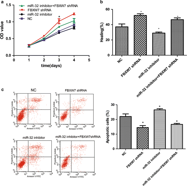
The effect of FBXW7 depletion on miR-32-mediated breast cancer chemoresistance. a The proliferation rate of MCF-7 cells were detected through MTT assay at different time periods after transfection of miR-32 inhibitor and FBXW7 shRNA independently or simultaneously compared with NC group. b Statistical analysis of the proportion of healing compared with NC group. c Annexin V/PI analysis was performed to determine cell apoptosis in the indicated groups. Data represents the mean of three independent assays ±SD. (*P < 0.05)

"V体育2025版" References

    1. Siegel RL, Miller KD, Jemal A. Cancer statistics, 2015. CA Cancer J Clin. 2015;65(1):5–29. doi: 10.3322/caac.21254. - DOI - PubMed
    1. Donepudi MS, Kondapalli K, Amos SJ, Venkanteshan P. Breast cancer statistics and markers. J Cancer Res Ther. 2014;10(3):506–511. - PubMed (VSports在线直播)
    1. Torre LA, Bray F, Siegel RL, Ferlay J, Lortet-Tieulent J, Jemal A. Global cancer statistics, 2012. CA Cancer J Clin. 2015;65(2):87–108. doi: 10.3322/caac.21262. - DOI - PubMed
    1. Duffy MJ, Maguire TM, Hill A, McDermott E, O’Higgins N. Metalloproteinases: role in breast carcinogenesis, invasion and metastasis. Breast Cancer Res. 2000;2(4):252–257. doi: 10.1186/bcr65. - DOI - PMC - PubMed
    1. Steeg PS. Molecular biology of breast cancer metastasis. ‘Has it spread?’: disarming one of the most terrifying questions. Breast Cancer Res. 2000;2(6):396–399. doi: 10.1186/bcr85. - DOI - PMC - PubMed

LinkOut - more resources