Skip to main page content
U.S. flag

An official website of the United States government

Dot gov

The . gov means it’s official. Federal government websites often end in . gov or . mil. Before sharing sensitive information, make sure you’re on a federal government site VSports app下载. .

Https

The site is secure. The https:// ensures that you are connecting to the official website and that any information you provide is encrypted and transmitted securely V体育官网. .

. 2017 Oct;66(10):1767-1778.
doi: 10.1136/gutjnl-2016-312094. Epub 2017 Jan 17.

Epithelial expression and function of trypsin-3 in irritable bowel syndrome

Affiliations

"VSports app下载" Epithelial expression and function of trypsin-3 in irritable bowel syndrome

V体育平台登录 - Claire Rolland-Fourcade et al. Gut. 2017 Oct.

VSports手机版 - Abstract

Objectives: Proteases are key mediators of pain and altered enteric neuronal signalling, although the types and sources of these important intestinal mediators are unknown VSports手机版. We hypothesised that intestinal epithelium is a major source of trypsin-like activity in patients with IBS and this activity signals to primary afferent and enteric nerves and induces visceral hypersensitivity. .

Design: Trypsin-like activity was determined in tissues from patients with IBS and in supernatants of Caco-2 cells stimulated or not V体育安卓版. These supernatants were also applied to cultures of primary afferents. mRNA isoforms of trypsin (PRSS1, 2 and 3) were detected by reverse transcription-PCR, and trypsin-3 protein expression was studied by western blot analysis and immunohistochemistry. Electrophysiological recordings and Ca2+ imaging in response to trypsin-3 were performed in mouse primary afferent and in human submucosal neurons, respectively. Visceromotor response to colorectal distension was recorded in mice administered intracolonically with trypsin-3. .

Results: We showed that stimulated intestinal epithelial cells released trypsin-like activity specifically from the basolateral side. This activity was able to activate sensory neurons V体育ios版. In colons of patients with IBS, increased trypsin-like activity was associated with the epithelium. We identified that trypsin-3 was the only form of trypsin upregulated in stimulated intestinal epithelial cells and in tissues from patients with IBS. Trypsin-3 was able to signal to human submucosal enteric neurons and mouse sensory neurons, and to induce visceral hypersensitivity in vivo, all by a protease-activated receptor-2-dependent mechanism. .

Conclusions: In IBS, the intestinal epithelium produces and releases the active protease trypsin-3, which is able to signal to enteric neurons and to induce visceral hypersensitivity VSports最新版本. .

Keywords: ABDOMINAL PAIN; IRRITABLE BOWEL SYNDROME; NERVE - GUT INTERACTIONS; TRYPSIN. V体育平台登录.


Conflict of interest statement

Competing interests: None declared.

Figures
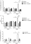
Figure 1
Figure 1
Conditioned medium from intestinal epithelial cells activates dorsal root ganglia (DRG) sensory neurons by the release of trypsin-like serine protease. Effect of the serine protease inhibitor FUT or the trypsin inhibitor leupeptin on the percentage of responsive neurons (A) and on the Fluo-4 (ΔF/F0) measured Ca2+ levels (B) stimulated by apical or basal supernatants recovered from control or lipopolysaccharide (LPS)-treated Caco-2 cell monolayers. (C) Percentage of sensory neurons dissociated from DRGs of wild-type (wt) or PAR2 knockout (KO) mice responding to apical or basal supernatants recovered from control or LPS-treated Caco-2 cell monolayers. Data are expressed as mean±SEM and were compared using a one-way analysis of variance followed by Bonferroni's post-test (n=6, 2 wells per condition for this experimentation) **p<0.01, ***p<0.001.
Figure 2
Figure 2
Intestinal epithelial cells released trypsin-like activity. (A) Trypsin-like activity measured in apical and basal supernatants recovered from control or lipopolysaccharide (LPS)-treated Caco-2 cells. Assembled data from seven independent experiments with 4–6 wells per test. Representative confocal photomicrographs of in situ zymography assays performed in colonic tissue slices from healthy controls and patients with IBS (scale bar: 50 μm), (B) from control (Ctrl) or water avoidance-induced hypersensitive rat (scale bar: 20 μm), (C) evidencing the level of trypsin-like activity. Graph representation of mean fluorescence intensity quantified from 6 to 12 patients per group. Data are expressed as mean±SEM and were compared using Student's t-test. *p<0.05, **p<0.01, ***p<0.005.
Figure 3
Figure 3
Gene expression of trypsinogens A. Relative gene expression of PRSS1 (cationic trypsin: trypsin-1 precursor), PRSS2 (anionic trypsin: trypsin-2 precursor) and PRSS3 (trypsin-3 precursor) in control, lipopolysaccharide (LPS)-treated or epinephrine-treated Caco-2 cell monolayers. Data are expressed as mean±SEM and were compared using one-way analysis of variance followed by Bonferroni's post-test. *p<0.05, ***p<0.001. (B) Analytical agarose-gel electrophoresis of RT-PCR products performed with RNA extracted from human colonic biopsies (healthy control), isolated colonic crypts or Caco-2 cells. Amplicons were amplified with oligonucleotides specific for PRSS1, PRSS2, PRSS3 and HPRT1. (C) Relative mRNA expression of PRSS3 within colonic biopsies from healthy control and different IBS subtypes: IBS-C (constipated), IBS-D (diarrhoea), IBS-M (mixed). Data are expressed as mean±SEM and were compared using Student's t-test. **p<0.01 vs control (Ctrl).
Figure 4
Figure 4
Intestinal epithelial cells upregulate trypsin-3 secretion in inflammatory condition and in IBS. (A) Relative trypsin-3 quantification by western blot analysis from protein extract of control or lipopolysaccharide (LPS)-treated Caco-2 cells. (B) Confocal photomicrographs of control and LPS-treated Caco-2 cell monolayer evidencing trypsin-3-immunoreactivity (green) and actin cytoskeleton (red), scale bar: 25 μm. (C) Relative trypsin-3 protein quantification from apical and basal supernatants from control or LPS-stimulated Caco-2 cell monolayers. (D) Representative confocal photomicrographs of colonic tissue slices from control (ctrl) and patients with IBS (scale bar: 20 μm). (E) Colonic tissues from control or cortagine-induced hypersensitive rat (scale bar: 20 μm), showing trypsin-3-immunoreactivity (green) for D and E, epithelial cell labelling (EpCAM-positive cells, red) and nuclei counterstain (cyan) for D. (D) Mean fluorescence intensity for trypsin-3-immunoreactivity quantified specifically in epithelial (EpCAM-positive) cells. (F) Confocal photomicrographs showing the transversal view of a colonic crypt labelled for trypsin-3 (green), EpCAM (red) and nuclei (cyan), scale bar: 25 μm. The representative profile at the bottom of the panel shows the polarisation of trypsin-3-immunoreactivity towards the basolateral side of intestinal epithelial cells. Data are expressed as mean±SEM and were analysed by Student's t-test in A and D, and a one-way analysis of variance followed by a Bonferroni's post-test in C.
Figure 5
Figure 5
Trypsin-3 increases cellular permeability in intestinal epithelial cells. (A) Dextran passage from apical to basal medium of Caco-2 cell monolayers after trypsin-3 exposure (0.5, 1 and 10 nM) on the basolateral compartment. Assembled data from three independent experiments with 4 wells per test. (B) Representative confocal photomicrographs of Caco-2 cell monolayers after trypsin-3 exposure on the basolateral compartment (0.5, 1 and 10 nM) showing immunodetection of cellular junction occludin (top) and zonula occludens (ZO)-1 (bottom) in green, scale bar: 50 μm. Data are expressed as mean±SEM and were analysed by one-way analysis of variance followed by Bonferroni's post-test, ***p<0.001.
Figure 6
Figure 6
Trypsin-3 evokes PAR2-dependent hyperexcitability of nociceptive dorsal root ganglia neurons and calcium signals in human submucosal neurons. (A) Representative traces of current clamp recordings showing the rheobase and the action potential discharge at twice the rheobase in control neurons (left panel), neurons incubated with trypsin-3 (10 nM) (middle panel) or neurons incubated with the PAR2 antagonist GB83 (10 μM) before trypsin-3 (right panel). (B) Mean data of the rheobase and the action potential number at twice rheobase for the control, and trypsin-3 with or without GB83 (10 μM). *p<0.05 and **p<0.01 compared versus control, ##p<0.01 compared versus trypsin-3. (C) Representative data showing the effect of trypsin and thrombin on the rheobase and action potential discharge is similar in magnitude to that observed with trypsin-3, shown in B. The PAR2 antagonist blocks the trypsin but not the thrombin effect on neuronal excitability. *p<0.05 compared with control, #p<0.05 compared with trypsin-1. One-way analysis of variance with Bonferroni's post-test. (D) Representative gray scale images of cultured human submucosal ganglia loaded with Fluo-4 and Ca2+ responses of three neurons (colour-coded numbers in the gray images) to trypsin-3 (10 nM). Matched colours are represented between cell numbering and traces. The identity of human neurons was confirmed by application of high-K+ solution (75 mM). Scale bar: 20 μm. (E) Average amplitude of trypsin-3 (0.5–10 nM)-induced [Ca2+]i rises in submucosal neurons in the presence or absence of the PAR2 antagonist GB83. Data are expressed as mean±SEM and were analysed by one-way analysis of variance followed by Bonferroni's post-test (n=6 subjects per group), *p<0.05, **p<0.01, °°p<0.01.
Figure 7
Figure 7
Colorectal administration of trypsin-3 induces visceral hypersensitivity. (A) Kinetic visceromotor response (VMR) to intracolonic administration of trypsin-3 (10 U/mouse) 1, 3, 6 and 9 hours after its administration. (B) Visceromotor response to intracolonic administration of trypsin-3 (0.1, 1 or 10 U/mouse) or vehicle, 3 hours after intracolonic administration in wild-type or PAR2 −/− mice (C). Data are expressed as mean±SEM and were analysed by two-way analysis of variance followed by Bonferroni's post-test (n=10 per group). *p<0.05, **p<0.01 and ***p<0.001.
Figure 8
Figure 8
Trypsin-3 causes internalisation of PAR2 in human submucosal neurons. Representative images showing that the expression of PAR2 in human submucosal neurons changes in the presence of trypsin-3 (10 nM). Upper panel shows the clustered staining of PAR2 (arrowheads, red) on the plasma membrane of submucosal neurons (asterisks, NF200, green). Trypsin-3 (2 hours) caused internalisation of PAR2 (red) inside the neuron (asterisk, NF200, green). PAR2 staining appeared diffuse or clustered (arrowheads) inside the neuron. Pre-incubation with GB83 (10 μM, 30 min before trypsin-3) inhibited the internalisation of PAR2, which was expressed on the plasma membrane (arrowheads, red) of neurons (asterisks, NF200, green). Right panels show merged images. Scale bars: 20 μm.

VSports最新版本 - Comment in

References

    1. Sperber AD, Dumitrascu D, Fukudo S, et al. . The global prevalence of IBS in adults remains elusive due to the heterogeneity of studies: a Rome Foundation working team literature review. Gut 2016. doi: 10.1136/gutjnl-2015-311240. [Epub ahead of print 27 Jan 2016] 10.1136/gutjnl-2015-311240 - DOI - PubMed
    1. Buhner S, Li Q, Vignali S, et al. . Activation of human enteric neurons by supernatants of colonic biopsy specimens from patients with irritable bowel syndrome. Gastroenterology 2009;137:1425–34. 10.1053/j.gastro.2009.07.005 - DOI - PubMed
    1. Cenac N, Bautzova T, Le Faouder P, et al. . Quantification and potential functions of endogenous agonists of transient receptor potential channels in patients with irritable bowel syndrome. Gastroenterology 2015;149:433–44.e7. 10.1053/j.gastro.2015.04.011 - DOI - PubMed
    1. Valdez-Morales EE, Overington J, Guerrero-Alba R, et al. . Sensitization of peripheral sensory nerves by mediators from colonic biopsies of diarrhea-predominant irritable bowel syndrome patients: a role for PAR2. Am J Gastroenterol 2013;108:1634–43. 10.1038/ajg.2013.241 - DOI - PubMed
    1. Hyun E, Andrade-Gordon P, Steinhoff M, et al. . Protease-activated receptor-2 activation: a major actor in intestinal inflammation. Gut 2008;57:1222–9. 10.1136/gut.2008.150722 - DOI (VSports手机版) - PubMed

Publication types

MeSH terms