Skip to main page content
U.S. flag

An official website of the United States government

Dot gov

The . gov means it’s official. Federal government websites often end in . gov or . mil. Before sharing sensitive information, make sure you’re on a federal government site. VSports app下载.

Https

The site is secure. The https:// ensures that you are connecting to the official website and that any information you provide is encrypted and transmitted securely V体育官网. .

. 2017 Sep;32(9):1587-1597.
doi: 10.1111/jgh.13731.

Prophylactic tributyrin treatment mitigates chronic-binge ethanol-induced intestinal barrier and liver injury

Affiliations

V体育官网入口 - Prophylactic tributyrin treatment mitigates chronic-binge ethanol-induced intestinal barrier and liver injury

Gail A Cresci et al. J Gastroenterol Hepatol. 2017 Sep.

Abstract

Background and aim: Impaired gut-liver axis is a potential factor contributing to alcoholic liver disease VSports手机版. Ethanol depletes intestinal integrity and causes gut dysbiosis. Butyrate, a fermentation byproduct of gut microbiota, is altered negatively following chronic ethanol exposure. This study aimed to determine whether prophylactic tributyrin could protect the intestinal barrier and liver in mice during combined chronic-binge ethanol exposure. .

Methods: C57BL/6J mice exposed to 5% v/v ethanol-containing diet for 10 days received a single ethanol gavage (5 g/kg) 9 h before euthanasia. Control mice were isocalorically pair-fed maltose dextrin for ethanol. Diets were supplemented (5 mM) with tributyrin or glycerol. Intestine and liver disease activity was assessed histologically. Protein and mRNA expression of tight junction (TJ) proteins, toll-like receptors, and tumor necrosis factor-alpha were assessed. Caco-2 monolayers with or without ethanol exposure and/or sodium butyrate were used to test butyrate's direct effects on intestinal integrity V体育安卓版. .

Results: Chronic-binge ethanol feeding impaired intestinal TJ protein co-localization staining; however, tributyrin co-treatment mitigated these effects. Ethanol depleted TJ and transepithelial electrical resistance in Caco-2 monolayers, but butyrate co-treatment reduced these effects. Hepatic toll-like receptor mRNA expression and tumor necrosis factor-alpha protein expression was induced by ethanol; however, the response was significantly dampened in mice co-treated with tributyrin. Tributyrin altered localization of both neutrophils and single hepatocyte death: Leukocytes and apoptotic hepatocytes localized predominantly around the portal tract in ethanol-only treated mice, whereas localization predominated around the central vein in ethanol-tributyrin mice. V体育ios版.

Conclusions: Prophylactic tributyrin supplementation mitigated effects of combined chronic-binge ethanol exposure on disruption of intestinal TJ localization and intestinal permeability and liver injury VSports最新版本. .

Keywords: alcoholic liver disease; chronic-binge ethanol; gut permeability; tight junction proteins; tributyrin V体育平台登录. .


V体育官网入口 - Conflict of interest statement

Disclosures:

No conflicts of interest, financial or otherwise, are declared by the authors.

Figures (VSports app下载)
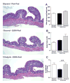
Figure 1
Figure 1. Histopathology of proximal colon in response to ethanol and tributyrin
Mice were fed an EtOH (5% v/v) containing liquid diet or pair-fed a diet with maltose-dextrin isocalorically substituted for ethanol for 10 days. Diets were supplemented with glycerol or tributyrin (5 mM). Mice were then treated with a single 5 g/kg gavage of ethanol the next day containing glycerol or tributyrin (2.5 mM). At 9 hr post-gavage, proximal colon was collected and used to prepare RNA or embedded in OCT for histology. Sections were stained with hematoxylin and eosin and bright-field images captured and analyzed. A) Number of goblet cells/villi were enumerated. B) Villus tip-to-crypt depth was measured (um); 10 crypts per section were evaluated. C) Expression of ITF mRNA was detected in proximal colon using qRT-PCR. D) Representative H&E histological sections of proximal colon from each treatment group are shown. The images are taken by Olympus BX41 microscope with 100× magnification (10×10). Images are representative of at least replicate images captured per mouse in 4–6 mice per treatment group. Data are the mean ± SEM.
Figure 2
Figure 2. Effects of tributyrin treatment on TJ protein expression in proximal colon
Mice were treated as described in Figure 1. Proximal colon was collected and used to prepare RNA or embedded in OCT for histology. A) Claudin-3 (green), occludin (red) and ZO-1(green) were visualized by immunohistochemistry in sections of proximal colon frozen in OCT. A selected area was cropped and enlarged. All images were acquired using a 40× objective. Images are representative of at least replicate images captured per mouse in 4–6 mice per treatment group. B–D) Expression of claudin-1, occludin, and ZO-1 mRNA was detected in the proximal colon of mice using qRT-PCR. Data are the mean ± SEM. Values with different alphabetical superscripts were significantly different from each other, p < 0.05.
Figure 3
Figure 3. Effects of tributyrin treatment on TJ protein expression in the ileum
Mice were treated as described in Figure 1. Ileum was collected and used to prepare RNA or embedded in OCT for histology. A) Occludin (red) and ZO-1(green) were visualized by immunohistochemistry in sections of ileum frozen in OCT. All images were acquired using a 40× objective. Images are representative of at least replicate images captured per mouse in 4–6 mice per treatment group. B & C) Expression of occludin and ZO-1 mRNA was detected in the ileum of mice using qRT-PCR. Data are the mean ± SEM. Values with different alphabetical superscripts were significantly different from each other, p < 0.05.
Figure 4
Figure 4. Effect of butyrate on stability of TJ proteins and TEER reduced by EtOH
Human intestinal epithelial cells (Caco2) were grown to confluency on inserts and then pretreated for 18 hrs with sodium butyrate (5 mM). Cells were then challenged with 40 mM ethanol for 3 h and expression of TJ proteins and transepithelial electrical resistance (TEER) was assessed. A) Expression of Occludin (red) and ZO-1(green) was visualized by immunocytochemistry in Caco2 cells fixed in ice-cold methanol. All images were acquired using a 40× objective. Images are representative of at least replicate images captured in three independent experiments. B) A World Precision epithelial volt/ohm meter electrical resistance system was used to measure TEER. TEER values represent relative fold changes compared to cells not challenged with ethanol and/or butyrate. Data are the mean ± SEM of three experiments performed in duplicate. Values with different alphabetical superscripts were significantly different from each other, p < 0.05.
Figure 5
Figure 5. Effects of tributyrin on hepatic expression of TLR and the inflammatory cytokine TNF-α following chronic-binge ethanol exposure
Mice were treated as described in Figure 1. At 9 hours post-gavage, liver was excised and used to prepare RNA or embedded in formalin for IHC. A–D) Expression of TLR2, TLR4 and TLR9 and TNFα mRNA was detected in mouse livers using qRT-PCR. E) TNFα protein was evaluated by immunohistochemistry in paraffin embedded liver tissue. All images were acquired using a 40× objective. TNF-α positive areas were quantified using Image-Pro Plus software and analyzed. Values represent means ±SEM. n = 4–6 mice per treatment group. Values with different alphabetical superscripts were significantly different from each other, p < 0.05.
Figure 6
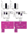
Figure 6. Effects of tributyrin on hepatic injury and zonation of neutrophilic infiltrate and acidophilic bodies following chronic-binge ethanol exposure
Mice were treated as described in Figure 1. Mice were euthanized 9-hours post-ethanol gavage. A & B) Plasma was separated from blood collected from the posterior vena cava. Activity of ALT was measured in plasma. Hepatic triglyceride content was measured in whole liver homogenates. C–E) Liver was excised, fixed in formalin, embedded in paraffin and later stained with hematoxylin and eosin for histologic analysis. Quantitative assessment of the neutrophilic infiltrate and acidophilic bodies are shown. Bars represent percentage of mice in each group exhibiting the designated number of foci of neutrophilic infiltrates per high power field, acidophilic bodies and their zonal distribution in the liver (zone 1, 2 or 3; azonal: no recognizable pattern; multiple zones, multifocal but possible throughout the tissue with inconsistent distribution). Representative H&E histological sections of livers are shown for the study groups. Yellow arrows indicate neutrophilic infiltrates; blue arrows indicate acidophil bodies. (H&E stain, 100×).

References

    1. Orman ES, Odena G, Bataller R. Alcoholic liver disease: pathogenesis, management, and novel targets for therapy. J Gastroenterol Hepatol. 2013;28(Suppl 1):77–84. - "VSports最新版本" PMC - PubMed
    1. Yan AW, Fouts DE, Brandl J, Stakel P, Torralba M, Schott E, et al. Enteric dysbiosis associated with a mouse model of alcoholic liver disease. Hepatology. 2011;53:96–105. - PMC - PubMed
    1. Bull-Otterson L, Feng W, Kirpich I, Wang Y, Quin X, Liu Y, et al. Metagenomic analysis of alcohol induced pathogenic alterations in the intestinal microbiome and the effect of Lactobacillus rhamnosus GG treatment. PLOS One. 2013;8:e53028. - PMC (VSports最新版本) - PubMed
    1. Ferrier LBF, Debrauwer L, Chabo C, Langella P, Bueno L, Fioramonti J. Impairment of the intestinal barrier by ethanol involves enteric microflora and mast cell activation in rodents. AmJ Pathol. 2006;168:1148–54. - PMC - PubMed
    1. Leclercq S, Matamoros S, Cani PD, Neyrinck AM, Jamar F, Starkel P, et al. Intestinal permeability, gut-bacterial dysbiosis, and behavioral markers of alcohol-dependence severity. Proc Natl Acad Sci USA. 2014;111:E4485–E4493. - PMC - PubMed

MeSH terms