Skip to main page content
U.S. flag

An official website of the United States government

Dot gov

The . gov means it’s official. Federal government websites often end in . gov or VSports app下载. mil. Before sharing sensitive information, make sure you’re on a federal government site. .

Https

The site is secure V体育官网. The https:// ensures that you are connecting to the official website and that any information you provide is encrypted and transmitted securely. .

. 2017 Jan 6:8:13824.
doi: 10.1038/ncomms13824.

BMP restricts stemness of intestinal Lgr5+ stem cells by directly suppressing their signature genes

Affiliations

BMP restricts stemness of intestinal Lgr5+ stem cells by directly suppressing their signature genes

Zhen Qi et al. Nat Commun. .

Abstract (VSports app下载)

The intestinal epithelium possesses a remarkable self-renewal ability, which is mediated by actively proliferating Lgr5+ stem cells. Bone morphogenetic protein (BMP) signalling represents one major counterforce that limits the hyperproliferation of intestinal epithelium, but the exact mechanism remains elusive. Here we demonstrate that epithelial BMP signalling plays an indispensable role in restricting Lgr5+ stem cell expansion to maintain intestinal homeostasis and prevent premalignant hyperproliferation on damage VSports手机版. Mechanistically, BMP inhibits stemness of Lgr5+ stem cells through Smad-mediated transcriptional repression of a large number of stem cell signature genes, including Lgr5, and this effect is independent of Wnt/β-catenin signalling. Smad1/Smad4 recruits histone deacetylase HDAC1 to the promoters to repress transcription, and knockout of Smad4 abolishes the negative effects of BMP on stem cells. Our findings therefore demonstrate that epithelial BMP constrains the Lgr5+ stem cell self-renewal via Smad-mediated repression of stem cell signature genes to ensure proper homeostatic renewal of intestinal epithelium. .


Figures
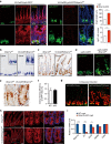
Figure 1
Figure 1. BMP restricts Lgr5+ stem cell expansion independently of Wnt/β-catenin during intestinal homeostasis.
(a) Vil-CreER;Lgr5-EGFP and Vil-CreER;Lgr5-EGFP;Bmpr1afl/fl mice were analysed 1 week after 5-day tamoxifen administration. Proximal jejunum sections were stained for p-Smad1/5/8 (for BMP signalling activity) and EGFP (for stem cells). Nuclei were counter-stained with 4′,6-diamidino-2-phenylindole (DAPI). The lower panels show enlargements of the upper panels. Images are representative of n=6 mice per genotype. Quantifications of crypt length and Lgr5+ cell number is shown aside, and the data represent mean±s.e.m. of n=6 mice per genotype. ***P<0.001 by Student's t-tests. (b,c) In situ hybridization of Olfm4 and immunohistochemical staining of Sox9 in Bmpr1afl/fl and Vil-CreER;Bmpr1afl/fl mice at day 12. Images are representative of n=6 mice per genotype. (d) Immunofluorescence images showing Lgr5+ stem cells 12 days after the first injection in Lgr5-CreERT2;Bmpr1afl/fl mice (n=5 mice per genotype). Dotted lines delineate the normal Lgr5+ stem cell pool. Images are representative of three independent experiments. (e) BrdU incorporation in ISCs and transient amplifying cells after a 2 h pulse in Vil-CreER;Bmpr1afl/fl and Bmpr1afl/fl mice at day 12. Images are representative of n=6 mice per genotype. The asterisks indicate the replicating CBC cells at the base of crypt. (f) In vitro colony formation assay of GFPhigh cells sorted from Vil-CreER;Bmpr1afl/fl and Bmpr1afl/fl mice at day 12. Data represent mean±s.e.m. of n=3 experiments. (g) Lineage tracing in the jejunum of Lgr5-CreERT2;td-Tomato mice and Lgr5-CreERT2;td-Tomato;Bmpr1afl/fl mice. Representative images of td-Tomato immunofluorescence at day 3.5 after induction were shown (n=4 mice per genotype). (h) β-catenin immunofluorescence staining of proximal jejunum sections from Bmpr1afl/fl and Vil-CreER;Bmpr1afl/fl mice at day 12. The right panels show enlargements of boxed areas. Nuclei were counter-stained with DAPI. Images are representative of n=6 mice per genotype. (i) Quantitative RT–PCR analysis of Bmpr1a, Id1, Axin2, EphB2 and Cd44 expression in intestinal crypts from Vil-CreER;Bmpr1afl/fl and control mice at day 12 after induction. Data represent mean±s.e.m. of n=5 mice per genotype. *P<0.05, ***P<0.001 by Student's t-tests. Scale bars, 50 μm. CBC, crypt columnar cell; EGFP, enhanced green fluorescent protein.
Figure 2
Figure 2. Loss of BMP accelerates crypt regeneration and drives premalignant hyperproliferation after exposure to ionizing radiation.
(a) Representative images of Lgr5-EGFP immunofluorescence and Ki67 staining in jejunum sections from Lgr5-EGFP and Vil-CreER;Lgr5-EGFP;Bmpr1afl/fl mice at different time points after 10 Gy abdominal X-ray radiation (n=5 mice per genotype). The mice were irradiated 2 weeks following 5-day tamoxifen induction. Dotted lines delineate the proliferating epithelial invaginations on villus. (b) Quantification of crypt number and Lgr5+ stem cell number in Lgr5-EGFP and Vil-CreER;Lgr5-EGFP;Bmpr1afl/fl mice at different time points after 10 Gy abdominal X-ray radiation. Data represent mean±s.e.m. of n=5 mice per genotype. Scale bars, 50 μm.
Figure 3
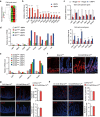
Figure 3. BMP directly suppresses the expression of Lgr5+ stem cell signature genes.
(a) BMP signalling was activated for 36 h in intestinal organoids through direct BMP4 treatment or Noggin withdrawal from the normal culture medium containing EGF, Noggin and R-spondin after cell passaging. Lgr5+ stem cells (Lgr5-EGFP) and proliferating cells (Ki67+) were examined in the organoids. Nuclei were counter-stained with 4′,6-diamidino-2-phenylindole. Images are representative of three independent experiments. (b) Quantitative RT–PCR analysis of Lgr5, Axin2 and Id1 in intestinal organoids after BMP stimulation for indicated time. (c) The organoids derived from Lgr5-EGFP mice were treated with 10 ng ml−1 BMP4 for 4 h, and then Lgr5+ stem cells were isolated by FACS and subjected to RNA-seq. Heatmap of RNA-seq data shows the downregulation of known ISC marker genes by BMP. (d) GO analysis of BMP-downregulated genes and BMP-upregulated genes in Lgr5+ stem cells, respectively. (e) GSEA revealed significant enrichment of ISC signature gene set with BMP-downregulated genes. ES, enrichment score; NES, normalized enrichment score. (f) Venn diagram showing the overlap between ISC signature genes with BMP-downregulated genes. (g) Quantitative RT–PCR analysis of stem cell signature gene expression in Lgr5+ stem cells sorted from mice with indicated genotypes at day 12. Data represent mean±s.e.m of n=5 mice. *P<0.05, **P<0.01, ***P<0.001 by Student's t-tests. Scale bars, 50 μm.
Figure 4
Figure 4. BMP activation has no direct inhibitory effects on Wnt signalling in Lgr5+ ISCs.
(a) Heatmap showing the expression levels of several canonical Wnt target genes in Lgr5+ stem cells on BMP stimulation. (b) Quantitative RT–PCR verification of BMP-regulated genes and Wnt target genes in Lgr5+ stem cells. Data represent mean±s.e.m of n=3 independent experiments. ***P<0.001 by Student's t-tests. (c) Immunoblots of cytoplasmic and nuclear proteins from intestinal organoids treated with or without BMP4 for 4 h. Data are representative of three independent experiments. (d) Quantitative RT–PCR analysis of gene expression in Apc mutant organoids derived from intestinal tumours in Vil-cre;Apcwt/fl mice that were treated with or without BMP for 4 h. Data represent mean±s.e.m of three independent experiments. *P<0.05, **P<0.01, ***P<0.001 by Student's t-tests. Scale bar, 50 μm.
Figure 5
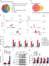
Figure 5. Smad proteins bind to the promoter regions of stem cell signature genes and recruit HDAC1 to repress transcription.
(a) Genome-wide distribution of Smad4-binding sites in Lgr5+ stem cells. TES, transcription end site; TSS, transcription start site. (b) Venn diagram showing the overlap between BMP-downregulated signature genes with Smad4 bound genes. P=6.00E-12. (c) ChIP-seq signals for Smad4 binding at the representative genomic loci. Id1 served as a positive control. (d) Lgr5+ stem cells were stimulated with or without BMP4 for 4 h and subjected to ChIP assays with anti-Smad1, anti-Smad4 antibodies or control IgG. Quantitative RT–PCR followed ChIP shows the enrichment of Smad1 or Smad4 in the promoter or enhancer regions of indicated genes. Smad7 served as positive control. Data represent mean±s.e.m of three independent experiments. **P<0.01 by Student's t-test. (e) HDAC1 ChIP assays in Lgr5+ stem cells with or without 4 h BMP4 treatment. Data represent mean±s.e.m of three independent experiments. **P<0.01 by Student's t-test. (f) Intestinal organoids were treated with 10 ng ml−1 BMP4 for 4 h before being collected for immunoprecipitation (IP) with control IgG or anti-Smad4 antibody followed by western blotting. Protein expression was determined with whole-cell lysates (WCL). (g) Quantitative RT–PCR analysis of Lgr5, Olfm4, Pdgfa, Cdca7 and Sox9 in intestinal organoids after BMP stimulation in the presence or absence of 300 nM trichostatin A (TSA) for indicated time. For TSA treatment, the organoids were first pretreated with 300 nM TSA for 2.5 h. Data represent mean±s.e.m of three independent experiments.
Figure 6
Figure 6. Low BMP activity restricts the self-renewal of Lgr5+ stem cells through modulating stem cell signature genes but not cell cycle arrest genes.
(a) BMP-regulated genes in Lgr5+ stem cells after 4 h BMP stimulation were grouped into cell cycle arrest cluster. (b) The expression of the selected genes in the cell cycle arrest cluster were validated by quantitative RT–PCR in Lgr5+ stem cells sorted from intestinal organoids. Data represent mean±s.e.m of three independent experiments. **P<0.01, ***P<0.001 by Student's t-test. (c) The expression of the stem cell signature genes and cell cycle arrest genes were measured by quantitative RT–PCR in intestinal organoids from indicated groups after passaging. Data represent mean±s.e.m of three independent experiments. *P<0.05, **P<0.01, ***P<0.001 by Student's t-test. (d) Relative level of FPKM of indicated genes from Lgr5-GFPhigh and Lgr5-GFPlow cells with or without BMP treatment for 4 h. (e) The expression of the stem cell signature genes and cell cycle arrest genes were measured by quantitative RT–PCR in EphB2high, EphB2medium and EphB2low cells sorted from intestinal organoids treated with or without BMP4 for 4 h. Data represent mean±s.e.m of three independent experiments. (f) Immunofluorescence staining of Ki67 in Bmpr1afl/fl and Vil-CreER;Bmpr1afl/fl mice at day 12. Dotted lines delineate the region of crypts. Nuclei were counter-stained with 4′,6-diamidino-2-phenylindole (DAPI). Images are representative of n=6 mice per genotype. (g,h) Muc2 immunofluorescence staining and chromogranin A immunofluorescence staining in intestine from Vil-creER;Bmpr1afl/fl and Bmpr1afl/fl mice at day 12 (g) and 1.5 month after induction (h). Nuclei were counter-stained with DAPI. Images are representative of n=6 mice per genotype. Quantifications of goblet cell and enteroendocrine cell number are shown aside. Data represent mean±s.e.m of n=5 mice per genotype. Scale bars, 50 μm.
Figure 7
Figure 7. Smad4 mediates the negative regulation of BMP on Lgr5+ stem cell self-renewal.
(a) Olfm4 in situ hybridization and Sox9 staining in intestines from 1-month-old Smad4fl/fl and Vil-Cre;Smad4fl/fl mice. Images are representative of n=5 mice per genotype. (b) Ki67 and lysozyme staining in intestines from 1-month-old Smad4fl/fl and Vil-Cre;Smad4fl/fl mice. Dotted lines delineate the region of crypts. Nuclei were counter-stained with 4′,6-diamidino-2-phenylindole (DAPI). Images are representative of n=5 mice per genotype. (c) Intestinal organoids derived from Smad4fl/fl and Smad4KO (Vil-Cre;Smad4fl/fl) mice were cultured in ENR (EGF, Noggin and R-spondin) or ER medium for 5 days and photographed. Images are representative of three independent experiments. (d) Intestinal organoids derived from Smad4fl/fl and Smad4KO (Vil-Cre;Smad4fl/fl) mice were treated with BMP4 for 36 h followed by Olfm4 in situ hybridization, Sox9 staining and Ki67 immunofluorescence staining. Nuclei were counter-stained with DAPI. Images are representative of three independent experiments. (e,f) Smad4fl/fl and Smad4KO (Vil-Cre;Smad4fl/fl) organoids were treated with BMP4 for 4 h and the mRNA level of indicated genes were measured by quantitative RT–PCR. Data represent mean±s.e.m of three independent experiments. *P<0.05, ***P<0.001 by Student's t-tests. Scale bars, 50 μm.

References

    1. Barker N. et al.. Identification of stem cells in small intestine and colon by marker gene Lgr5. Nature 449, 1003–1007 (2007). - V体育官网 - PubMed
    1. Vermeulen L. & Snippert H. J. Stem cell dynamics in homeostasis and cancer of the intestine. Nat. Rev. Cancer 14, 468–480 (2014). - PubMed
    1. van der Flier L. G. & Clevers H. Stem cells, self-renewal, and differentiation in the intestinal epithelium. Annu. Rev. Physiol. 71, 241–260 (2009). - PubMed
    1. Sancho E., Batlle E. & Clevers H. Signaling pathways in intestinal development and cancer. Annu. Rev. Cell Dev. Biol. 20, 695–723 (2004). - PubMed
    1. Clevers H. The intestinal crypt, a prototype stem cell compartment. Cell 154, 274–284 (2013). - PubMed

Publication types

MeSH terms