Skip to main page content
U.S. flag

An official website of the United States government

Dot gov

The . gov means it’s official. Federal government websites often end in . gov or . mil. Before sharing sensitive information, make sure you’re on a federal government site VSports app下载. .

Https

The site is secure V体育官网. The https:// ensures that you are connecting to the official website and that any information you provide is encrypted and transmitted securely. .

. 2017 Feb;23(2):223-234.
doi: 10.1038/nm.4245. Epub 2016 Dec 19.

Dissociation of muscle insulin sensitivity from exercise endurance in mice by HDAC3 depletion

Affiliations

Dissociation of muscle insulin sensitivity from exercise endurance in mice by HDAC3 depletion (VSports app下载)

"V体育2025版" Sungguan Hong et al. Nat Med. 2017 Feb.

"VSports最新版本" Abstract

Type 2 diabetes and insulin resistance are associated with reduced glucose utilization in the muscle and poor exercise performance. Here we find that depletion of the epigenome modifier histone deacetylase 3 (HDAC3) specifically in skeletal muscle causes severe systemic insulin resistance in mice but markedly enhances endurance and resistance to muscle fatigue, despite reducing muscle force VSports手机版. This seemingly paradoxical phenotype is due to lower glucose utilization and greater lipid oxidation in HDAC3-depleted muscles, a fuel switch caused by the activation of anaplerotic reactions driven by AMP deaminase 3 (Ampd3) and catabolism of branched-chain amino acids. These findings highlight the pivotal role of amino acid catabolism in muscle fatigue and type 2 diabetes pathogenesis. Further, as genome occupancy of HDAC3 in skeletal muscle is controlled by the circadian clock, these results delineate an epigenomic regulatory mechanism through which the circadian clock governs skeletal muscle bioenergetics. These findings suggest that physical exercise at certain times of the day or pharmacological targeting of HDAC3 could potentially be harnessed to alter systemic fuel metabolism and exercise performance. .


Conflict of interest statement

COMPETING FINANCIAL INTERESTS STATEMENT

The authors disclose no competing financial conflict of interest.

VSports手机版 - Figures
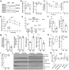
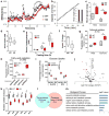
Figure 1
Figure 1. Skeletal muscle HDAC3 deletion reduces glucose uptake and insulin sensitivity independently of the upstream insulin signalling
(a) Western blot analysis (n = 2) of quadriceps femoris (Quads), gastrocnemius (Gastro), and cardiac muscles. (b) Basal insulin and glucose levels, n = 8. (c) Insulin tolerance test (ITT) and body weight on chow at the age of 4 months, n = 8. (d) Glucose tolerance test (GTT) at the age of 5 months after feeding high fat diet for two weeks, n = 8. (e) Body composition measured by nuclear magnetic resonance (NMR), n = 5. (f,g) Hyperinsulinemic euglycemic clamp analysis, GIR, glucose infusion rate; Rd, rate of disposition; EGP, endogenous glucose production; WAT, white adipose tissue. n = 5. (h) Mass spectrometry-based lipidomics analysis of diacylglycerol (DAG) and ceramides in quadriceps muscles, n = 5. (i) Total muscle triglycerides (TG) measurement, n = 5. (j) Western blot analysis (n = 2) of molecular insulin signaling in quadriceps muscles harvested at 20 min after a bolus insulin injection. Phosphorylation on Akt (pAkt), glycogen synthase kinase (GSK3β), S6 ribosomal protein (S6), and insulin receptor substrate (IRS1) at indicated sites were shown. (k) Quantification of the western blot densities in the insulin treatment condition, n = 7. Box-plots center line, limits, and whiskers represent median, quartile, and minimum/maximum values respectively. All other graphs were presented as the mean ± S.E.M. *P < 0.05 between groups by 2-sided t-test.
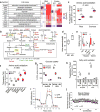
Figure 2
Figure 2. HDAC3 deletion reduces glucose utilization during muscle contractions, but enhances exercise endurance and oxidative metabolism
(a,b) 13C-enrichment in metabolites from serum or muscles after infusion of U-13C-glucose through jugular vein catheter while mice are running on treadmill, n = 6 for WT, n = 5 for KO. Total number of labelled carbon atoms in a given metabolite is indicated by color. G6P, glucose-6-phosphate, F1,6,P, fructose-1, 6-phosphate, DHA, dihydroxyacetone phosphate, 3PG, 3-phosphoglycerate, G3P, glycerol-3-phosphate, Lac, lactate, and Pyr, pyruvate. (c) Diagram of glycolysis and TCA cycle pathways with detected intermediates highlighted. (d) Relative utilization rate of circulating glucose. Fractional contribution (FC) is derived from muscle pyruvate labeling after correction with blood precursor labeling. (e) 13C-enrichment in muscle TCA intermediates. Cit, citrate, Suc, succinate, Mal, malate, Glu, glutamate, and Asp, aspartate. (f) Treadmill speed profile and real-time shock detection during running, n = 10. (g) Distance run when mice received 50 shocks and body weight, n = 10. (h,i) Muscle fatigue and recovery in ex vivo contraction study with extensor digitorum longus (EDL) muscles, n = 8. Scale bar, 50 µm. (j) Representative immunofluorescence staining (n = 8 images) of tibialis anterior (TA) with indicated MHC isoform-specific antibodies, and quantification of staining in TA and soleus, n = 8 animals. (k) Western blot analysis (n = 3) of mitochondrial OXPHOS complexes in quadriceps complexes. (l) Respirometry analysis in fully-differentiated C2C12 myotubes after treatment with adenoviral vectors for HDAC3 knockdown, n = 6 wells of cells. Oligomycin, inhibitor of ATP synthase. FCCP, uncoupling agent. Antimycin A (AA) and rotenone, inhibitors of electron transport. Box-plots center line, limits, and whiskers represent median, quartile, and minimum/maximum values respectively. All other graphs were presented as the mean ± S.E.M. *P < 0.05 between genotypes under the same condition by 2-sided t-test.
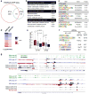
Figure 3
Figure 3. HDAC3 regulates muscle fuel preference and controls amino acid metabolism
(a) Respiratory exchange ratio (RER) in mice running on treadmill with increasing speed to exhaustion, n = 6. (b) Speed profile of the treadmill and body weight. (c) Muscle glycogen at rest or immediately after exercise, n = 5. (d) Blood lactate after exercise, n = 8. (e) Blood ketone body after fasting, n = 8. (f) Liver triglyceride (TG) after fasting, n = 8. (g) Fatty acid oxidation rate measured by 3H-H2O generation in blood at 20 min after i.p. injection of 3H-palmitate, n = 5. (h) Fatty acid oxidation in differentiated C2C12 myotubes treated with adenovirus to knock down HDAC3, n = 6 wells of cells. EPS, electric pulse stimulation. (i) Glucose uptake assay in differentiated C2C12 myotubes treated with adenovirus. Cytochalasin B (CB), an inhibitor of glucose transport, n = 6 wells of cells. (j) Volcano plot of metabolomics profiling in quadriceps muscles harvested at rest. Altered amino acids highlighted in red. (k) Amino acid contents from metabolomics profiling in quadriceps muscles, n = 5. (l) Venn diagram of differentially expressed genes and proteins (KO versus WT) as identified by RNA-seq (q<0.1, fold change>1.5) and proteomics profiling using nano-liquid chromatography mass spectrometry (nano-LC-MS) with label-free and tandem mass tag (TMT) quantitation (p<0.05 in label-free quantification and p<0.1 in TMT quantification). (m) Enriched biological pathway of overlapping genes/proteins. Box-plots center line, limits, and whiskers represent median, quartile, and minimum/maximum values respectively. All other graphs were presented as the mean ± S.E.M. *P < 0.05 between genotypes under the same condition by 2-sided t-test.
Figure 4
Figure 4. Enhanced amino acid catabolism in HDAC3-depleted muscles underlies the fuel switch
(a) Heat map of altered genes or proteins from RNA-seq and proteomics analysis. Data for Asns protein is missing. (b) Metabolic pathway with genes upregulated (red) or downregulated (green) in HDAC3-depleted muscles (KO) versus WT control. (c) Aspartate catabolism measured by 3H-H2O accumulation in blood at 20 min after i.p. injection of 3H-aspartate, n = 6. (d) Urine ammonia measurement, n = 8. (e) In vivo metabolites measurement by LC-MS/MS after exercise, n = 5. (f) Aspartate catabolism assay in C2C12 myotubes treated with indicated adenovirus, n = 6 wells of cells. (g) Glucose uptake assay in C2C12 myotubes after overexpression of Ampd3, n = 6 wells of cells. (h) Fatty acid oxidation assay in C2C12 myotubes, n = 6 wells of cells. (i) Respirometry analysis of mitochondrial oxidative capacity in C2C12 myotubes. OCR, oxygen consumption rate; n = 6 wells of cells. (j) Ex vivo contraction analysis of EDL muscles fatigue resistance in the presence of AMP deaminase inhibitor deoxycoformycin (DCF), n = 8. Box-plots center line, limits, and whiskers represent median, quartile, and minimum/maximum values respectively. All other graphs were presented as the mean ± S.E.M. *P < 0.05 between genotypes under the same condition by 2-sided t-test.
Figure 5
Figure 5. Nonbiased identification of the circadian clock as an upstream regulator of muscle HDAC3
(a) Venn diagram of HDAC3 ChIP-seq binding sites in mouse quadriceps muscles at Zeitgeber time 10 (ZT10) versus ZT22 (>1rpm, filtered by HDAC3-KO, >1bp overlap for common sites). (b) Top enriched biological processes of genes near HDAC3 binding sites. (c) Top enriched motif in HDAC3 binding sites at ZT10 versus ZT22. (d) Heat map correlation of RNA-seq and GRO-seq performed at ZT10 in HDAC3-depleted quadriceps muscle versus WT control. Red indicates upregulation (KO versus WT), and blue indicate downregulation. (e) HDAC3 binding at upregulated (red), unchanged (gray), or downregulated (blue) bidirectional eRNAs in KO versus WT. p value by t-test. (f) Top enriched motif in bidirectional eRNAs that are up- or down- regulated in HDAC3-depleted muscles. (g) Browser tracks showing HDAC3 ChIP-seq at ZT10 and ZT22 in WT muscles as well as RNA-seq and GRO-seq in WT and KO muscles at ZT10. Same scales were used for each type of experiment.
Figure 6
Figure 6. HDAC3 couples circadian cues with the regulation of genes in anaplerotic reactions and amino acid catabolism
(a) Enrichment of circadian expressed genes in HDAC3 targets. HDAC3 target genes are defined by their upregulation in KO versus WT in RNA-seq and the presence of HDAC3 binding sites within 50 kb of their transcription start sites. (b) Enrichment of HDAC3 target genes in circadian expressed genes in skeletal muscle. (c) ChIP-qPCR analysis at ZT10 and ZT22 in WT quadriceps muscles, n = 6. (d) Western blot analysis in WT quadriceps harvested at the indicated time points. (e) HDAC3 ChIP-qPCR analysis in Rev-erbα-KO quadriceps muscles at ZT10, n = 6. (f) RT-qPCR analysis in Rev-erbα-KO quadriceps muscles at ZT14, n = 4. (g) Circadian expression of Ampd3 and Tbc1d4 pre-mRNA and mRNA in WT quadriceps muscles, n = 6. (h) Western blot analysis of Ampd3 in HDAC3-depleted quadriceps muscles at ZT10 and ZT22. (i) Summary model of how HDAC3 connects circadian cues to muscle metabolism. Box-plots center line, limits, and whiskers represent median, quartile, and minimum/maximum values respectively. All other graphs were presented as the mean ± S.E.M. *P < 0.05 between genotypes by 2-sided t-test.

Comment in

References

    1. DeFronzo RA, Tripathy D. Skeletal muscle insulin resistance is the primary defect in type 2 diabetes. Diabetes Care. 2009;32(Suppl 2):S157–163. - PMC - PubMed
    1. Samuel VT, Shulman GI. Mechanisms for insulin resistance: common threads and missing links. Cell. 2012;148:852–871. - PMC - PubMed
    1. Dubé JJ, et al. Exercise-induced alterations in intramyocellular lipids and insulin resistance: the athlete’s paradox revisited. Am. J. Physiol. Endocrinol. Metab. 2008;294:E882–888. - PMC - PubMed
    1. Brüning JC, et al. A muscle-specific insulin receptor knockout exhibits features of the metabolic syndrome of NIDDM without altering glucose tolerance. Mol. Cell. 1998;2:559–569. - PubMed
    1. Kim JK, et al. Redistribution of substrates to adipose tissue promotes obesity in mice with selective insulin resistance in muscle. J. Clin. Invest. 2000;105:1791–1797. - PMC - PubMed

MeSH terms